Caso de Yersinia Eterocolitica
5
357 OPIS PRZYPADKU /CASE REPORT Endokrynologia Polska/P olish Journal of Endocrinology Tom/Volume 62; Numer/Number 4/2011 ISSN 0423–104X Liver abscess due to Yersinia bacteremia in a well-controlled type I diabetic patient Ropień wątroby w następstwie bakteriemii spowodowanej przez pałeczki Yersinia u chorego z dobrze kontrolowaną cukrzycą typu 1 Meral Mert 1 , Gonenc Kocabay 2 , Tamer Öz ülker 3 , Mustafa Temizel 3 , Hakan Yanar 3 , Özlem Uygun 4 , Filiz Özülker 5 , Yücel Arman 3 , Ertan Cevizci 3 , Ali Çetin Ölek 3 1 Department of Endocrinology and Metabolism, Okmeydani Trainin g and R esearch Hospital, Istanbul, Turkey 2 Department of Internal Medicine, Istanbul Medical School, Istanbul University, Istanbul, Turkey 3 Department of Internal Medicine, Okmeydani Tra ining and Research Hospital, Istanbul, Turkey 4 Department of Radiology, Okmeydani T raining and Research Hospital, Istanbul, Turkey 5 Department of Nuclear Medicine, Okmeydani T raining and Research Hospital, Istanbul, Turkey Abstract Y ersiniae enterocolitica , a gram negative rod-like organism, causes terminal ileitis and mesenteric adenitis in adolescents and adults. Some forms present with liver and spleen abscesses and have worse prognosis. We report a type 1 diabetic patient with a liver abscess mimicking metastatic liver disease who was successfully treated with percutane- ous drainage and antibiotic administration; culture from blood was positive for Y ersinia enterocolitica , but drainage material from the liver abscess did not yield a positive result for Y ersinia enterocolitica . Although the prognosis is not good in such cases, with high mortality rates, our patient recovered from the disease with appropriate treatment. (Pol J Endocrinol 2011; 62 (4): 357–360) Key words: Type I diabetes mellitus, liver abscess, Ye rsinia enterocolitica Streszczenie Gram-ujemna pałeczka Y ersiniae enterocolitica powoduje zapalenie końcowego odcinka jelita cienkiego i zapalenie węzłów chłonnych krezki u młodzieży i dorosłych. Czasami obserwuje się przypadki ropni wątroby lub śledziony , które wiążą się z gorszym rokowaniem. Autorzy opisują przypadek chorego na cukrzycę typu 1 z ropniem wątroby imitującym guz przerzutowy wątroby , u którego zastosowano skuteczne leczenie obejmujące drenaż przezskórny i antybiotykoterapię. W posiewie krwi wyhodowano Yersinia Enterocolitica , jednak z treści uzyskanej w wyniku drenażu ropnia nie uzyskano potwierdzenia obecno ści tych bakterii. Mimo że w takich przypadkach rokowanie jest niepomyślne i notuje się wysoki odsetek zgonów , dzięki odpowiedniemu leczeniu pacjent powrócił do zdrowia. (Endokrynol Pol 2011; 62 (4): 357–360) Słowa kluczowe: cukrzyca typu 1, ropień wątroby, Yersinia Enterocolitica Gonenc Kocabay, MD, Department of Internal Medicine, Istanbul Medical Shool, Istanbul University, Istanbul, Turkey, tel: 00905325180035, e-mail: gonenckocabay @yahoo.com Introduction Y ersinia enterocolitica (YE) is a gram negative rod-like organism which causes an infection that presents itself in different clinical forms in children, adolescents and adults and is generally associated with predisposing factors like cirrhosis and haemochromatosis. While the adult form can be seen with erythema nodosum, polyarthritis and Reiter’s syndrome, it may also present itself with liver and spleen abscesses which have a poor prognosis [1]. Most cases are sporadic. Multiple serotypes from sporadic cases have been isolated and serotype O:8 has been reported most commonly. The true prevalence rate of Y ersinia enterocolitica infection is not known, but a study has reported it as 2.8% [1]. Fatality rates of 34–50% have been reported in cases of bacteriemia, death is not frequently encountered. The incidence is equal among males and females. Several se- rologic methods like tube agglutination , enzyme-linked immunosorbent assay and radioimmunoassay have been used for diagnosis. When cultures do not yield positive results for YE, serologic tests should be inter- preted carefully . It must be kept in mind that antibodies can remain positive for years and antibody titres should be followed . A gglutini n levels which rise 1–2 weeks after infection and peak at up to 1:1,200 may be diagnostic. When an abscess is detected, surgical drainage and ap- propriate antibiotic therapy is necessar y. Patients should
-
Upload
alesandro-de-la-paz -
Category
Documents
-
view
218 -
download
0
Transcript of Caso de Yersinia Eterocolitica
7/28/2019 Caso de Yersinia Eterocolitica
http://slidepdf.com/reader/full/caso-de-yersinia-eterocolitica 1/4
357
OPIS PRZYPADKU /CASE REPORT
Endokrynologia Polska/Polish Journal of EndocrinologyTom/Volume 62; Numer/Number 4/2011
ISSN 0423–104X
Liver abscess due to Yersinia bacteremia in a well-controlled
type I diabetic patient Ropień wątroby w następstwie bakteriemii spowodowanej przez pałeczki Yersinia u chorego z dobrze kontrolowaną cukrzycą typu 1
Meral Mert 1 , Gonenc Kocabay 2 , Tamer Özülker 3 , Mustafa Temizel 3 , Hakan Yanar 3 , Özlem Uygun 4 ,Filiz Özülker 5 , Yücel Arman 3 , Ertan Cevizci 3 , Ali Çetin Ölek 3
1Department of Endocrinology and Metabolism, Okmeydani Training and Research Hospital, Istanbul, Turkey2Department of Internal Medicine, Istanbul Medical School, Istanbul University, Istanbul, Turkey3Department of Internal Medicine, Okmeydani Training and Research Hospital, Istanbul, Turkey
4Department of Radiology, Okmeydani Training and Research Hospital, Istanbul, Turkey5Department of Nuclear Medicine, Okmeydani Training and Research Hospital, Istanbul, Turkey
AbstractYersiniae enterocolitica, a gram negative rod-like organism, causes terminal ileitis and mesenteric adenitis in adolescents and adults. Someforms present with liver and spleen abscesses and have worse prognosis.We report a type 1 diabetic patient with a liver abscess mimicking metastatic liver disease who was successfully treated with percutane-ous drainage and antibiotic administration; culture from blood was positive for Yersinia enterocolitica, but drainage material from the liverabscess did not yield a positive result for Yersinia enterocolitica. Although the prognosis is not good in such cases, with high mortality rates,our patient recovered from the disease with appropriate treatment. (Pol J Endocrinol 2011; 62 (4): 357–360)
Key words: Type I diabetes mellitus, liver abscess, Yersinia enterocolitica
StreszczenieGram-ujemna pałeczka Yersiniae enterocolitica powoduje zapalenie końcowego odcinka jelita cienkiego i zapalenie węzłów chłonnychkrezki u młodzieży i dorosłych. Czasami obserwuje się przypadki ropni wątroby lub śledziony, które wiążą się z gorszym rokowaniem.Autorzy opisują przypadek chorego na cukrzycę typu 1 z ropniem wątroby imitującym guz przerzutowy wątroby, u którego zastosowano
skuteczne leczenie obejmujące drenaż przezskórny i antybiotykoterapię. W posiewie krwi wyhodowano Yersinia Enterocolitica, jednak z treściuzyskanej w wyniku drenażu ropnia nie uzyskano potwierdzenia obecności tych bakterii. Mimo że w takich przypadkach rokowanie jestniepomyślne i notuje się wysoki odsetek zgonów, dzięki odpowiedniemu leczeniu pacjent powrócił do zdrowia.(Endokrynol Pol 2011; 62 (4): 357–360)
Słowa kluczowe: cukrzyca typu 1, ropień wątroby, Yersinia Enterocolitica
Gonenc Kocabay, MD, Department of Internal Medicine, Istanbul Medical Shool, Istanbul University, Istanbul, Turkey,
tel: 00905325180035, e-mail: [email protected]
Introduction
Yersinia enterocolitica (YE) is a gram negative rod-like
organism which causes an infection that presents itself
in different clinical forms in children, adolescents and
adults and is generally associated with predisposing factors like cirrhosis and haemochromatosis. While
the adult form can be seen with erythema nodosum,
polyarthritis and Reiter’s syndrome, it may also present
itself with liver and spleen abscesses which have
a poor prognosis [1]. Most cases are sporadic. Multiple
serotypes from sporadic cases have been isolated and
serotype O:8 has been reported most commonly. The
true prevalence rate of Yersinia enterocolitica infection
is not known, but a study has reported it as 2.8% [1].
Fatality rates of 34–50% have been reported in cases of
bacteriemia, death is not frequently encountered. The
incidence is equal among males and females. Several se-
rologic methods like tube agglutination, enzyme-linked
immunosorbent assay and radioimmunoassay have been used for diagnosis. When cultures do not yield
positive results for YE, serologic tests should be inter-
preted carefully. It must be kept in mind that antibodies
can remain positive for years and antibody titres should
be followed. Agglutinin levels which rise 1–2 weeks after
infection and peak at up to 1:1,200 may be diagnostic.
When an abscess is detected, surgical drainage and ap-
propriate antibiotic therapy is necessary. Patients should
7/28/2019 Caso de Yersinia Eterocolitica
http://slidepdf.com/reader/full/caso-de-yersinia-eterocolitica 2/4
358
Liver abscess due to Yersinia bacteremia Meral Mert et al.
O P I S P R Z Y P A D K
U
be kept under observation for signs of septicemia, and
supportive measures should be taken.
Case report
A 34 year-old male patient was admitted to our cliniccomplaining of fever, nausea, vomiting and chills of six
weeks’ duration. Sweating, palpitations, poor appetite
and loss of 10 kg of body weight were also noted. He
had had type 1 diabetes mellitus (DM) for 11 years,
which was well controlled with intensive multiple
daily insulin injections (regular insulin three times and
glargin insulin); his haemoglobin A1c level was 7.1.
He had background retinopathy and neuropathy. He
had no history of travel, exposure to animals, medical
invasive procedures, or ingestion of contaminated food,
water or milk.
On physical examination, he was conscious, coop-erated and oriented with a body temperature of 38.5,
pulse rate of 96 bpm, and blood pressure of 100/70 mm
Hg. (BMI: 21 kg/m2). Breath sounds were coarse on
auscultation and decreased in the right base with dull-
ness on percussion.
Abdominal examination revealed widespread ten-
derness by palpation without defence or rebound. The
rest of the physical examination was normal. Blood
tests showed mean fasting glucose levels 138 (70–105)
mg/dl, glucose levels two hours post-prandially 165 mg/
dl, BUN 21 mg/dl, creatinin 1.4 (normal: 0.5–1.3) mg/dl,
aspartate aminotransferase (AST) 423 (normal: 5–34)U/L, alanine aminotransferase (ALT) 158 (normal: 0–55)
U/L, alkaline phosphatase (ALP) 561 (normal: 40–150)
U/L, gamma-glutamyl transpeptidase 264 (normal:
9–36) U/L, lactate dehydrogenase 861 (normal: 125–243)
U/L, albumin 4.3 g/dl, globulin 4.5 (normal: 2.2–3.5)
g/dl, potassium (K) 5.8 mmol/L, calcium 8 (normal:
8.4–10) mg/dl, phosphorus 6.2 (normal: 3–4.5) mg/dl,
alpha-fetoprotein (AFP) 3.18 (normal: 0.0–9.0) ng/ml,
carcinoembryonic antigen (CEA) 1.89 (normal: 0.0–4)
ng/ml, C-reactive protein (CRP) 149 (normal: < 0.8)
mg/L, leukocyte 38,480/mm3, platelet 388.000/mm3,
erythrocyte 405 × 10,000/mm3
, haemoglobin 9.4 g/dl,haematocrit 37,2%, international normalised ratio (INR)
1.22, and aPTT 46.5 seconds.
Gastroscopy was performed to evaluate any ma-
lignancy, and histopathology of the biopsy material
showed chronic active gastritis without any evidence of
malignant disease. High Resolution Computed Tomog-
raphy (HRCT) of the thorax showed a massive pleural
effusion at the right hemithorax extending to the major
fissure and parenchymal atelectatic areas. Thoracentesis
was undertaken and analysis of the pleural effusion
showed only neutrophile, macrophage and mesothelial
cells without any atypical cells. There was no pathologi-
cal finding in bone scintigraphy.
Portal system colour Doppler ultrasonography
revealed a blunt liver contour with an increase in size
and detected a hypoechoic mass lesion measuring
153 × 110 mm at the right posterior lobe of the liver,showing internal septations and indentations into the
liver parenchyme, in some parts not well discriminated
from the adjacent normal liver. These findings were
consistent with liver abscess.
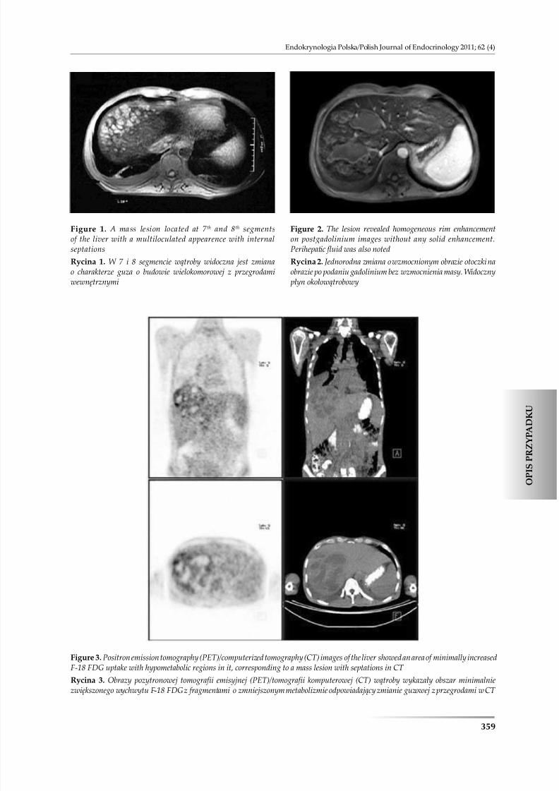
MR showed a mass lesion located at the 7th and 8th
segments of the liver, which had a multiloculated ap-
pearance with internal septations; the largest diameter
of the lesion was 6 cm. The lesion had decreased signal
intensity compared with that of liver parenchyma on
T1-weighted images and increased signal intensity on
T2-weighted images (Figure 1). The lesion revealed
homogeneous rim enhancement on postgadoliniumimages without any solid enhancement, which was
strongly suggestive of liver abscess (Figure 2). Perihe-
patic and perisplenic fluid was also noted on images.
The patient underwent a whole body Fluorine-18
fluorodeoxyglucose (F-18 FDG) positron emission to-
mography (PET)/CT (F-18 FDG-PET/CT) scan to rule
out any possible malignancy. After ten hours of fasting,
and having serum glucose 137 mg/dl, the patient was
injected with 481 MBq (13 mCi) of F-18 FDG intrave-
nously. After 50 minutes of waiting in a semi-reclined
chair, the patient was imaged using an integrated
PET/CT scanner which consisted of a full-ring HI-REZLSO PET and a 6-slice CT (Siemens Biograph 6, Chicago,
IL, USA). The CT portion of the study was done without
an iv contrast medium, just for defining anatomical
landmarks and making attenuation correction on PET
images. F-18 FDG-PET/CT images of the liver showed
an area of minimally increased F-18 FDG uptake with
hypometabolic regions in it, corresponding to a mass
lesion with septations in CT (Figure 3). Blood culture
demonstrated the presence of Yersinia enterocolitica.
Piperacillin/tazobactam (3 × 13.5 gr/day) therapy
was initiated before the planned abscess drainage. On
the fourth
day of antibiotherapy, after the patient’s feverhad subsided and blood sugar levels had become stable,
percutaneous drainage was performed. After drainage,
the patient was discharged and further treated with
tavanic 1 × 1 (500 gr/day/po) for an additional ten days
as an outpatient.
Discussion
Given the gradual rise in the number of cases with YE,
the fall in mortality rates due to development of new
antibiotics, and the difficulty of distinguishing from
metastatic lesions, diagnosing abscesses due to YE using
7/28/2019 Caso de Yersinia Eterocolitica
http://slidepdf.com/reader/full/caso-de-yersinia-eterocolitica 3/4
359
Endokrynologia Polska/Polish Journal of Endocrinology 2011; 62 (4)
O P I S P R Z Y P A D K
U
Figure 1. A mass lesion located at 7 th and 8th segmentsof the liver with a multiloculated appearence with internalseptations
Rycina 1. W 7 i 8 segmencie wątroby widoczna jest zmianao charakterze guza o budowie wielokomorowej z przegrodamiwewnętrznymi
Figure 2. The lesion revealed homogeneous rim enhancementon postgadolinium images without any solid enhancement.Perihepatic fluid was also noted
Rycina 2. Jednorodna zmiana o wzmocnionym obrazie otoczki naobrazie po podaniu gadolinium bez wzmocnienia masy. Widocznypłyn okołowątrobowy
Figure 3. Positron emission tomography (PET)/computerized tomography (CT) images of the liver showed an area of minimally increasedF-18 FDG uptake with hypometabolic regions in it, corresponding to a mass lesion with septations in CT
Rycina 3. Obrazy pozytronowej tomografii emisyjnej (PET)/tomografii komputerowej (CT) wątroby wykazały obszar minimalnie
zwiększonego wychwytu F-18 FDG z fragmentami o zmniejszonym metabolizmie odpowiadający zmianie guzowej z przegrodami w CT
7/28/2019 Caso de Yersinia Eterocolitica
http://slidepdf.com/reader/full/caso-de-yersinia-eterocolitica 4/4
360
Liver abscess due to Yersinia bacteremia Meral Mert et al.
O P I S P R Z Y P A D K
U
laboratory tests and imaging modalities has growing
importance. In a review of the literature, we found
that nearly 50 cases have been reported with hepatic
abscess secondary to YE [2–5]. One study showed that
45 cases of hepatic abscess secondary to YE have been
registered. Of the 45 reported cases, 64% had underlying haemochromatosis and 29% had diabetes mellitus. The
overall mortality was 31%. Mortality prior to 1987 was
60% (n = 20); since 1987, it has been 8% (n = 25) [2].
While investigating the multiple noncharacteristic
lesions in the liver and distinguishing between an ab-
scess and neoplastic disease is the issue, metastasis from
colon carcinoma, ovarian carcinoma, lung carcinoma,
carcinoid tumours, malignant melanoma and sarcomas
are among possible reasons. The MRI images of the
foci of fungal abscesses usually present as multiple foci
of lesions less than 1 cm in diameter. There are often
coexisting lesions in the spleen [6–12]. Our patient’sMRI images showed a lesion with irregular, heterog-
enous and gradually increasing contrast enhancement
circumferentially and peripheric oedema, rather than
early uptake and following wash-out. Imaging and
clinical findings were consistent with abscess rather
than malignancy or metastasis.
F-18 FDG PET/CT imaging has been well accepted
in oncology as an effective modality in the diagnosis,
staging, restaging and therapy response evaluation for
a variety of malignancies. F-18 FDG accumulates in
cancer cells due to an increased glucose metabolism of
the cancer cells, but it is not a tumour-specific agent. F-18FDG is also known to accumulate in inflammatory cells
infiltrating various inflammatory and infectious lesions,
such as lymphocytes, neutrophils, and macrophages,
which have elevated glucose requirements [13, 14]. FDG
uptake by infectious and inflammatory lesions may also
cause false-positive results by resembling metastases
when exploring the metastatic sites in cancer patients. It
is important to be aware of this pitfall when interpret-
ing PET/CT images in patients with malignant diseases.
The cases reported in the literature have mostly
been associated with immune deficiency and unregu-
lated DM [15–17]. In our case, HbA1c, fasting bloodsugar and postprandial blood sugar levels showed
that it was a well controlled DM case without any
findings which would raise suspicion of ketoacidosis
on admission. The patient’s serum iron, iron binding
capacity and ferritin levels were normal, ruling out
any predisposing iron overload due to diseases like
haemochromatosis. When the initial symptoms are
taken into account, the patient was supposed to have
been infected via the alimentary tract.
Mortality rates due to YE are decreasing with the
development of new antibiotics. Following parenteraland oral antibiotherapy, together with percutaneous
drainage of the abscess, the symptoms of the patient
improved and recovery in the general status of the
patient was observed. Insulin and fluid replacement
was done since the course of the blood glucose levels
of the patient was labile.
It should be kept in mind that even if it is well con-
trolled, DM is a predisposing condition for infections,
and glycaemic regulation might also be disturbed be-
cause of infection.
References1. Khanna R, Levendoglu H. Liver abscess due to Yersinia Enterocolitica:
case report and review of the literature. Digestive Diseases and Sciences1989; 34: 636–639.
2. Bergmann TK, Vinding K, Hey H. Multiple hepatic abscesses due toYersinia enterocolitica infection secondary to primary haemochromato-sis. Scandinavian Journal of Gastroenterology 2001; 36: 891–895.
3. Ismail MHA, Hodkinson HJ, Patel M et al. Multiple liver abscesses caused by Yersinia Enterocolitica. A case report. S Afr Med J 1987; 72: 291.
4. Alberti-Flor JJ, Jeffers LJ, Iskandarani M et al. Successful medical manage-ment of a Yersinia Enterocolitica liver abscess. Digestion 1984; 29: 250.
5. Hopwood AH, Riddle BW. Yersinia Enterocolitica hepatic abscesses. JK Med Assoc 1986; 84: 13.
6. Baici NC, Sirvanci M. MR imaging of infective liver lesions. Magn ResonImaging Clin N Am 2002; 10: 121–135.
7. Baici NC, Semelka RC, Noone TC et al. Pyogenic hepatic abscesses MRIfindings on T1 and T2 weighted and serial gadolinium-enhanced gradientecho images. Magn Reson Imaging 1999; 9: 285–290.
8. Mendez RJ, Schiebelar ML, Outwater EK et al. Hepatic abscesses: MRimaging findings. Radiology 1994; 190: 431–436.
9. Jeffrey RB, Tolantino CS, Chang FL et al. CT of small pyogenic hepaticabscesses: the cluster sign. AJR Am J Roentgenol 1988; 151: 487–489.
10. Danet IM, Semelka RL, Leonardou P et al. Spectrum of MRI appearancesof untreated metastases of the liver. AJR Am J Roentgenol 2003; 181:809–817.
11. Imam K, Bluemke DA. MRI imaging in the evaluation of hepatic metas-tases. Magn Reson Imaging Clin N Am 2000; 8: 741–756.
12. Lewis KH, Chezmar JL. Hepatic metastases. Magn Reson Imaging. ClinN Am 1997; 5: 19–330.
13. Weisdorf DJ, Craddock PR, Jacob HS. Glycogenolysis versus glucosetransport in human granulocytes: differential activation in phagocytosisand chemotaxis. Blood 1982; 60: 888–893.
14. Fantone JC, Ward PA. Role of oxygen-derived free radicals and metabolitesin leukocyte-dependent inflammatory reactions. Am J Pathol 1982; 107:395–418.
15. Siewko K , Szelachowska M, Popławska-Kita A et al. The C-peptide asa risk factor of development of type 1 diabetes in the first degree rela-tives of the autoimmunological diabetic patients. Endokrynol Pol 2009;60: 357–362.
16. Piatkiewicz P, Czech A, Tatoń J et al. Investigations of cellular glucosetransport and its regulation under the influence of insulin in humanperipheral blood lymphocytes. Endokrynol Pol 2010; 61: 182–187.
17. Matuszek B, Lenart-Lipińska M, Duma D et al. Evaluation of concentra-tions of FGF-21 — a new adipocytokine in type 2 diabetes. EndokrynolPol 2010; 61: 50–54.