W J C C World Journal of · sity of Rio Grande do Norte, 59012-300 Natal - Rio Grande do Norte,...
5
Appendicitis in double cecal appendix: Case report José Roberto Alves, Ícaro Godeiro de Oliveira Maranhão, Patrick Vanttinny Vieira de Oliveira José Roberto Alves, Department of Integrated Medicine, Uni- versity Hospital Onofre Lopes, Federal University of Rio Grande do Norte, 59012-300 Natal - Rio Grande do Norte, Brazil Ícaro Godeiro de Oliveira Maranhão, Patrick Vanttinny Vieira de Oliveira, University Hospital Onofre Lopes, Federal Univer- sity of Rio Grande do Norte, 59012-300 Natal - Rio Grande do Norte, Brazil Author contributions: Alves JR conducted the patient care in the emergency, surgery and photography service during the intra- operative period (Figure 1) and the postoperative medical care, was in charge of general supervision of students, writing, transla- tion, final review and article submission; Maranhão IGO, Oliveira PVV performed the literature review on the anatomical variations of the cecal appendix, and are co-authors of the manuscript. All the authors read and approved the final manuscript. Correspondence to: José Roberto Alves, PhD, Department of Integrated Medicine, University Hospital Onofre Lopes, Fed- eral University of Rio Grande do Norte. Av. Nilo Peçanha, 620 - Petrópolis, 59012-300 Natal - Rio Grande do Norte, Brazil. [email protected] Telephone: +55-84-81661115 Fax: +55-84-32153270 Received: March 9, 2014 Revised: April 16, 2014 Accepted: May 25, 2014 Published online: August 16, 2014 Abstract Double cecal appendix is a rare anatomical variation. Approximately 100 cases have been reported world- wide. It is usually diagnosed incidentally during emer- gency appendectomies due to inflammatory processes in the cecal appendix. Case presentation: male, white, 36 years old, obese, presenting with pain in the lower abdomen for 24 h followed by nausea, vomiting and mild fever. He was subjected to additional tests, with the leukogram showing leukocytosis and abdominal ul- trasonography depicting cecal appendix with thickened wall, locally associated with small quantities of liquid and intestinal loop obstruction. He underwent laparoto- my, revealing acute appendicitis. Another intestinal loop obstruction was identified next to the ileum, leading to recognizing another cecal appendix after local dissec- tion. Double appendectomy and segmental iliectomy were performed although not needed. Results of the anatomopathological examination of the surgical sam- ples showed acute inflammation in the two cecal appen- dices. So, performing a routine retroperitoneal release and a complete cecum evaluation during such surgical procedures is recommended and suggested due to the possibility of not identifying a second cecal appendix. © 2014 Baishideng Publishing Group Inc. All rights reserved. Key words: Appendix; Anatomic variation; Appendicitis; Appendectomy; General surgery Core tip: Double cecal appendix is a rare (about 100 cases reported worldwide) anatomical variation often incidentally diagnosed in the face of inflammation in the organ. The current paper presents the first case re- ported in South America. The case is extremely impor- tant for the study of this possible anatomical variation since the lack of a diagnosis in a second cecal appendix can cause further complications for the patient and the physician. Moreover, it is associated with the presence of other anatomical variations, such as intestinal, geni- tourinary and bone. Such variations will be investigated in cases of the aforementioned diagnosis. Alves JR, Maranhão IGO, Oliveira PVV. Appendicitis in double ce- cal appendix: Case report. World J Clin Cases 2014; 2(8): 391-394 Available from: URL: http://www.wjgnet.com/2307-8960/full/v2/ i8/391.htm DOI: http://dx.doi.org/10.12998/wjcc.v2.i8.391 INTRODUCTION Double cecal appendix is a rare anatomical variation, found in 0.004% [1] to 0.009% [2] of performed appendec- tomies. Approximately 100 cases of double cecal appen- dix [3-5] have been described worldwide so far, with no case reports in South America [2,3,6-37] . CASE REPORT A male, white, 36 years old, slightly obese [body mass CASE REPORT Submit a Manuscript: http://www.wjgnet.com/esps/ Help Desk: http://www.wjgnet.com/esps/helpdesk.aspx DOI: 10.12998/wjcc.v2.i8.391 World J Clin Cases 2014 August 16; 2(8): 391-394 ISSN 2307-8960 (online) © 2014 Baishideng Publishing Group Inc. All rights reserved. World Journal of Clinical Cases W JC C August 16, 2014|Volume 2|Issue 8| WJCC|www.wjgnet.com 391
Transcript of W J C C World Journal of · sity of Rio Grande do Norte, 59012-300 Natal - Rio Grande do Norte,...
Appendicitis in double cecal appendix: Case report
José Roberto Alves, Ícaro Godeiro de Oliveira Maranhão, Patrick Vanttinny Vieira de Oliveira
José Roberto Alves, Department of Integrated Medicine, Uni-versity Hospital Onofre Lopes, Federal University of Rio Grande do Norte, 59012-300 Natal - Rio Grande do Norte, Brazil Ícaro Godeiro de Oliveira Maranhão, Patrick Vanttinny Vieira de Oliveira, University Hospital Onofre Lopes, Federal Univer-sity of Rio Grande do Norte, 59012-300 Natal - Rio Grande do Norte, BrazilAuthor contributions: Alves JR conducted the patient care in the emergency, surgery and photography service during the intra-operative period (Figure 1) and the postoperative medical care, was in charge of general supervision of students, writing, transla-tion, final review and article submission; Maranhão IGO, Oliveira PVV performed the literature review on the anatomical variations of the cecal appendix, and are co-authors of the manuscript. All the authors read and approved the final manuscript.Correspondence to: José Roberto Alves, PhD, Department of Integrated Medicine, University Hospital Onofre Lopes, Fed-eral University of Rio Grande do Norte. Av. Nilo Peçanha, 620 - Petrópolis, 59012-300 Natal - Rio Grande do Norte, Brazil. [email protected]: +55-84-81661115 Fax: +55-84-32153270Received: March 9, 2014 Revised: April 16, 2014Accepted: May 25, 2014Published online: August 16, 2014
AbstractDouble cecal appendix is a rare anatomical variation. Approximately 100 cases have been reported world-wide. It is usually diagnosed incidentally during emer-gency appendectomies due to inflammatory processes in the cecal appendix. Case presentation: male, white, 36 years old, obese, presenting with pain in the lower abdomen for 24 h followed by nausea, vomiting and mild fever. He was subjected to additional tests, with the leukogram showing leukocytosis and abdominal ul-trasonography depicting cecal appendix with thickened wall, locally associated with small quantities of liquid and intestinal loop obstruction. He underwent laparoto-my, revealing acute appendicitis. Another intestinal loop obstruction was identified next to the ileum, leading to recognizing another cecal appendix after local dissec-tion. Double appendectomy and segmental iliectomy were performed although not needed. Results of the
anatomopathological examination of the surgical sam-ples showed acute inflammation in the two cecal appen-dices. So, performing a routine retroperitoneal release and a complete cecum evaluation during such surgical procedures is recommended and suggested due to the possibility of not identifying a second cecal appendix.
© 2014 Baishideng Publishing Group Inc. All rights reserved.
Key words: Appendix; Anatomic variation; Appendicitis; Appendectomy; General surgery
Core tip: Double cecal appendix is a rare (about 100 cases reported worldwide) anatomical variation often incidentally diagnosed in the face of inflammation in the organ. The current paper presents the first case re-ported in South America. The case is extremely impor-tant for the study of this possible anatomical variation since the lack of a diagnosis in a second cecal appendix can cause further complications for the patient and the physician. Moreover, it is associated with the presence of other anatomical variations, such as intestinal, geni-tourinary and bone. Such variations will be investigated in cases of the aforementioned diagnosis.
Alves JR, Maranhão IGO, Oliveira PVV. Appendicitis in double ce-cal appendix: Case report. World J Clin Cases 2014; 2(8): 391-394 Available from: URL: http://www.wjgnet.com/2307-8960/full/v2/i8/391.htm DOI: http://dx.doi.org/10.12998/wjcc.v2.i8.391
INTRODUCTIONDouble cecal appendix is a rare anatomical variation, found in 0.004%[1] to 0.009%[2] of performed appendec-tomies. Approximately 100 cases of double cecal appen-dix[3-5] have been described worldwide so far, with no case reports in South America[2,3,6-37].
CASE REPORTA male, white, 36 years old, slightly obese [body mass
CASE REPORT
Submit a Manuscript: http://www.wjgnet.com/esps/Help Desk: http://www.wjgnet.com/esps/helpdesk.aspxDOI: 10.12998/wjcc.v2.i8.391
World J Clin Cases 2014 August 16; 2(8): 391-394 ISSN 2307-8960 (online)
© 2014 Baishideng Publishing Group Inc. All rights reserved.
World Journal ofClinical CasesW J C C
August 16, 2014|Volume 2|Issue 8|WJCC|www.wjgnet.com 391
index (BMI) = 31.1 kg/m2], presented with abdominal pain in the lower abdomen for 24 h, followed by nausea, vomiting and mild fever (axillary temperature = 37.9 ℃). He was subjected to blood tests that only showed leukocytosis without left shift. In addition, abdominal ultrasonography depicted cecal appendix with thickened wall, locally associated with small quantities of intra-abdominal fluid and local obstruction of intestinal loops.
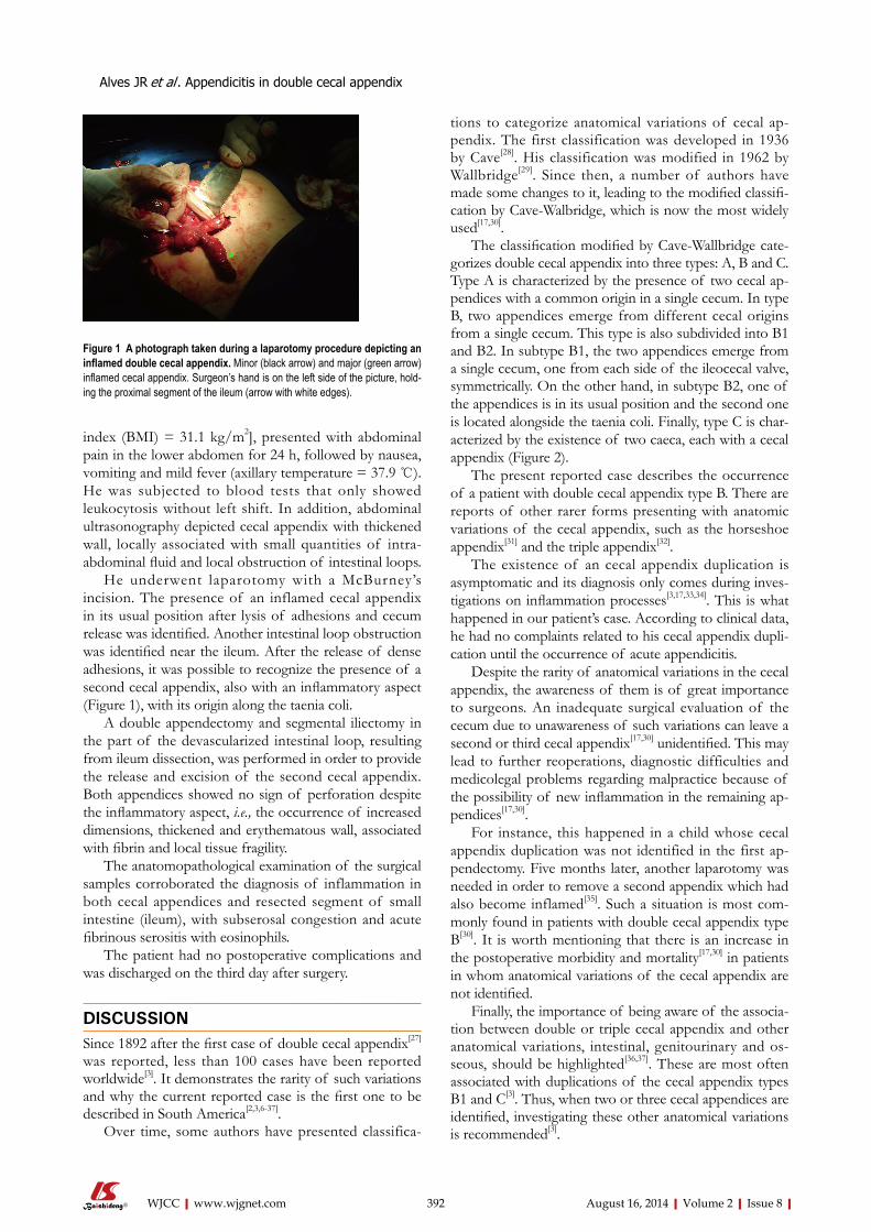
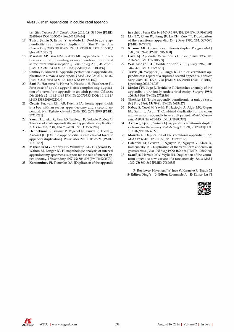
He underwent laparotomy with a McBurney’s incision. The presence of an inflamed cecal appendix in its usual position after lysis of adhesions and cecum release was identified. Another intestinal loop obstruction was identified near the ileum. After the release of dense adhesions, it was possible to recognize the presence of a second cecal appendix, also with an inflammatory aspect (Figure 1), with its origin along the taenia coli.
A double appendectomy and segmental iliectomy in the part of the devascularized intestinal loop, resulting from ileum dissection, was performed in order to provide the release and excision of the second cecal appendix. Both appendices showed no sign of perforation despite the inflammatory aspect, i.e., the occurrence of increased dimensions, thickened and erythematous wall, associated with fibrin and local tissue fragility.
The anatomopathological examination of the surgical samples corroborated the diagnosis of inflammation in both cecal appendices and resected segment of small intestine (ileum), with subserosal congestion and acute fibrinous serositis with eosinophils.
The patient had no postoperative complications and was discharged on the third day after surgery.
DISCUSSIONSince 1892 after the first case of double cecal appendix[27] was reported, less than 100 cases have been reported worldwide[3]. It demonstrates the rarity of such variations and why the current reported case is the first one to be described in South America[2,3,6-37].
Over time, some authors have presented classifica-
tions to categorize anatomical variations of cecal ap-pendix. The first classification was developed in 1936 by Cave[28]. His classification was modified in 1962 by Wallbridge[29]. Since then, a number of authors have made some changes to it, leading to the modified classifi-cation by Cave-Walbridge, which is now the most widely used[17,30].
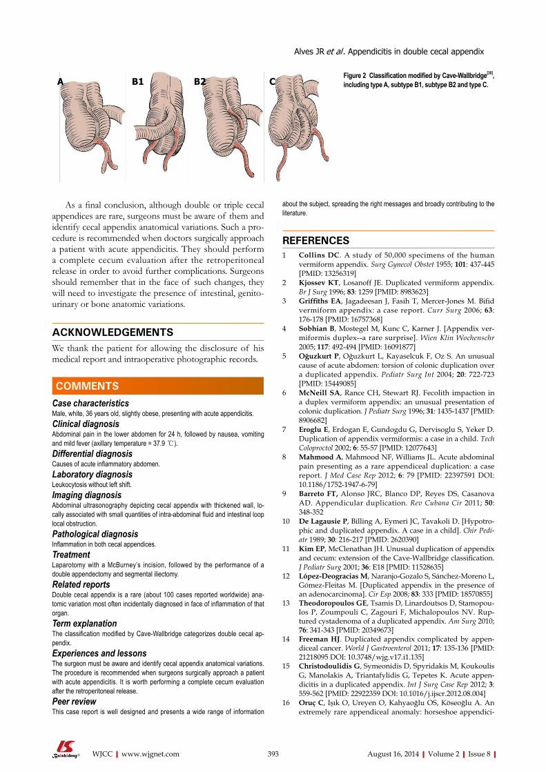
The classification modified by Cave-Wallbridge cate-gorizes double cecal appendix into three types: A, B and C. Type A is characterized by the presence of two cecal ap-pendices with a common origin in a single cecum. In type B, two appendices emerge from different cecal origins from a single cecum. This type is also subdivided into B1 and B2. In subtype B1, the two appendices emerge from a single cecum, one from each side of the ileocecal valve, symmetrically. On the other hand, in subtype B2, one of the appendices is in its usual position and the second one is located alongside the taenia coli. Finally, type C is char-acterized by the existence of two caeca, each with a cecal appendix (Figure 2).
The present reported case describes the occurrence of a patient with double cecal appendix type B. There are reports of other rarer forms presenting with anatomic variations of the cecal appendix, such as the horseshoe appendix[31] and the triple appendix[32].
The existence of an cecal appendix duplication is asymptomatic and its diagnosis only comes during inves-tigations on inflammation processes[3,17,33,34]. This is what happened in our patient’s case. According to clinical data, he had no complaints related to his cecal appendix dupli-cation until the occurrence of acute appendicitis.
Despite the rarity of anatomical variations in the cecal appendix, the awareness of them is of great importance to surgeons. An inadequate surgical evaluation of the cecum due to unawareness of such variations can leave a second or third cecal appendix[17,30] unidentified. This may lead to further reoperations, diagnostic difficulties and medicolegal problems regarding malpractice because of the possibility of new inflammation in the remaining ap-pendices[17,30].
For instance, this happened in a child whose cecal appendix duplication was not identified in the first ap-pendectomy. Five months later, another laparotomy was needed in order to remove a second appendix which had also become inflamed[35]. Such a situation is most com-monly found in patients with double cecal appendix type B[30]. It is worth mentioning that there is an increase in the postoperative morbidity and mortality[17,30] in patients in whom anatomical variations of the cecal appendix are not identified.
Finally, the importance of being aware of the associa-tion between double or triple cecal appendix and other anatomical variations, intestinal, genitourinary and os-seous, should be highlighted[36,37]. These are most often associated with duplications of the cecal appendix types B1 and C[3]. Thus, when two or three cecal appendices are identified, investigating these other anatomical variations is recommended[3].
Alves JR et al . Appendicitis in double cecal appendix
August 16, 2014|Volume 2|Issue 8|WJCC|www.wjgnet.com 392
Figure 1 A photograph taken during a laparotomy procedure depicting an inflamed double cecal appendix. Minor (black arrow) and major (green arrow) inflamed cecal appendix. Surgeon’s hand is on the left side of the picture, hold-ing the proximal segment of the ileum (arrow with white edges).
As a final conclusion, although double or triple cecal appendices are rare, surgeons must be aware of them and identify cecal appendix anatomical variations. Such a pro-cedure is recommended when doctors surgically approach a patient with acute appendicitis. They should perform a complete cecum evaluation after the retroperitoneal release in order to avoid further complications. Surgeons should remember that in the face of such changes, they will need to investigate the presence of intestinal, genito-urinary or bone anatomic variations.
ACKNOWLEDGEMENTSWe thank the patient for allowing the disclosure of his medical report and intraoperative photographic records.
COMMENTSCase characteristicsMale, white, 36 years old, slightly obese, presenting with acute appendicitis.Clinical diagnosisAbdominal pain in the lower abdomen for 24 h, followed by nausea, vomiting and mild fever (axillary temperature = 37.9 ℃).Differential diagnosisCauses of acute inflammatory abdomen.Laboratory diagnosisLeukocytosis without left shift. Imaging diagnosisAbdominal ultrasonography depicting cecal appendix with thickened wall, lo-cally associated with small quantities of intra-abdominal fluid and intestinal loop local obstruction.Pathological diagnosisInflammation in both cecal appendices.TreatmentLaparotomy with a McBurney’s incision, followed by the performance of a double appendectomy and segmental iliectomy.Related reportsDouble cecal appendix is a rare (about 100 cases reported worldwide) ana-tomic variation most often incidentally diagnosed in face of inflammation of that organ. Term explanation The classification modified by Cave-Wallbridge categorizes double cecal ap-pendix.Experiences and lessonsThe surgeon must be aware and identify cecal appendix anatomical variations. The procedure is recommended when surgeons surgically approach a patient with acute appendicitis. It is worth performing a complete cecum evaluation after the retroperitoneal release.Peer reviewThis case report is well designed and presents a wide range of information
about the subject, spreading the right messages and broadly contributing to the literature.
REFERENCES 1 Collins DC. A study of 50,000 specimens of the human
vermiform appendix. Surg Gynecol Obstet 1955; 101: 437-445 [PMID: 13256319]
2 Kjossev KT, Losanoff JE. Duplicated vermiform appendix. Br J Surg 1996; 83: 1259 [PMID: 8983623]
3 Griffiths EA, Jagadeesan J, Fasih T, Mercer-Jones M. Bifid vermiform appendix: a case report. Curr Surg 2006; 63: 176-178 [PMID: 16757368]
4 Sobhian B, Mostegel M, Kunc C, Karner J. [Appendix ver-miformis duplex--a rare surprise]. Wien Klin Wochenschr 2005; 117: 492-494 [PMID: 16091877]
5 Oğuzkurt P, Oğuzkurt L, Kayaselcuk F, Oz S. An unusual cause of acute abdomen: torsion of colonic duplication over a duplicated appendix. Pediatr Surg Int 2004; 20: 722-723 [PMID: 15449085]
6 McNeill SA, Rance CH, Stewart RJ. Fecolith impaction in a duplex vermiform appendix: an unusual presentation of colonic duplication. J Pediatr Surg 1996; 31: 1435-1437 [PMID: 8906682]
7 Eroglu E, Erdogan E, Gundogdu G, Dervisoglu S, Yeker D. Duplication of appendix vermiformis: a case in a child. Tech Coloproctol 2002; 6: 55-57 [PMID: 12077643]
8 Mahmood A, Mahmood NF, Williams JL. Acute abdominal pain presenting as a rare appendiceal duplication: a case report. J Med Case Rep 2012; 6: 79 [PMID: 22397591 DOI: 10.1186/1752-1947-6-79]
9 Barreto FT, Alonso JRC, Blanco DP, Reyes DS, Casanova AD. Appendicular duplication. Rev Cubana Cir 2011; 50: 348-352
10 De Lagausie P, Billing A, Eymeri JC, Tavakoli D. [Hypotro-phic and duplicated appendix. A case in a child]. Chir Pedi-atr 1989; 30: 216-217 [PMID: 2620390]
11 Kim EP, McClenathan JH. Unusual duplication of appendix and cecum: extension of the Cave-Wallbridge classification. J Pediatr Surg 2001; 36: E18 [PMID: 11528635]
12 López-Deogracias M, Naranjo-Gozalo S, Sánchez-Moreno L, Gómez-Fleitas M. [Duplicated appendix in the presence of an adenocarcinoma]. Cir Esp 2008; 83: 333 [PMID: 18570855]
13 Theodoropoulos GE, Tsamis D, Linardoutsos D, Stamopou-los P, Zoumpouli C, Zagouri F, Michalopoulos NV. Rup-tured cystadenoma of a duplicated appendix. Am Surg 2010; 76: 341-343 [PMID: 20349673]
14 Freeman HJ. Duplicated appendix complicated by appen-diceal cancer. World J Gastroenterol 2011; 17: 135-136 [PMID: 21218095 DOI: 10.3748/wjg.v17.i1.135]
15 Christodoulidis G, Symeonidis D, Spyridakis M, Koukoulis G, Manolakis A, Triantafylidis G, Tepetes K. Acute appen-dicitis in a duplicated appendix. Int J Surg Case Rep 2012; 3: 559-562 [PMID: 22922359 DOI: 10.1016/j.ijscr.2012.08.004]
16 Oruç C, Işık O, Ureyen O, Kahyaoğlu OS, Köseoğlu A. An extremely rare appendiceal anomaly: horseshoe appendici-
August 16, 2014|Volume 2|Issue 8|WJCC|www.wjgnet.com 393
Figure 2 Classification modified by Cave-Wallbridge[30], including type A, subtype B1, subtype B2 and type C.A B1 B2 C
COMMENTS
Alves JR et al . Appendicitis in double cecal appendix
tis. Ulus Travma Acil Cerrahi Derg 2013; 19: 385-386 [PMID: 23884686 DOI: 10.5505/tjtes.2013.67424]
17 Tutcu Şahin S, Erhan Y, Aydede H. Double acute ap-pendicitis in appendical duplication. Ulus Travma Acil Cerrahi Derg 2013; 19: 83-85 [PMID: 23588988 DOI: 10.5505/tjtes.2013.80557]
18 Marshall AP, Issar NM, Blakely ML. Appendiceal duplica-tion in children presenting as an appendiceal tumor and as recurrent intussusception. J Pediatr Surg 2013; 48: e9-e12 [PMID: 23583164 DOI: 10.1016/j.jpedsurg.2013.01.036]
19 Canbay E, Akman E. Appendix perforation in appendix du-plication in a man: a case report. J Med Case Rep 2011; 5: 162 [PMID: 21513538 DOI: 10.1186/1752-1947-5-162]
20 Sani R, Harouna Y, Hama Y, Nouhou H, Faucheron JL. First case of double appendicitis complicating duplica-tion of a vermiform appendix in an adult patient. Colorectal Dis 2010; 12: 1162-1163 [PMID: 20070333 DOI: 10.1111/j.1463-1318.2010.02200.x]
21 Geurts BA, van Rijn AB, Koelma IA. [Acute appendicitis in a boy with an earlier appendectomy and a second ap-pendix]. Ned Tijdschr Geneeskd 2006; 150: 2876-2879 [PMID: 17319221]
22 Yanar H, Ertekin C, Unal ES, Taviloglu K, Guloglu R, Mete O. The case of acute appendicitis and appendiceal duplication. Acta Chir Belg 2004; 104: 736-738 [PMID: 15663287]
23 Hennekinne S, Pessaux P, Regenet N, Fauvet R, Tuech JJ, Arnaud JP. [Double appendicitis: a rare clinical form in appendix duplication]. Presse Med 2001; 30: 23-24 [PMID: 11210582]
24 Mazziotti MV, Marley EF, Winthrop AL, Fitzgerald PG, Walton M, Langer JC. Histopathologic analysis of interval appendectomy specimens: support for the role of interval ap-pendectomy. J Pediatr Surg 1997; 32: 806-809 [PMID: 9200074]
25 Konstantinov PI, Titarenko IaA. [Duplication of the appendix
in a child]. Vestn Khir Im I I Grek 1997; 156: 109 [PMID: 9163180]26 Lin BC, Chen RJ, Fang JF, Lo TH, Kuo TT. Duplication
of the vermiform appendix. Eur J Surg 1996; 162: 589-591 [PMID: 8874171]
27 Khanna AK. Appendix vermiformis duplex. Postgrad Med J 1983; 59: 69-70 [PMID: 6866880]
28 Cave AJ. Appendix Vermiformis Duplex. J Anat 1936; 70: 283-292 [PMID: 17104589]
29 Wallbridge PH. Double appendix. Br J Surg 1962; 50: 346-347 [PMID: 13998581]
30 Travis JR, Weppner JL, Paugh JC. Duplex vermiform ap-pendix: case report of a ruptured second appendix. J Pediatr Surg 2008; 43: 1726-1728 [PMID: 18779015 DOI: 10.1016/j.jpedsurg.2008.04.023]
31 Mesko TW, Lugo R, Breitholtz T. Horseshoe anomaly of the appendix: a previously undescribed entity. Surgery 1989; 106: 563-566 [PMID: 2772830]
32 Tinckler LF. Triple appendix vermiformis--a unique case. Br J Surg 1968; 55: 79-81 [PMID: 5635427]
33 Kabay S, Yucel M, Yaylak F, Hacioglu A, Algin MC, Olgun EG, Sahin L, Aydin T. Combined duplication of the colon and vermiform appendix in an adult patient. World J Gastro-enterol 2008; 14: 641-643 [PMID: 18203303]
34 Akhtar J, Ejaz T, Guiney EJ. Appendix vermiformis duplex - a lesson for the unwary. Pediatr Surg Int 1994; 9: 429-30 [DOI: 10.1007/BF01686027]
35 Maizels G. Duplication of the vermiform appendix. S Afr Med J 1966; 40: 1123-1125 [PMID: 5957812]
36 Gilchrist BF, Scriven R, Nguyen M, Nguyen V, Klotz D, Ramenofsky ML. Duplication of the vermiform appendix in gastroschisis. J Am Coll Surg 1999; 189: 426 [PMID: 10509468]
37 Scarff JE, Harrold MW, Wylie JH. Duplication of the vermi-form appendix: new variant of a rare anomaly. South Med J 1982; 75: 860-862 [PMID: 7089658]
P- Reviewer: Haveman JW, Ince V, Karateke F, Tsuda M S- Editor: Ding Y L- Editor: Roemmele A E- Editor: Lu YJ
August 16, 2014|Volume 2|Issue 8|WJCC|www.wjgnet.com 394
Alves JR et al . Appendicitis in double cecal appendix

© 2014 Baishideng Publishing Group Inc. All rights reserved.
Published by Baishideng Publishing Group Inc8226 Regency Drive, Pleasanton, CA 94588, USA
Telephone: +1-925-223-8242Fax: +1-925-223-8243
E-mail: [email protected] Desk: http://www.wjgnet.com/esps/helpdesk.aspx
http://www.wjgnet.com