Research Article Neopterin and Beta-2 Microglobulin...
9
Research Article Neopterin and Beta-2 Microglobulin Relations to Immunity and Inflammatory Status in Nonischemic Dilated Cardiomyopathy Patients Celina Wojciechowska, 1 Jan Wodniecki, 1 Romuald Wojnicz, 2 Ewa Romuk, 3 Wojciech JacheT, 1 Andrzej Tomasik, 1 BronisBawa Skrzep-Poloczek, 3 Beata Spinczyk, 2 and Ewa Nowalany-Kozielska 1 1 Second Department of Cardiology, School of Medicine with the Division of Dentistry, Medical University of Silesia, Ulica M.C. Skłodowskiej 10, 41-800 Zabrze, Poland 2 Department of Histology and Embryology, School of Medicine with the Division of Dentistry, Medical University of Silesia, 41-800 Zabrze, Poland 3 Department of Biochemistry, School of Medicine with the Division of Dentistry, Medical University of Silesia, 41-800 Zabrze, Poland Correspondence should be addressed to Celina Wojciechowska; [email protected] Received 6 May 2014; Accepted 29 July 2014; Published 18 August 2014 Academic Editor: Jan G. C. van Amsterdam Copyright © 2014 Celina Wojciechowska et al. is is an open access article distributed under the Creative Commons Attribution License, which permits unrestricted use, distribution, and reproduction in any medium, provided the original work is properly cited. Background. e aim of the study was to assess the relationships among serum neopterin (NPT), 2-microglobulin (2-M) levels, clinical status, and endomyocardial biopsy results of dilated cardiomyopathy patients (DCM). Methods. Serum NPT and -2 M were determined in 172 nonischaemic DCM patients who underwent right ventricular endomyocardial biopsy and 30 healthy subjects (ELISA test). e cryostat biopsy specimens were assessed using histology, immunohistology, and immunochemistry methods (HLA ABC, HLA DR expression, CD3 + lymphocytes, and macrophages counts). Results. e strong increase of HLA ABC or HLA DR expression was detected in 27.2% patients—group A—being low in 72.8% patients—group B. Neopterin level was increased in patients in group A compared to healthy controls 8.11 (4.50–12.57) versus 4.99 (2.66–8.28) nmol/L ( < 0.05). -2 microglobulin level was higher in DCM groups A (2.60 (1.71–3.58)) and B (2.52 (1.51–3.72)) than in the control group 1.75 (1.28–1.96) mg/L, < 0.001. Neopterin correlated positively with the number of macrophages in biopsy specimens ( < 0.05) acute phase proteins: C-reactive proteins ( < 0.05); fibrinogen ( < 0.01); and NYHA functional class ( < 0.05) and negatively with leſt ventricular ejection fraction ( < 0.05). Conclusions. Neopterin but not -2 microglobulin concentration reflected immune response in biopsy specimens. Neopterin correlated with acute phase proteins and stage of heart failure and may indicate a general immune and inflammatory activation in heart failure. 1. Introduction Inflammatory dilated cardiomyopathy results from myo- carditis induced by infectious and noninfectious triggers. Most of the evidence which suggests that myocarditis and inflammatory cardiomyopathy are autoimmune diseases come from animal models [1, 2]. e macrophages and T lymphocytes were found to be directly engaged in cell- mediated immune mechanism in experimental autoimmune myocarditis (EAM) [3–5]. Endomyocardial biopsy (EMB) is considered the diagnostic “gold standard” for myocarditis and both cells mentioned above might be assessed in it [6, 7]. To increase the diagnostic sensitivity EBM immuno- histochemistry may be used for the identification of the Hindawi Publishing Corporation Mediators of Inflammation Volume 2014, Article ID 585067, 8 pages http://dx.doi.org/10.1155/2014/585067
Transcript of Research Article Neopterin and Beta-2 Microglobulin...
Research ArticleNeopterin and Beta-2 MicroglobulinRelations to Immunity and Inflammatory Status in NonischemicDilated Cardiomyopathy Patients
Celina Wojciechowska,1 Jan Wodniecki,1 Romuald Wojnicz,2
Ewa Romuk,3 Wojciech JacheT,1 Andrzej Tomasik,1 BronisBawa Skrzep-Poloczek,3
Beata Spinczyk,2 and Ewa Nowalany-Kozielska1
1 Second Department of Cardiology, School of Medicine with the Division of Dentistry, Medical University of Silesia,Ulica M.C. Skłodowskiej 10, 41-800 Zabrze, Poland
2Department of Histology and Embryology, School of Medicine with the Division of Dentistry, Medical University of Silesia,41-800 Zabrze, Poland
3Department of Biochemistry, School of Medicine with the Division of Dentistry, Medical University of Silesia,41-800 Zabrze, Poland
Correspondence should be addressed to Celina Wojciechowska; [email protected]
Received 6 May 2014; Accepted 29 July 2014; Published 18 August 2014
Academic Editor: Jan G. C. van Amsterdam
Copyright © 2014 Celina Wojciechowska et al. This is an open access article distributed under the Creative Commons AttributionLicense, which permits unrestricted use, distribution, and reproduction in any medium, provided the original work is properlycited.
Background. The aim of the study was to assess the relationships among serum neopterin (NPT), 𝛽2-microglobulin (𝛽2-M) levels,clinical status, and endomyocardial biopsy results of dilated cardiomyopathy patients (DCM).Methods. SerumNPT and𝛽-2Mweredetermined in 172 nonischaemic DCM patients who underwent right ventricular endomyocardial biopsy and 30 healthy subjects(ELISA test). The cryostat biopsy specimens were assessed using histology, immunohistology, and immunochemistry methods(HLAABC, HLADR expression, CD3 + lymphocytes, andmacrophages counts). Results.The strong increase of HLAABC or HLADR expression was detected in 27.2% patients—group A—being low in 72.8% patients—group B. Neopterin level was increased inpatients in group A compared to healthy controls 8.11 (4.50–12.57) versus 4.99 (2.66–8.28) nmol/L (𝑃 < 0.05). 𝛽-2 microglobulinlevel was higher in DCM groups A (2.60 (1.71–3.58)) and B (2.52 (1.51–3.72)) than in the control group 1.75 (1.28–1.96) mg/L,𝑃 < 0.001. Neopterin correlated positively with the number of macrophages in biopsy specimens (𝑃 < 0.05) acute phase proteins:C-reactive proteins (𝑃 < 0.05); fibrinogen (𝑃 < 0.01); and NYHA functional class (𝑃 < 0.05) and negatively with left ventricularejection fraction (𝑃 < 0.05). Conclusions. Neopterin but not 𝛽-2 microglobulin concentration reflected immune response in biopsyspecimens. Neopterin correlated with acute phase proteins and stage of heart failure and may indicate a general immune andinflammatory activation in heart failure.
1. Introduction
Inflammatory dilated cardiomyopathy results from myo-carditis induced by infectious and noninfectious triggers.Most of the evidence which suggests that myocarditisand inflammatory cardiomyopathy are autoimmune diseasescome from animal models [1, 2]. The macrophages and
T lymphocytes were found to be directly engaged in cell-mediated immune mechanism in experimental autoimmunemyocarditis (EAM) [3–5]. Endomyocardial biopsy (EMB) isconsidered the diagnostic “gold standard” for myocarditisand both cells mentioned above might be assessed in it[6, 7]. To increase the diagnostic sensitivity EBM immuno-histochemistry may be used for the identification of the
Hindawi Publishing CorporationMediators of InflammationVolume 2014, Article ID 585067, 8 pageshttp://dx.doi.org/10.1155/2014/585067

2 Mediators of Inflammation
inflammatory infiltration and for the detection of HLA-DRupregulation [8]. Progression from myocarditis to dilatedcardiomyopathy (DCM) seems to occur predominantly inpatients with histologically confirmed persistent (chronic)inflammation [9]. Besides cell-mediated myocytes damage,various cytokines including interleukin-1𝛽 (IL-1𝛽), inter-leukin IL-2 (IL-2), interleukin IL-6 (IL-6), interferon-𝛾 (IFN-𝛾), and tumor necrosis factor-𝛼 (TNF-𝛼) have multiplebiological activities and modulate immune response [10, 11].
Neopterin (NPT) arising from guanosine triphosphate(GTP) is mainly secreted by activated macrophages. IFN-𝛾and TNF-𝛼were found to be themost frequent inducers of itssynthesis [12]. Elevated NTP level was observed in infectiousdiseases, allograft rejection, disorders with potential autoim-mune pathogenesis, and atherosclerosis [13–15]. Despite NPTbeing produced locally, serum level of NPT can reflect theextent and activity of disease. 𝛽-2 microglobulin (𝛽-2M) isa light chain of HLA class I and its elevated serum concen-tration indicates cell surface expression of HLA. Increasedlevel of 𝛽-2M was reported in viral infections and in chronicinflammatory and lymphoproliferative disorders [16, 17].
Accordingly, the aim of this studywas to assess the useful-ness of NPT and 𝛽-2M serum concentration as the potentialbiomarkers that may reflect upregulation of immune systemin DCM patients.
2. Study Group and Methods
2.1. Patients. We recruited 172 patients hospitalized due todilated cardiomyopathy and diagnosed according to theWHO criteria [18] who underwent endomyocardial biopsy.Chronic heart failure was recognized if dyspnea or fatigue atrest or on exertion in association with an ejection fraction(EF) ≤45% was present. All patients were clinically stable andreceived optimal conventional heart failure therapy includingACE inhibitors, 𝛽-blockers, digitalis, and diuretics for atleast 6 months. Patients were excluded from the study ifthey had at least one the following: any changes in coronaryvessels as assessed by coronary angiography, valvular diseaseexcept relativemitral regurgitation, connective tissue disease,endocrine disorders, renal insufficiency (estimated glomeru-lar filtration rate usingModification of Diet in Renal Disease-MDRD formula <60mL/min/1.73m2), infectious disease,malignancy, moderate or severe hypertension, and alcoholabuse.
2.2. Clinical Assessements. Noninvasive clinical assessmentincluded the following: physical examination, ECG, andechocardiography. Echocardiographic images were acquiredin standard views as recommended by the American Soci-ety of Echocardiography Committee. Left ventricular end-diastolic volume (EDV) and end-systolic volume (ESV) wereobtained from the apical 4- and 2-chamber views bymodifiedSimpson’s method. Left ventricular ejection fraction (EF) wascalculated in a standard manner as follows: EDV − ESV ×100/EDV to assess ventricular systolic function. The NewYork Heart Association (NYHA) classification was used to
assess functional capacity. To rule out coronary artery diseaseall patients underwent coronary angiography.
2.3. Biochemical Methods. Blood samples for laboratory as-sessments were obtained from the patients at time of thebiopsy. Serum was separated by centrifugation at 1500 g for10 minutes and was frozen at −70∘C and protected fromlight. Neopterin and 𝛽-2 microglobulin concentrations weredetermined in serum by ELISA method using commerciallyavailable kit manufactured by IBL, Hamburg (Germany).The limit of detection for neopterin was 0.7 nmol/L. Theintra- and interassay coefficient of variation were <5.5% and<10.3%. Serum for 𝛽-2 microglobulin assay was diluted ina ratio of 1 : 50. The minimum detectable concentration of𝛽-2 microglobulin was 0.1mg/L. The intra- and interassaycoefficient of variation were <11.6% and <15.5%. Additionally,we also determined serum creatinine, fibrinogen, and hs-CRP concentrations using routine techniques.
2.4. Endomyocardial Biopsy. At endomyocardial biopsy pro-cedure a minimum of four specimens was obtained fromthe right side of the ventricular septum. Specimens wereroutinely distributed for histological, immunohistological,and immunohistochemical studies. Histopathological exam-ination was performed by light microscopy according to Dal-las classification [6] and immunohistologically as describedpreviously [19]. For immunohistochemistry, frozen sectionswere incubated with murine monoclonal antihuman anti-bodies: anti-HLA class II (DR antigens), Alpha chain (cloneTAL.1B5), anti-HLA class I (ABC antigens, clone W6/32),anti-CD3 for T lymphocytes (Clone T3-4B5), and anti-macrophages (clone EBM11). The bound primary antibodywas detected using the EnVision method (DAKO EnVisionKit/DAKO A/S). Each specimen was evaluated qualitativelyand quantitatively for CD 3 lymphocytes and macrophagescount and semiquantitatively for HLA expression on thepreviously defined and presented below immunoreactivityscoring system IR:
0 lack of focal staining on the endothelial and inter-stitial cells,1+ focal staining on the endothelial and other intersti-tial cells,2+ multifocal staining restricted to interstitial cells,3+ diffuse endothelial and concomitant focal cardiacmyocyte staining,4+ diffuse endothelial and myocyte staining [8].
IR ≥ 3+ was assessed as positive for major histocompat-ibility complex. For the purpose of this study patients weredivided into two groups. Group A is patients with positiveIR ≥ 3+ for HLA ABC or HLA DR, considered as havingstrong immune mediated inflammation. Group B is patientswith weak immunoreactivity, staining of HLA complex IRof 0–2+. Group C is control group for neopterin and 𝛽-2microglobulin assay comprised of healthy volunteers fromhospital staff (8 male and 22 female, aged 39.03± 9.86 years).Approval of Local Ethic Committee was obtained.
Mediators of Inflammation 3
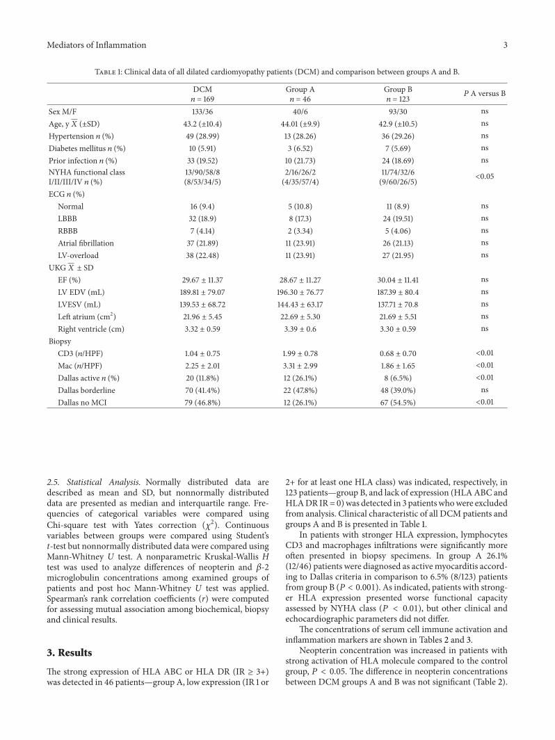
Table 1: Clinical data of all dilated cardiomyopathy patients (DCM) and comparison between groups A and B.
DCM𝑛 = 169
Group A𝑛 = 46
Group B𝑛 = 123 𝑃 A versus B
Sex M/F 133/36 40/6 93/30 nsAge, y𝑋 (±SD) 43.2 (±10.4) 44.01 (±9.9) 42.9 (±10.5) nsHypertension 𝑛 (%) 49 (28.99) 13 (28.26) 36 (29.26) nsDiabetes mellitus 𝑛 (%) 10 (5.91) 3 (6.52) 7 (5.69) nsPrior infection 𝑛 (%) 33 (19.52) 10 (21.73) 24 (18.69) nsNYHA functional classI/II/III/IV 𝑛 (%)
13/90/58/8(8/53/34/5)
2/16/26/2(4/35/57/4)
11/74/32/6(9/60/26/5) <0.05
ECG 𝑛 (%)Normal 16 (9.4) 5 (10.8) 11 (8.9) nsLBBB 32 (18.9) 8 (17.3) 24 (19.51) nsRBBB 7 (4.14) 2 (3.34) 5 (4.06) nsAtrial fibrillation 37 (21.89) 11 (23.91) 26 (21.13) nsLV-overload 38 (22.48) 11 (23.91) 27 (21.95) ns
UKG𝑋 ± SDEF (%) 29.67 ± 11.37 28.67 ± 11.27 30.04 ± 11.41 nsLV EDV (mL) 189.81 ± 79.07 196.30 ± 76.77 187.39 ± 80.4 nsLVESV (mL) 139.53 ± 68.72 144.43 ± 63.17 137.71 ± 70.8 nsLeft atrium (cm2) 21.96 ± 5.45 22.69 ± 5.30 21.69 ± 5.51 nsRight ventricle (cm) 3.32 ± 0.59 3.39 ± 0.6 3.30 ± 0.59 ns
BiopsyCD3 (𝑛/HPF) 1.04 ± 0.75 1.99 ± 0.78 0.68 ± 0.70 <0.01Mac (𝑛/HPF) 2.25 ± 2.01 3.31 ± 2.99 1.86 ± 1.65 <0.01Dallas active 𝑛 (%) 20 (11.8%) 12 (26.1%) 8 (6.5%) <0.01Dallas borderline 70 (41.4%) 22 (47.8%) 48 (39.0%) nsDallas no MCI 79 (46.8%) 12 (26.1%) 67 (54.5%) <0.01
2.5. Statistical Analysis. Normally distributed data aredescribed as mean and SD, but nonnormally distributeddata are presented as median and interquartile range. Fre-quencies of categorical variables were compared usingChi-square test with Yates correction (𝜒2). Continuousvariables between groups were compared using Student’st-test but nonnormally distributed data were compared usingMann-Whitney U test. A nonparametric Kruskal-Wallis Htest was used to analyze differences of neopterin and 𝛽-2microglobulin concentrations among examined groups ofpatients and post hoc Mann-Whitney U test was applied.Spearman’s rank correlation coefficients (r) were computedfor assessing mutual association among biochemical, biopsyand clinical results.
3. Results
The strong expression of HLA ABC or HLA DR (IR ≥ 3+)was detected in 46 patients—group A, low expression (IR 1 or
2+ for at least one HLA class) was indicated, respectively, in123 patients—group B, and lack of expression (HLAABC andHLADR IR= 0)was detected in 3 patientswhowere excludedfrom analysis. Clinical characteristic of all DCM patients andgroups A and B is presented in Table 1.
In patients with stronger HLA expression, lymphocytesCD3 and macrophages infiltrations were significantly moreoften presented in biopsy specimens. In group A 26.1%(12/46) patients were diagnosed as activemyocarditis accord-ing to Dallas criteria in comparison to 6.5% (8/123) patientsfrom group B (𝑃 < 0.001). As indicated, patients with strong-er HLA expression presented worse functional capacityassessed by NYHA class (𝑃 < 0.01), but other clinical andechocardiographic parameters did not differ.
The concentrations of serum cell immune activation andinflammation markers are shown in Tables 2 and 3.
Neopterin concentration was increased in patients withstrong activation of HLA molecule compared to the controlgroup, 𝑃 < 0.05. The difference in neopterin concentrationsbetween DCM groups A and B was not significant (Table 2).
4 Mediators of Inflammation
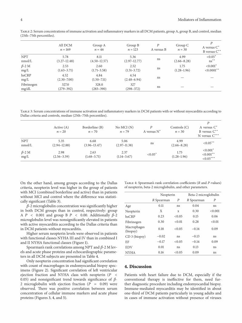
Table 2: Serum concentrations of immune activation and inflammatory markers in all DCM patients, group A, group B, and control, median(25th–75th percentiles).
All DCM𝑛 = 169
Group A𝑛 = 46
Group B𝑛 = 123
𝑃A versus B
Group C𝑛 = 30
𝑃A versus C∗B versus C∗∗
NPTnmol/L
5.78(3.27–12.48)
8.11(4.50–12.57)
5.36(2.97–12.77) ns 4.99
(2.66–8.28)<0.05∗ns∗∗
𝛽-2 Mmg/L
2.53(1.63–3.71)
2.60(1.71–3.58)
2.52(1.51–3.72) ns 1.75
(1.28–1.96)<0.001∗<0.0001∗∗
hsCRPmg/L
4.52(2.30–7.00)
4.84(1.50–7.11)
4.54(2.48–6.94) ns — —
Fibrinogenmg/dL
327.0(279–392)
328.0(283–390)
327(298–372) ns — —
Table 3: Serum concentrations of immune activation and inflammatory markers in DCM patients with or without myocarditis according toDallas criteria and controls, median (25th–75th percentiles).
Active (A)𝑛 = 20
Borderline (B)𝑛 = 70
No MCI (N)𝑛 = 79
𝑃A versus N∗
Controls (C)𝑛 = 30
𝑃A versus C∗B versus C∗∗N versus C∗∗∗
NPTnmol/L
5.35(2.94–12.88)
6.68(3.96–13.47)
5.06(2.97–11.38) ns 4.99
(2.66–8.28) <0.05∗∗
𝛽-2 Mmg/L
2.98(2.56–3.59)
2.63(1.68–3.71)
2.37(1.14–3.67) <0.05∗ 1.75
(1.28–1.96)
<0.001∗<0.001∗∗<0.05∗∗∗
On the other hand, among groups according to the Dallascriteria, neopterin level was higher in the group of patientswith MCI (combined borderline and active) than in patientswithout MCI and control where the difference was statisti-cally significant (Table 3).𝛽-2 microglobulin concentration was significantly higher
in both DCM groups than in control, respectively: groupA 𝑃 < 0.001 and group B 𝑃 < 0.00. Additionally 𝛽-2microglobulin level was nonsignificantly elevated in patientswith active myocarditis according to the Dallas criteria thanin DCM patients without myocarditis.
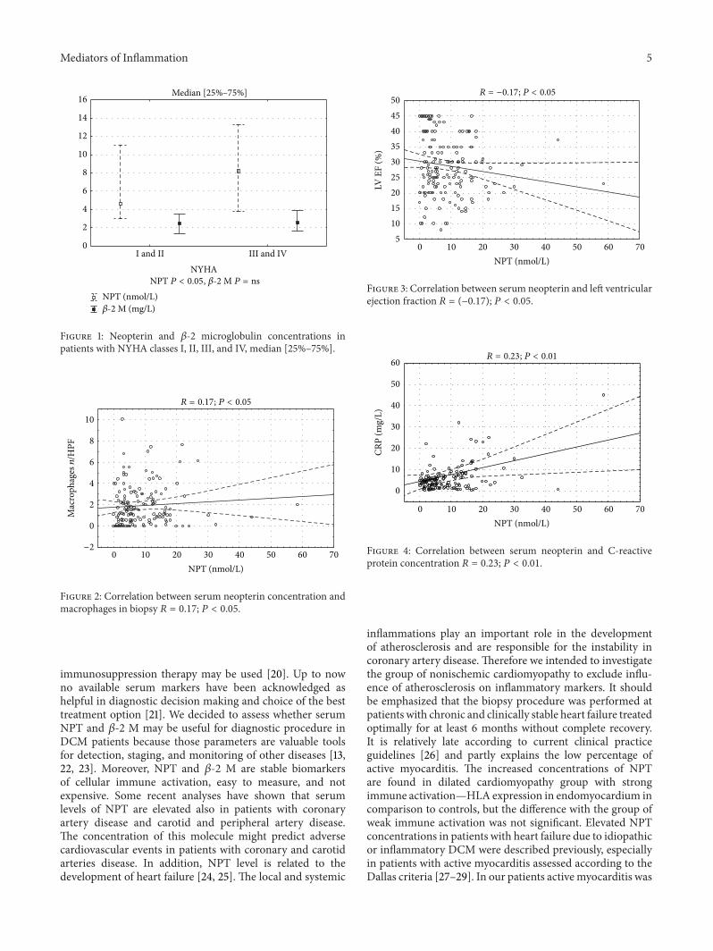
Higher serum neopterin levels were observed in patientswith functional classes NYHA III and IV than in combined Iand II NYHA functional classes (Figure 1).
Spearman’s rank correlations among NPT and 𝛽-2 M lev-els and acute phase proteins and echocardiographic parame-ters in all DCM subjects are presented in Table 4.
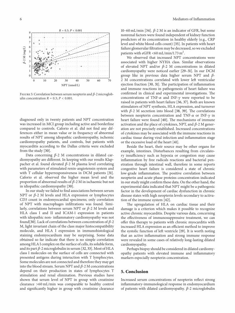
Only neopterin concentration had significant correlationwith count of macrophages in endomyocardial biopsy spec-imens (Figure 2). Significant correlation of left ventricularejection fraction and NYHA class with neopterin (𝑃 <0.05) and nonsignificant trend towards significance of 𝛽-2 microglobulin with ejection fraction (𝑃 = 0.09) wereobserved. There was positive correlation between serumconcentration of cellular immune markers and acute phaseproteins (Figures 3, 4, and 5).
Table 4: Spearman’s rank correlation coefficients (𝑅 and 𝑃-values)of neopterin, beta-2 microglobulin, and other parameters.
Neopterin Beta-2 microglobulin𝑅 Spearman 𝑃 𝑅 Spearman 𝑃
Age 0.11 ns 0.04 nsNeopterin X x 0.30 <0.001hsCRP 0.23 <0.05 0.15 0.06Fibrinogen 0.30 <0.01 0.28 <0.01Macrophages(biopsy) 0.18 <0.05 −0.14 0.09
CD 3 (biopsy) −0.02 ns −0.13 nsEF −0.17 <0.05 −0.14 0.09EDV 0.01 ns 0.13 nsNYHA 0.16 <0.05 0.09 ns
4. Discussion
Patients with heart failure due to DCM, especially if theconventional therapy is ineffective for them, need fur-ther diagnostic procedure including endomyocardial biopsy.Immune-mediated myocarditis may be identified in aboutone-third of DCM patients particularly in young adults andin cases of immune activation without presence of viruses
Mediators of Inflammation 5
0
2
4
6
8
10
12
14
16
I and II III and IV
NPT (nmol/L)
NYHA
𝛽-2 M (mg/L)
Median [25%–75%]
NPT P < 0.05, 𝛽-2 M P = ns
Figure 1: Neopterin and 𝛽-2 microglobulin concentrations inpatients with NYHA classes I, II, III, and IV, median [25%–75%].
0 10 20 30 40 50 60 70
NPT (nmol/L)
−2
0
2
4
6
8
10
Mac
roph
agesn
/HPF
R = 0.17; P < 0.05
Figure 2: Correlation between serum neopterin concentration andmacrophages in biopsy 𝑅 = 0.17; 𝑃 < 0.05.
immunosuppression therapy may be used [20]. Up to nowno available serum markers have been acknowledged ashelpful in diagnostic decision making and choice of the besttreatment option [21]. We decided to assess whether serumNPT and 𝛽-2 M may be useful for diagnostic procedure inDCM patients because those parameters are valuable toolsfor detection, staging, and monitoring of other diseases [13,22, 23]. Moreover, NPT and 𝛽-2 M are stable biomarkersof cellular immune activation, easy to measure, and notexpensive. Some recent analyses have shown that serumlevels of NPT are elevated also in patients with coronaryartery disease and carotid and peripheral artery disease.The concentration of this molecule might predict adversecardiovascular events in patients with coronary and carotidarteries disease. In addition, NPT level is related to thedevelopment of heart failure [24, 25]. The local and systemic
0 10 20 30 40 50 60 70
NPT (nmol/L)
5
10
15
20
25
30
35
40
45
50
LV E
F (%
)
R = −0.17; P < 0.05
Figure 3: Correlation between serum neopterin and left ventricularejection fraction 𝑅 = (−0.17); 𝑃 < 0.05.
0 10 20 30 40 50 60 70
NPT (nmol/L)
0
10
20
30
40
50
60
CRP
(mg/
L)
R = 0.23; P < 0.01
Figure 4: Correlation between serum neopterin and C-reactiveprotein concentration 𝑅 = 0.23; 𝑃 < 0.01.
inflammations play an important role in the developmentof atherosclerosis and are responsible for the instability incoronary artery disease. Therefore we intended to investigatethe group of nonischemic cardiomyopathy to exclude influ-ence of atherosclerosis on inflammatory markers. It shouldbe emphasized that the biopsy procedure was performed atpatients with chronic and clinically stable heart failure treatedoptimally for at least 6 months without complete recovery.It is relatively late according to current clinical practiceguidelines [26] and partly explains the low percentage ofactive myocarditis. The increased concentrations of NPTare found in dilated cardiomyopathy group with strongimmune activation—HLA expression in endomyocardium incomparison to controls, but the difference with the group ofweak immune activation was not significant. Elevated NPTconcentrations in patients with heart failure due to idiopathicor inflammatory DCM were described previously, especiallyin patients with active myocarditis assessed according to theDallas criteria [27–29]. In our patients activemyocarditis was
6 Mediators of Inflammation
0 10 20 30 40 50 60 70
NPT (nmol/L)
0
1
2
3
4
5
6
7
8
9R = 0.3; P < 0.001
𝛽-2
M (m
g/L)
Figure 5: Correlation between serumneopterin and𝛽-2microglob-ulin concentration 𝑅 = 0.3; 𝑃 < 0.001.
diagnosed only in twenty patients and NPT concentrationwas increased in MCI group including active and borderlinecompared to controls. Caforio et al. did not find any dif-ferences either in mean value or in frequency of abnormalresults of NPT among idiopathic cardiomyopathy, ischemiccardiomyopathy patients, and controls, but patients withmyocarditis according to the Dallas criteria were excludedfrom the study [30].
Data concerning 𝛽-2 M concentrations in dilated car-diomyopathy are different. In keeping with our results Klap-pacher et al. found elevated 𝛽-2 M plasma level correlatingwith parameters of stimulated renin-angiotensin system andwith T cellular hyperresponsiveness in DCM patients [31].Caforio et al. observed the higher mean level and theproportion of abnormal results of 𝛽-2M in ischaemic but notin idiopathic cardiomyopathy [30].
In our study we failed to find associations between serumNPT or 𝛽-2 M levels and HLA expression or lymphocytesCD3 count in endomyocardial specimens; only correlationof NPT with macrophages infiltrations was found. Simi-larly, correlations between serum NPT or 𝛽-2 M levels andHLA class I and II and ICAM-1 expression in patientswith idiopathic non- inflammatory cardiomyopathy was notfound[30]. Lack of correlations between concentration of 𝛽-2M, light invariant chain of the class major histocompatibilitymolecule, and HLA I expression in immunohistologicalstaining endomyocardium may be surprising. Some dataobtained so far indicate that there is no simple correlationamongHLA I complex on the surface of cells, its soluble form,and its part𝛽-2microglobulin in serum [32, 33].Most ofHLAclass I molecules on the surface of cells are connected withpresented antigens during interaction with T lymphocytes.Somemolecules are not connected and therefore theymay getinto the blood stream. SerumNPT and 𝛽-2M concentrationsdepend on their production in states of lymphocytes Tstimulation and renal elimination. Previous studies haveshown that serum level of NPT in group with creatinineclearance >60mL/min was comparable to healthy controland significantly higher in group with creatinine clearance
10–60mL/min [34]. 𝛽-2 M is an indicator of GFR, but somenonrenal factors were found independent of kidney functionpredictors of its concentration in healthy elderly (e.g., CRPlevel and white blood cells count) [35]. In patients with heartfailure glomerular filtrationmay be decreased, sowe excludedpatients with eGFR <60mL/min/1.73m2.
We observed that increased NPT concentrations wereassociated with higher NYHA class. Similar observationsof elevated NPT and/or 𝛽-2 M concentrations in dilatedcardiomyopathy were noticed earlier [29–31]. In our DCMgroup like in previous data higher serum NPT and 𝛽-2 M concentrations correlated with lower left ventricularejection fraction [30, 31]. The participation of inflammationand immune reactions in pathogenesis of heart failure wasconfirmed in clinical and experimental investigations. Theconcentrations of TNF-𝛼 and INF-𝛾 were reported to beraised in patients with heart failure [36, 37]. Both are knownstimulators of NPT synthesis, HLA expression, and turnoverwith 𝛽-2 M secretion into blood [38, 39]. The correlationsbetween neopterin concentration and TNF-𝛼 or INF-𝛾 inheart failure were found [40]. The mechanisms of immuneactivation and the place of cytokines, NPT, and 𝛽-2 M gener-ation are not precisely established. Increased concentrationsof cytokines may be associated with the immune reactions incardiac tissue during viral infection and inflammation stageor the excessive load of the heart [41].
Beside the heart, their source may be other organs forexample, intestines. Disturbances resulting from circulato-ry insufficiency such as hypoxia or congestion may causeinflammation by free radicals reactions and bacterial pen-etration through intestinal wall, therefore in some reportscongestive heart failure is considered a state of chroniclow-grade inflammation. The positive correlation betweenneopterin and acute phase proteins concentration indicatedin our studymight confirm these data. On the other hand, theexperimental data indicated that NPT might be a pathogenicfactor in the development of cardiac dysfunction in chronicdisease states with high neopterin levels secondary to activa-tion of the immune system [42].
The upregulation of HLA on cardiac tissue and theirdamage is a criterion which makes it possible to recognizeactive chronic myocarditis. Despite various data, concerningthe effectiveness of immunosuppressive treatment, we canoffer this therapy to patients with chronic myocarditis withincreased HLA expression as an efficient method to improvethe systolic function of left ventricle [19]. It is worth notingthat an active inflammation and strong immune responsewere revealed in some cases of relatively long-lasting dilatedcardiomyopathy.
Perhaps biopsy should be considered in dilated cardiomy-opathy patients with elevated immune and inflammatorymarkers especially neopterin concentration.
5. Conclusion
Increased serum concentrations of neopterin reflect stronginflammatory-immunological response in endomyocardiumof patients with dilated cardiomyopathy. 𝛽-2 microglobulin
Mediators of Inflammation 7
concentration is elevated in all patients with DCM indepen-dently on immune activation in myocardium.
Correlations between above markers of cellular immuneactivation with acute phase proteins level and left ventricleejection fraction suggest the participation of low gradeinflammatory state in pathophysiology of heart failure poten-tially related to its severity.
Conflict of Interests
The authors declare that there is no conflict of interestsregarding the publication of this paper.
References
[1] R. Watanabe, R. W. Azuma, J. I. Suzuki et al., “Inhibition ofNF-𝜅B activation by a novel IKK inhibitor reduces the severityof experimental autoimmune myocarditis via suppression ofT-cell activation,” American Journal of Physiology Heart andCirculatory Physiology, vol. 305, no. 12, pp. H1761–H1771, 2013.
[2] P. Chen, G. C. Baldeviano, D. L. Ligons et al., “Susceptibility toautoimmunemyocarditis is associated with intrinsic differencesin CD4+ T cells,” Clinical and Experimental Immunology, vol.169, no. 2, pp. 79–88, 2012.
[3] M. L. Tarrio, N. Grabie, D. Bu, A. H. Sharpe, and A. H. Licht-man, “PD-1 protects against inflammation andmyocyte damagein T cell-mediated myocarditis,” Journal of Immunology, vol.188, no. 10, pp. 4876–4884, 2012.
[4] P. Blyszczuk, C. Berthonneche, S. Behnke et al., “Nitric oxidesynthase 2 is required for conversion of pro-fibrogenic inflam-matoryCD133+ progenitors into F4/80+macrophages in exper-imental autoimmune myocarditis,” Cardiovascular Research,vol. 97, no. 2, pp. 219–229, 2013.
[5] K. Li, W. Xu, Q. Guo et al., “Differential macrophage polariza-tion in male and female BALB/c mice infected with coxsack-ievirus B3 defines susceptibility to viral myocarditis,” Circula-tion Research, vol. 105, no. 4, pp. 353–364, 2009.
[6] H. T.Aretz, “Myocarditis: theDallas criteria,”HumanPathology,vol. 18, no. 6, pp. 619–624, 1987.
[7] B. Maisch, I. Portig, A. Ristic, G. Hufnagel, and S. Pankuweit,“Definition of inflammatory cardiomyopathy (myocarditis): onthe way to consensus,” Herz, vol. 25, no. 3, pp. 200–209, 2000.
[8] R. Wojnicz, E. Nowalany-Kozielska, J. Wodniecki et al.,“Immunohistological diagnosis of myocarditis. Potential roleof sarcolemmal induction of the MHC and ICAM-1 in thedetection of autoimmune mediated myocyte injury,” EuropeanHeart Journal, vol. 19, no. 10, pp. 1564–1572, 1998.
[9] I. Kindermann, M. Kindermann, R. Kandolf et al., “Predictorsof outcome in patients with suspectedmyocarditis,”Circulation,vol. 118, no. 6, pp. 639–648, 2008.
[10] P. F. Cosper, P. A. Harvey, and L. A. Leinwand, “Interferon-𝛾 causes cardiac myocyte atrophy via selective degradation ofmyosin heavy chain in a model of chronic myocarditis,” TheAmerican Journal of Pathology, vol. 181, no. 6, pp. 2038–2046,2012.
[11] A. Henke, C. Mohr, H. Sprenger et al., “Coxsackievirus B3-induced production of tumor necrosis factor-alpha, IL-1 beta,and IL-6 in humanmonocytes,”The Journal of Immunology, vol.148, no. 7, pp. 2270–2277, 1992.
[12] C. Huber, J. R. Batchelor, D. Fuchs et al., “Immune response-associated production of neopterin. Release from macrophages
primarily under control of interferon-gamma,” Journal of Exper-imental Medicine, vol. 160, no. 1, pp. 310–316, 1984.
[13] D. Arshadi, B. Nikbin, Y. Shakiba, A. Kiani, A. R. Jamshidi, andM. T. Boroushaki, “Plasma level of neopterin as a marker ofdisease activity in treated rheumatoid arthritis patients: asso-ciation with gender, disease activity and anti-CCP antibody,”International Immunopharmacology, vol. 17, no. 3, pp. 763–767,2013.
[14] A. K. Yadav, V. Sharma, and V. Jha, “Association between serumneopterin and inflammatory activation in chronic kidney dis-ease,”Mediators of Inflammation, vol. 2012, Article ID 476979, 6pages, 2012.
[15] M. Erren, H. Reinecke, R. Junker et al., “Systemic inflammatoryparameters in patients with atherosclerosis of the coronary andperipheral arteries,” Arteriosclerosis, Thrombosis, and VascularBiology, vol. 19, no. 10, pp. 2355–2363, 1999.
[16] F. Puppo, S. Brenci, P. Contini et al., “Increased 𝛽2-microglobulin-free HLA class I heavy chain serum levelsin the course of immune responses to viral antigens and tomismatched HLA antigens,” Tissue Antigens, vol. 55, no. 4, pp.333–341, 2000.
[17] K. L. Bourantas, E. C. Hatzimichael, A. C. Makis et al., “Serumbeta-2-microglobulin, TNF-𝛼 and interleukins in myeloprolif-erative disorders,” European Journal of Haematology, vol. 63, no.1, pp. 19–25, 1999.
[18] P. Richardson, R. W. McKenna, M. Bristow et al., “Report ofthe 1995 World Health Organization/International Society andFederation of Cardiology Task Force on the definition andclassification of cardiomyopathies,” Circulation, vol. 93, no. 5,pp. 841–842, 1996.
[19] R. Wojnicz, E. Nowalany-Kozielska, C. Wojciechowska et al.,“Randomized, placebo- controlled study for immunosuppres-sive treatment of inflammatory dilated cardiomyopathy: two-year follow-up results,” Circulation, vol. 104, no. 1, pp. 39–45,2001.
[20] H. Schultheiss, U. Khl, and L. T. Cooper, “The management ofmyocarditis,” European Heart Journal, vol. 32, no. 21, pp. 2616–2625, 2011.
[21] A. L. Caforio, S. Pankuweit, E. Arbustini et al., “EuropeanSociety of Cardiology Working Group on Myocardial andPericardial Diseases. Current state of knowledge on aetiology ,diagnosis, management, and therapy of myocarditis: a positionstatement of the European Society of Cardiology WorkingGrouponMyocardial andPericardialDiseases,”EuropeanHeartJournal, vol. 34, no. 33, pp. 2636–2648, 2013.
[22] G. F. Oxenkrug, P. J. Requintina, D. L. Mikolich et al.,“Neopterin as a marker of response to antiviral therapy inhepatitis C virus patients,” Hepatitis Research and Treatment,vol. 2012, Article ID 619609, 4 pages, 2012.
[23] M. G. Alexandrakis, P. Roussou, C. A. Pappa et al., “Relation-ship between circulating BAFF serum levels with proliferatingmarkers in patients with multiple myeloma,” BioMed ResearchInternational, vol. 2013, Article ID 389579, 6 pages, 2013.
[24] K. K. Ray, D. A. Morrow, M. S. Sabatine et al., “Long-termprognostic value of neopterin: a novel marker of monocyte acti-vation in patients with acute coronary syndrome,” Circulation,vol. 115, no. 24, pp. 3071–3078, 2007.
[25] T. B. Grammer, D. Fuchs, B. O. Boehm, B. R. Winkelmann,andW.Maerz, “Neopterin as a predictor of total and cardiovas-cular mortality in individuals undergoing angiography in theLudwigshafen Risk and Cardiovascular Health study,” ClinicalChemistry, vol. 55, no. 6, pp. 1135–1146, 2009.
8 Mediators of Inflammation
[26] L. T. Cooper, K. L. Baughman, A. M. Feldman et al., “The roleof endomyocardial biopsy in themanagement of cardiovasculardisease: a scientific statement from the American Heart Associ-ation, the American College of Cardiology, and the EuropeanSociety of Cardiology,” Circulation, vol. 116, no. 19, pp. 2216–2233, 2007.
[27] D. S. Fuchs, M. Samsonov, G. P. Tilz et al., “Stimulated cellularimmune system in patients with congestive heart failure,” Euro-pean Journal of Clinical Chemistry and Clinical Biochemistry,vol. 31, no. 3, pp. 111–114, 1993.
[28] E. Nassonov, M. Samsonow, V. Masenkow et al., “Serumneopterin in dilated cardiomyopathy,” Terapevticheskii Arkhiv,vol. 95, pp. 72–74, 1990.
[29] M. Samsonov, D. Fuchs, G. Reibnegger, J. N. Belenkov, E. L.Nassonov, and H. Wachter, “Patterns of serological markers forcellular immune activation in patients with dilated cardiomy-opathy and chronicmyocarditis,”Clinical Chemistry, vol. 38, no.5, pp. 678–680, 1992.
[30] A. L. P. Caforio, J. H. Goldman, M. K. Baig et al., “Elevatedserum levels of soluble interleukin-2 receptor, neopterin and 𝛽-2-microglobulin in idiopathic dilated cardiomyopathy: relationto disease severity and autoimmune pathogenesis,” EuropeanJournal of Heart Failure, vol. 3, no. 2, pp. 155–163, 2001.
[31] G. Klappacher, G. Mundigler, A. Papousek et al., “Elevatedcirculating levels of beta 2-microglobulin in patients withidiopathic dilated cardiomyopathy,” American Journal of Cardi-ology, vol. 71, no. 1, pp. 119–122, 1993.
[32] E. T. Everett, J. C. Scornik, G. Davis, and K. J. Kao, “Inductionof erythrocyte HLA expression during interferon treatment andHIV infection,” Human Immunology, vol. 29, no. 1, pp. 14–22,1990.
[33] K. Sakaguchi, N. Koide, T. Takenami et al., “Soluble HLAclass I antigens in sera of patients with chronic hepatitis,”Gastroenterologia Japonica, vol. 27, no. 2, pp. 206–211, 1992.
[34] K. Yokoyama, M. Tajima, H. Yoshida et al., “Plasma pteridineconcentrations in patients with chronic renal failure,” Nephrol-ogy Dialysis Transplantation, vol. 17, no. 6, pp. 1032–1036, 2002.
[35] Z. Stanga, S.Nock, P.Medina-Escobar,U. E.Nydegger,M.Risch,and L. Risch, “Factors other than the glomerular filtration ratethat determine the serum beta-2-microglobulin level,” PLoSONE, vol. 8, no. 8, Article ID e72073, 2013.
[36] I. C. Steele, A. M. Nugent, S. Maguire et al., “Cytokine profile inchronic cardiac failure,” European Journal of Clinical Investiga-tion, vol. 26, no. 11, pp. 1018–1022, 1996.
[37] B. Levine, J. Kalman, L. Mayer, H. M. Fillit, and M. Packer,“Elevated circulating levels of tumor necrosis factor in severechronic heart failure,”TheNewEngland Journal ofMedicine, vol.323, no. 4, pp. 236–241, 1990.
[38] J. McMurray, I. Abdullah, H. J. Dargie, and D. Shapiro,“Increased concentrations of tumour necrosis factor in “cachec-tic” patients with severe chronic heart failure,” British HeartJournal, vol. 66, no. 5, pp. 356–358, 1991.
[39] M. A. Freni, A. Ajello, A. Spadaro et al., “Class I HLA antigenshepatic display and beta-2-microglobulin serum values inchronic hepatitis C: effect of treatment with recombinant alphainterferon,” Hepatogastroenterology, vol. 44, no. 17, pp. 1295–1301, 1997.
[40] G. Torre-Amione, S. Kapadia, J. Lee et al., “Tumor necrosisfactor-𝛼 and tumor necrosis factor receptors in the failinghuman heart,” Circulation, vol. 93, no. 4, pp. 704–711, 1996.
[41] C. J. Wiedermann, H. Beimpold, M. Herold, E. Knapp, andH. Braunsteiner, “Increased levels of serum neopterin and
decreased production of neutrophil superoxide anions inchronic heart failure with elevated levels of tumor necrosisfactor-alpha,” Journal of the American College of Cardiology, vol.22, no. 7, pp. 1897–1901, 1993.
[42] A. Balogh, M. Mittermayr, A. Schlager et al., “Mechanismof neopterin-induced myocardial dysfunction in the isolatedperfused rat heart,” Biochimica et Biophysica Acta—GeneralSubjects, vol. 1724, no. 1-2, pp. 17–22, 2005.

Submit your manuscripts athttp://www.hindawi.com
Stem CellsInternational
Hindawi Publishing Corporationhttp://www.hindawi.com Volume 2014
Hindawi Publishing Corporationhttp://www.hindawi.com Volume 2014
MEDIATORSINFLAMMATION
of
Hindawi Publishing Corporationhttp://www.hindawi.com Volume 2014
Behavioural Neurology
EndocrinologyInternational Journal of
Hindawi Publishing Corporationhttp://www.hindawi.com Volume 2014
Hindawi Publishing Corporationhttp://www.hindawi.com Volume 2014
Disease Markers
Hindawi Publishing Corporationhttp://www.hindawi.com Volume 2014
BioMed Research International
OncologyJournal of
Hindawi Publishing Corporationhttp://www.hindawi.com Volume 2014
Hindawi Publishing Corporationhttp://www.hindawi.com Volume 2014
Oxidative Medicine and Cellular Longevity
Hindawi Publishing Corporationhttp://www.hindawi.com Volume 2014
PPAR Research
The Scientific World JournalHindawi Publishing Corporation http://www.hindawi.com Volume 2014
Immunology ResearchHindawi Publishing Corporationhttp://www.hindawi.com Volume 2014
Journal of
ObesityJournal of
Hindawi Publishing Corporationhttp://www.hindawi.com Volume 2014
Hindawi Publishing Corporationhttp://www.hindawi.com Volume 2014
Computational and Mathematical Methods in Medicine
OphthalmologyJournal of
Hindawi Publishing Corporationhttp://www.hindawi.com Volume 2014
Diabetes ResearchJournal of
Hindawi Publishing Corporationhttp://www.hindawi.com Volume 2014
Hindawi Publishing Corporationhttp://www.hindawi.com Volume 2014
Research and TreatmentAIDS
Hindawi Publishing Corporationhttp://www.hindawi.com Volume 2014
Gastroenterology Research and Practice
Hindawi Publishing Corporationhttp://www.hindawi.com Volume 2014
Parkinson’s Disease
Evidence-Based Complementary and Alternative Medicine
Volume 2014Hindawi Publishing Corporationhttp://www.hindawi.com