Propionibacterium acnes pathogenicity and possible role in ... · PDF filePaweł Szmygin,...
11
HEALTH AND WELLNESS 2/2014 WELLNESS AND HEALTH CHAPTER XI Department of Neurosurgery, Medical University of Lublin Katedra i Klinika Neurochirurgii i Neurochirurgii Dziecięcej Uniwersytet Medyczny w Lublinie PAWEŁ SZMYGIN, ELŻBIETA BARTOŃ, KRZYSZTOF TUROWSKI Propionibacterium acnes – pathogenicity and possible role in intervertebral disc herniation Propionibacterium acnes – chorobotwórczość i rola w przepuklinie krążka międzykręgowego Key words: Propionibacterium acnes, lower back pain, disc degeneration disease, lumbar disc herniation Słowa kluczowe: Propionibacterium acnes, dolegliwości bólowe lędzwiowego od- cinka kręgosłupa, choroba degeneracyjna krążka międzykręgowego, przepuklina krążka międzykręgowego PROPIONIBACTERIUM ACNES – CHARACTERISTICS OF THE SPECIES Propionibacterium acnes is an anaerobic Gram-positive rod-shaped bacterium. It is slow-growing and belongs to skin microbiome, but is also the pathogenic factor of acne vulgaris. Its older names include Bacillus acnes and Corynebacterium ac- nes. Within the species P. acnes, around 100 strains have been described; the ge- nome of many has been completely sequenced. A single bacterium is 0,4-0,5 μm wide and 0,8-0,9 μm long [1]. Under light microscope it can most often be seen in groups of two or bigger chains. It has no flagella, thus no capability of active movement and it does not form spores [2]. Both in vitro and in vivo it forms biofilm, in anaerobic conditions it can survive 8 month. The fact that the bacteria can be found in macrophages of alveoli, proves that it persists within phagocytes [3].
Transcript of Propionibacterium acnes pathogenicity and possible role in ... · PDF filePaweł Szmygin,...
HEALTH AND WELLNESS 2/2014
WELLNESS AND HEALTH
CHAPTER XI
Department of Neurosurgery, Medical University of Lublin
Katedra i Klinika Neurochirurgii i Neurochirurgii Dziecięcej
Uniwersytet Medyczny w Lublinie
PAWEŁ SZMYGIN, ELŻBIETA BARTOŃ, KRZYSZTOF TUROWSKI
Propionibacterium acnes – pathogenicity and possible role
in intervertebral disc herniation
Propionibacterium acnes – chorobotwórczość i rola
w przepuklinie krążka międzykręgowego
Key words: Propionibacterium acnes, lower back pain, disc degeneration disease,
lumbar disc herniation
Słowa kluczowe: Propionibacterium acnes, dolegliwości bólowe lędzwiowego od-
cinka kręgosłupa, choroba degeneracyjna krążka międzykręgowego, przepuklina
krążka międzykręgowego
PROPIONIBACTERIUM ACNES – CHARACTERISTICS OF THE SPECIES
Propionibacterium acnes is an anaerobic Gram-positive rod-shaped bacterium.
It is slow-growing and belongs to skin microbiome, but is also the pathogenic factor
of acne vulgaris. Its older names include Bacillus acnes and Corynebacterium ac-
nes. Within the species P. acnes, around 100 strains have been described; the ge-
nome of many has been completely sequenced. A single bacterium is 0,4-0,5 μm
wide and 0,8-0,9 μm long [1]. Under light microscope it can most often be seen in
groups of two or bigger chains. It has no flagella, thus no capability of active
movement and it does not form spores [2]. Both in vitro and in vivo it forms biofilm,
in anaerobic conditions it can survive 8 month. The fact that the bacteria can be
found in macrophages of alveoli, proves that it persists within phagocytes [3].

HEALTH AND WELLNESS 2/2014
Wellness and health
148
The bacterium lives mainly in the sebaceous glands of the hair follicle, but its
presence has also been reported in gastro-intestinal tract. Pathogenic strains cause
release of cytokines from sebocytes and inflammation [4]. Study of various polysac-
charide chains in the cell wall have led to subdivision of P. acnes species into the
following groups: IA, IB, IC, II and III. Group IA is responsible for acne vulgaris,
other groups are isolated from foci of opportunistic inflammation, e.g. postoperative
infection of hip joint [5].
On solid culture media, incubated 4-5 days anaerobically it forms round, elevat-
ed colonies, 1,5 to 5 mm in diameter. Initially white, in time they turn light pink.
Propionibacterium acnes is aerotolerant anaerobic bacteria, it survives in oxygen
but its growth is retarded. Its optimal growing temperature is 37°C, in room temper-
ature the growth slows down, at 45°C – stops altogether. The bacterium prefers
neutral pH, but even with optimal conditions the growth proceeds at a slow pace. It
uses organic compounds (sugars, fibers, pectins) to produce energy in the process of
fermentation, the products being, among others: propionic acid, acetic acid and car-
bon dioxide. Propionibacterium genome sequencing, completed in 2004 revealed
that the genetic material of the bacterium consists of 2560 base pairs, with high
percentage of guanine and cytosine [6].
The presence of some enzymes, that break down certain substrates is used for the
identification of P. acnes in biochemical test. The enzymes are catalase, nitrite re-
ductase (NADH), tryptophanase. Some of the strains are capable of beta-hemolysis
[7]. Propionibacterium has considerable demands for medium nutrients. Most fre-
quently used are: bovine broth, casein peptones, yeast extract, KH2PO4, cysteine,
hemin, some vitamins, glucose and sodium thioglycolate.
PATHOGENICITY
Propionibacterium acnes is associated with the pathogenesis of acne vulgaris.
The bacteria proliferate in comedones, where the conditions are moderately anaero-
bic. Using lipase they break down components of sebum to produce energy. Addi-
tionally, inflammatory factors (IL-1β, IL-8, IL-12, TNF-α) are produced and stimu-
late chemotaxis of leukocytes, that die in the tissue forming pustules [8].
Moreover, studies have shown that P. acnes can be found in granulomas in sar-
coidosis, keratitis, discitis, endocarditis, osteomyelitis and endophtalmitis [9]. In
cases of sarcoidosis, where the bacteria have been identified, remission has been
observed after minocycline treatment (broad-spectrum tetracycline antibiotic widely
used for acne [10]). However, there are two hypothesis explaining the efficiency of
this therapy – antimicrobial and immunoregulative action of minocycline is empha-
sized. P. acnes is also associated with SAPHO (Synovitis, Acne, Pustulosis, Hyper-
ostosis, Osteitis) syndrome, where immunological complexes antibody-P. acnes
antigen are suspected to deposit in bone and joint [11]. After Staphylococci P. acnes
is the most frequent cause of infection of cerebrospinal liquid shunts (9% of all cas-
es), where they form biofilm. It is recommended to obtain material for anaerobic
culture from the infected shunt [12]. The bacterium is also reported to cause cerebral
abscesses as a late postoperative complication of neurosurgeries.
Paweł Szmygin, Elżbieta Bartoń, Krzysztof Turowski
Propionibacterium acnes – pathogenicity and possible role
in intervertebral disc herniation
149
P. acnes has been isolated from the canal of tooth root, dental pulp and periodon-
tium [13]. Cases of endocarditis caused by P. acnes are rare and most often occur
around foreign material (artificial valve, annuloplasty rings, pacemaker electrodes).
In 79% of endocarditis cases analyzed by Sohail and Gray, prosthetic material was
the source of the infection [14]. Bacterial inflammation of the eye ball, a very seri-
ous complication after a lens replacement operation in patients with cataract, are
almost always caused by Gram-positive bacteria (91%); P. acnes is sometimes the
responsible pathogen, as it is for corneal ulcerations and conjunctivitis. When diag-
nosing inflammations of hip joint endoprothesis, it is recommended to use ultra-
sound to break up the biofilm from the infected prosthesis, than to incubate the sam-
ples for time long enough for the slow-growing bacterial species to form colonies.
As is presented below, P. acnes is the most frequently identified anaerobic agent in
spondylodyscitis, postoperative or after an invasive procedure, i.e. epidural anesthe-
sia.
ANTIBIOTIC TREATMENT
Usually Propionibacterium acnes is susceptible to a large number of antibiotics,
and its infections are efficiently cured with simple therapeutic schemes. The bacte-
rium is resistant to metronidazole and partly to aminoglycosides. In recent years, a
decrease in efficiency of clindamycin, erythromycin and minocycline is being ob-
served due to their excessive use in acne [15].
LUMBAR DISC HERNIATION – SHORT CHARACTERISTICS
Low back pain (LBP) is familiar to almost every adult [16]. Lumbar disc herni-
ation (LDH) is a condition affecting the spine as consequence of trauma, injury or
without evident cause (idiopathic). Tears in the outer layer (annulus fibrosus) lead to
bulging out of the soft, central part of the disc (nucleus pulposus). In the ventral part
of the spine annulus fibrosus is reinforced by the ligamentum longitudinale anterior,
in the dorsal part, from the inside of the spinal canal – by the ligamentum longitudi-
nale posterior. Consequently, the posterior-lateral portion of the ring is the locus
minoris resistantiae, where herniation most commonly occurs.
Intervertebral disc is a bradytrophic tissue – it is not supplied by capillary ves-
sels, but through diffusion. Nucleus pulposus is a mix of water and aggrecan-
proteoglycan gel, suspended on a matrix made of type II collagen and elastin fibers.
Annulus fibrosus is composed of 15-25 laminae made of type I collagen [17]. If the
nucleus (comprising in 80% of water) loses its elasticity (so-called ‘black disc dis-
ease’) its function as shock-absorber decreases. The causes are: genetic predisposi-
tion, asymmetric load, weak paravertebral muscles, pregnancy. Modern lifestyle –
little activity, sitting work, postural deformities, addictions – have a share in the
occurrence of the condition. Possibly, childhood infections, when the intervertebral
disc are supplied by capillaries, play a part in the degeneration [18]. The sequence
leading to the disc degeneration: until the disc is supplied by circulation (age of 20),
daily activity causes transitory dehydration, during sleep it is replenished. At 30-40
HEALTH AND WELLNESS 2/2014
Wellness and health
150
years, dehydration proceeds irreversibly, the disc loses elasticity, tears occur in the
annulus. The final stage is stabilization through ossification, thickening of the discs
and decreased concentration of inflammatory proteins [19]. Thus, 30-40 years-old
patients are more likely to suffer from LBP, than 60-year-olds. Disc degeneration
detected in diagnostic imaging does not have to be painful. MRI based studies
showed that 30% of young people with bulging disc had no clinical symptoms [20].
Disc herniation (prolapsus disci intervertebralis) advances gradually, previous
stage being protrusion of the nuclear masses into the fissures in the ring. Finally,
degenerated disc becomes fibrous and scarred, intervertebral space becomes narrow-
er, osteophytes grow at the edges of the vertebrae and the whole motor unit becomes
stiff [21]. The direct cause of pain is the compression of the spinal cord, spinal
nerves, release of inflammatory factors and acidic metabolites of the nucleus. The
disc itself is not enervated, unlike posterior longitudinal ligament, periosteum, facet
joints (ramus meningeus nervi spinalis) [22].
Symptoms differ depending on localization and affected tissues – from symp-
tomless through severe pain radiating into the regions enervated by the compressed
nerves to Cauda Equina Syndrome, requiring quick surgical intervention. Other
complaints are: diffuse pain in thighs, knees, feet, paresthesia, tingling, tickling,
burning, numbness, increased muscle tonus, dystonia. The onset is usually rapid,
connected with an abrupt movement of bending or rotating of the spine. It can also
be caused by static load, such as prolonged sitting. Professional drivers can suffer
from LBP, due to vibrations of the car (4-5 Hz) [23]. The pain is especially intense,
when patient is sitting or at the beginning of walking, increases at coughing, sneez-
ing and any increase of abdominal pressure. In examination, reflexes are below
normal and stretch tests for nerve roots L5-S1 (Lasegue’s, Bragard’s Fajersztajn-
Krzemicki’s sign) or L4 (Mackiewicz’s sign) are positive [24]. In the last one (also
referred to as ‘reverse Lasegue’) knee flexion is performed, patient lying face down.
Pain in the knee or front part of the thigh suggests pathology in the femoral nerve.
Pain of the lumbar spine at pressing the chin to the chest is known as Negrie’s sign.
Disc herniation it the cause in 90% of sciatica [25]. It occurs when compression
affects L3-L5 lumbar or S1-S3 sacral nerve roots or the sciatic nerve. Lumbar pain
radiates to the buttock and lower limb impeding walking. In such cases we speak of
claudicatio caudae equinae, if pain occurs after a short distance (similarly to vascu-
lar claudicatio intermittens [26]). Particularly severe, central disc herniation is also
the most common cause of the Cauda Equina Syndrome. Cauda Equina is bundle of
nerve roots L2 to coccygeal nerve, that hangs in the spinal canal. Segments S2-S4 of
the spinal cord form pudendal nerve, which is a part of sacral plexus. This mixed
nerve carries motor, sensory, parasympathetic and sympathetic fibers; when it is
compressed erectile dysfunction, urinary retention and incontinence, bowel dysfunc-
tion result.
The first diagnostic tool in degenerative disc disease should be lumbar spine x-
ray. It can rule spondylolisthesis and bone neoplasm out. More precise information
about bone structures is delivered by computed tomography (CT), and magnetic
resonance imaging (MRI) is best for nervous tissue.
Paweł Szmygin, Elżbieta Bartoń, Krzysztof Turowski
Propionibacterium acnes – pathogenicity and possible role
in intervertebral disc herniation
151
PROPIONIBACTERIUM ACNES IN INTERVERTEBRAL DISC -
LITERATURE REVIEW
Reports about postoperative spondylodiscitis due to Propionibacterium acnes
were published over 30 years ago. In 1983, Burki and al. described a case of L4-L5
disc degeneration (confirmed in x-ray, CT and increased radioisotope up-take in
scintigraphy) in a 49-year-old female patient treated with epidural corticosteroid
infiltrations for sciatica. Microbiologic culture and serologic antigen identification
revealed bacterial origin of the inflammation. Identified pathogens were Peptococ-
cus Constellatus and Propionibacterium acnes [27]. In 1987 Noble and Overman
presented a case of a male patient who developed similar symptoms 4 weeks after
discectomy. The authors present a literature review and summarize P. acnes related
bone infections. Nobel and Overman distinguish four categories: infection after a
surgery or an invasive procedure, infection in an immunodeficient patient, infection
in a patient without obvious predisposition and infection where P. acnes as causative
agent is uncertain. The first category predominates [28].
A controversy was provoked by the study Association between sciatica and Pro-
pionibacter acnes, by Stirling and al. published in 2001 in The Lancet. It concerned
36 patients, with no history of an infection in the previous 6 months, who had
discectomy. In 19 out of 36 (52,7%) patients the obtained disc samples gave positive
microbiological cultures (Propionibacterium acnes – x16, coagulase-negative
Staphylococcus - x 2, Corynebacterium propinquum- x1). None of the patients in the
control group (operated for other spine conditions: scoliosis, trauma, myeloma) were
tested positive (0 out of 14 patients – 0%). Additionally, the authors used ELISA to
detect lipid S (Gram-positive bacteria cell wall component) in the serum of the pa-
tients. Both methods put together gave the following results:
positive culture + positive serology - 7/36 (19,4%) patients
positive culture + negative serology - 12/36 (33,3%) patients
negative culture + positive serology - 1/36 (2,8%) patients
negative culture + negative serology - 16/36 (44,4%) patients
Neither living nor dead bacteria could be found in Gram-staining of interverte-
bral disc samples and microscopic examination [29].
Another article in favor of the role of P. acnes in the inflammation around herni-
ated disc, published in 2013, reports the results of the study of a Danish-English
research group (Rollason, McDowell, Albert and al.). Under strict aseptic regime, 5
disc samples were harvested from 64 patients who underwent discectomy. After
incubation in anaerobic conditions, P. acnes was detected in 24 patients (37,5%).
Using polymerase chain reaction (PCR), recA gene (P. acnes housekeeping gene,
necessary for vital functions of the bacterium, continuously expressed) was se-
quenced. Additionally, scientist used immunofluorescent microscopy and mouse
monoclonal antibodies QUBPa1 and QUBPa2, that attach to strains IA, II and partly
IC, but do not adhere to IB and III. Both techniques allowed to determine the phylo-
genetic groups, that the isolated P. acnes populations belonged to. Two conclusions
drawn by Rollason and al. speak against contamination as the source of the bacteria.
HEALTH AND WELLNESS 2/2014
Wellness and health
152
Firstly, in 16 patients P. acnes was identified in at least 2 samples of the disc (ac-
cording to the Infectious Diseases Society of America (IDSA) criteria, it proves the
infection [30]). Secondly, the populations had the following distribution: II (52%),
IA (28%), III (11%), IB(9%), IC (0%), so the phylogroups rarely isolated from acne
and skin, and frequently found in blood, soft tissues and prosthetic material (IB, II,
III) were widely represented. The authors remark that P. acnes could enter the
bloodstream during teeth brushing or a dental procedure. Neovascularization taking
place around the herniated disc allows the colonization of that region [31].
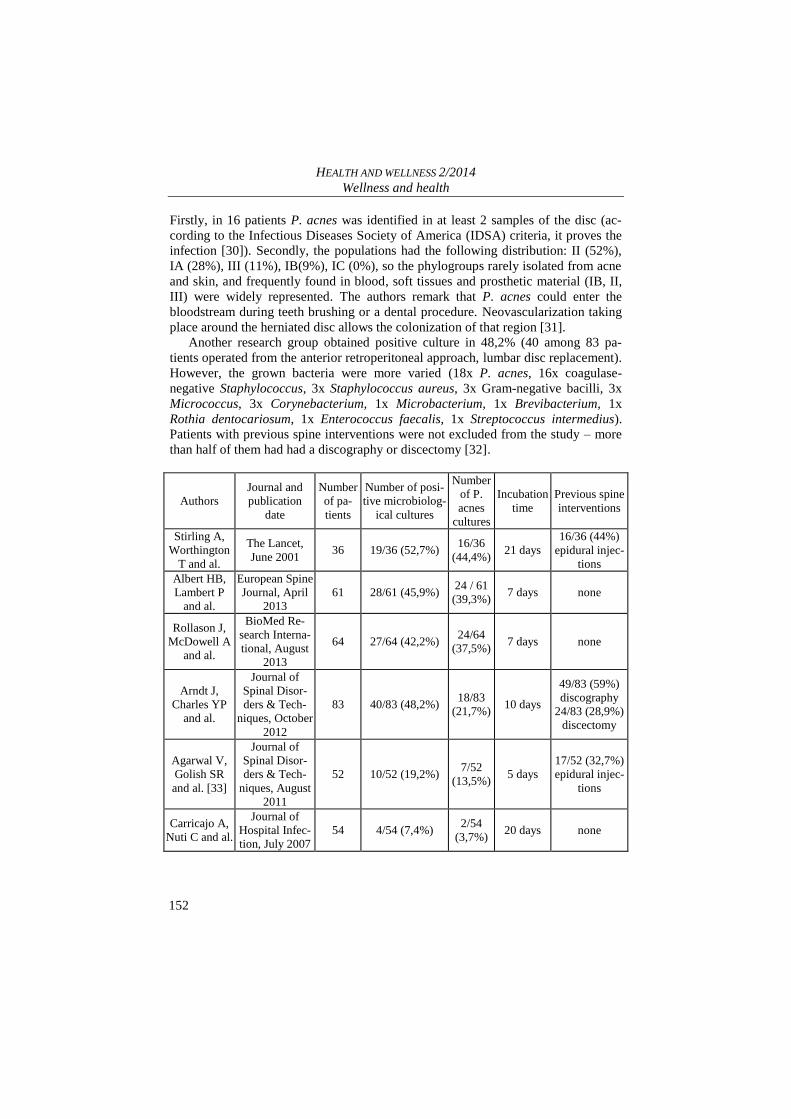
Another research group obtained positive culture in 48,2% (40 among 83 pa-
tients operated from the anterior retroperitoneal approach, lumbar disc replacement).
However, the grown bacteria were more varied (18x P. acnes, 16x coagulase-
negative Staphylococcus, 3x Staphylococcus aureus, 3x Gram-negative bacilli, 3x
Micrococcus, 3x Corynebacterium, 1x Microbacterium, 1x Brevibacterium, 1x
Rothia dentocariosum, 1x Enterococcus faecalis, 1x Streptococcus intermedius).
Patients with previous spine interventions were not excluded from the study – more
than half of them had had a discography or discectomy [32].
Authors
Journal and
publication
date
Number
of pa-
tients
Number of posi-
tive microbiolog-
ical cultures
Number
of P.
acnes
cultures
Incubation
time
Previous spine
interventions
Stirling A,
Worthington
T and al.
The Lancet,
June 2001 36 19/36 (52,7%)
16/36
(44,4%) 21 days
16/36 (44%)
epidural injec-
tions
Albert HB,
Lambert P
and al.
European Spine
Journal, April
2013
61 28/61 (45,9%) 24 / 61
(39,3%) 7 days none
Rollason J,
McDowell A
and al.
BioMed Re-
search Interna-
tional, August
2013
64 27/64 (42,2%) 24/64
(37,5%) 7 days none
Arndt J,
Charles YP
and al.
Journal of
Spinal Disor-
ders & Tech-
niques, October
2012
83 40/83 (48,2%) 18/83
(21,7%) 10 days
49/83 (59%)
discography
24/83 (28,9%)
discectomy
Agarwal V,
Golish SR
and al. [33]
Journal of
Spinal Disor-
ders & Tech-
niques, August
2011
52 10/52 (19,2%) 7/52
(13,5%) 5 days
17/52 (32,7%)
epidural injec-
tions
Carricajo A,
Nuti C and al.
Journal of
Hospital Infec-
tion, July 2007
54 4/54 (7,4%) 2/54
(3,7%) 20 days none

Paweł Szmygin, Elżbieta Bartoń, Krzysztof Turowski
Propionibacterium acnes – pathogenicity and possible role
in intervertebral disc herniation
153
Other scientists try to find a connection between P. acnes and edema in the end-
plates of the vertebrae adjacent to the degenerated disc – so called Modic changes.
They were first described in 1988 by Michael T. Modic; they can be observed in T1
and T2-weighted MRI (hypo- and hyperdense foci respectively) and they correlate
with LBP [34,35]. Trials of antibiotic treatments were conducted (100-day therapy
amoxycillin-clavulanic acid (500 mg/ 125 mg) every 8 hours) [36].
A very important contribution in the discussion about the causative role of low-
virulence bacteria in LBP, are studies which show the high probability of contami-
nation of the tissue samples during the surgery. French scientific group from CHU
Bellevue Saint-Etienne clinical hospital, led by Anne Carricajo, designed their ex-
periment similarly to the studies mentioned before, but additionally used air moni-
toring equipment and harvested tissue samples from ligamentum flavum and back
muscles (Musculus erector spinae). The study group was 54 patients undergoing
lumbar discectomy, with no previous epidural or spinal surgeries. The results were
as follows:
2 out of 54 (3,7%) patients – positive P. acnes cultures from disc tissue
4 out of 54 patients (7,4%) – positive microbiological cultures from disc
tissue (P. acnes x 2, Anaerobic Streptococci x 1, Actinomyces sp. +
Coagulase-negative Staphylococci x 1).
12 out of 54 (22,2%) patients - Ligamentum flavum and muscle tissue
samples positive cultures (P. acnes; importantly, all patients with positive
disc culture had positive muscle/ligament tissue samples, as well)
4 out of 54 (7,4%) patients - laminar flow controls positive cultures (3x P.
acnes, 1x Anaerobic Streptococci) The authors point out, that two facts speak against the inflectional hypothesis:
absence of inflammation markers (CRP, leukocytes) and absence of conditions pre-
disposing to low-virulence bacterial infection (compromised immunity or foreign
material, e.g. fixateur externe)[37].
CONCLUSIONS
Propionibacterium acnes is a commensal bacterium, also identified in
various pathologic conditions, although contamination or coincidental
presence (so-called innocent bystander) cannot be dismissed.
Although P. acnes is usually susceptible to the majority of standard anti-
biotics, growing resistance is being observed.
The analyzed literature reports positive P. acnes cultures in 3,7% to
44,4% of examined disc tissue samples.
Causative role of P. acnes in lower back pain (LBP) and Modic changes
has been put forward

HEALTH AND WELLNESS 2/2014
Wellness and health
154
REFERENCES
1. Douglas HC, Gunter SE: The taxonomic position of Corynebacterium acnes,
Journal of bacteriology, 1946, 52(7): 15–23.
2. Madigan MT, Martinko JM, Parker J: Brock Mikrobiologie, Spektrum Akademi-
scher Verlag GmbH, Heidelberg/Berlin 2000, p. 572–574.
3. Hightower JA, Welsh MG, Jackson RA, Gangemi JD: An ultrastructural exami-
nation of murine alveolar macrophages following intranasal administration of
propionibacterium acnes, Histology and Histopathology. 1987 Jul;2(3):217-22.
4. Nagy I, Pivarcsi A: Propionibacterium acnes and lipopolysaccharide induce the
expression of antimicrobial peptides and proinflammatory cytokines/chemokines
in human sebocytes. Microbes and infection / Institut Pasteur, 8(8), July
2006: 2195–2205.
5. Lomholt HB, Kilian M: Population genetic analysis of Propionibacterium acnes
identifies a subpopulation and epidemic clones associated with acne, PloS one.
5(8), 2010, S. e12277, Internet source: http://www.plosone.org
6. Propionibacterium acnes KPA171202, Internet source:
http://www.genomesonline.org
7. Lomholt HB, Kilian M: ibid.
8. Hof H, Dörries R: Duale Reihe: Medizinische Mikrobiologie, Thieme Verlag,
Stuttgart 2005, p. 335–336.
9. Perry AL, Lambert PA: Propionibacterium acnes, Letters in applied microbiolo-
gy, 42(3), March 2006, p. 185–188.
10. Strauss, et al. (2007). "Guidelines of care for acne vulgaris management." Jour-
nal of the American Academy of Dermatology 56 (4): 651–63.
11. Gutzmer R, Herbst RA, Kapp A, Weiß J: Das SAPHO-Syndrom - Fallbeschrei-
bung von drei Patienten mit Akne conglobata und osteoartikulären Symptomen,
Der Hautarzt (48), Springer-Verlag 1997, p. 186–190.
12. Conen A, Walti LN, Merlo A, Fluckiger U, Battegay M, Trampuz A: Character-
istics and treatment outcome of cerebrospinal fluid shunt-associated infections in
adults: a retrospective analysis over an 11-year period, July 2008, 47(1): p. 73-
82.
13. Niazi SA, Clarke D, Do T, Gilbert SC, Mannocci F, Beighton D: Propionibacte-
rium acnes and Staphylococcus epidermidis isolated from refractory endodontic
lesions are opportunistic pathogens, Journal of Clinical Microbiology . Novem-
ber 2010;48(11):3859-69.
14. Sohail MR, Gray AL, Baddour LM, Tleyjeh IM, Virk A: Infective endocarditis
due to Propionibacterium species, Clinical Microbiology and Infection, April
2009;15(4):387-94.
Paweł Szmygin, Elżbieta Bartoń, Krzysztof Turowski
Propionibacterium acnes – pathogenicity and possible role
in intervertebral disc herniation
155
15. Eady EA, Gloor M, Leyden JJ: Propionibacterium acnes resistance: a worldwide
problem, Dermatology, 2003;206(1):54-6.
16. Deyo R.A., Cherkin D.: Cost, controversy, crisis: low back pain and the health of
the public. „Annu Rev Public Health”. 12, 141-56, 1991.
17. Goel VK, Kim YE: Effects of injury on the spinal motion segment mechanics in
the axial compression mode. Clinical Biomechanics. 1989;4(3):161–167.
18. Alpantaki K., Katonis P.: Herpes virus infections can cause intervertebral disc
degeneration: a causal relationship? , The Journal of bone and joint surgery. Brit-
ish Volume 2011, 93(9): 1253-8, źródło internetowe: PubMed
http://www.ncbi.nlm.nih.gov.
19. Kirkaldy-Wills WH, et al. Pathology and pathogenesis of lumbar spondylosis
and stenosis. Spine 1978;3(4), p. 319-28.
20. Vanharanta H, et al. Pain provocation and disc deterioration by age. A CT-
discography study in low-back pain population. Spine. 1989; 14: p. 420-3.
21. Ząbek M. (red.): Zarys neurochirurgii, PZWL, Warszawa 1999, p. 503.
22. Bochenek A., Reicher M.: Anatomia człowieka. Tom V, PZWL, Warszawa
2010, p. 17.
23. Manish K., Gaurav G., Singh L. R.: Epidemiology, Pathophysiology and Symp-
tomatic Treatment of Sciatica: A Review, International Journal of Pharmaceuti-
cal & Biological Archives 2011; 2(4):1050-1061, źródło internetowe: PubMed
http://www.ncbi.nlm.nih.gov.
24. Drużdż A. (red.): Neurologia w medycynie ratunkowej, Uniwersytet Medyczny
im. Karola Marcinkowskiego w Poznaniu, Poznań 2011, p. 164.
25. Valat J.P., Genevay, S.: Sciatica. Best practice & research. Clinical rheumatolo-
gy 24 (2): 241-52.
26. Prusiński A.: Neurologia praktyczna, PZWL, Warszawa 2005, p. 438.
27. Burki F, Treves R, Desproges-Gotteron R, Denis F: A case of spondylodiscitis
caused by Propionibacterium acnes and Peptococcus constellatus, Revue du
rheumatisme et des maladies osteo-articulaires, 1983; 50(7):541-3.
28. Noble RC, Overman SB: Propionibacterium acnes osteomyelitis: case report and
review of the literature, Journal of Clinical Microbiology, 1987, 25(2):251-4.
29. Stirling A, Worthington T, Rafiq M, Lambert PA, Elliott TS: Association be-
tween sciatica and Propionibacter acnes, The Lancet, 2001, 357(9273):2024-5.
30. Osmon DR, Berbari EF, Berendt AR, Lew D, Zimmerli W, Steckelberg JM, Rao
N, Hanssen A, Wilson WR: Diagnosis and Management of Prosthetic Joint In-
fection: Clinical Practice Guidelines by the Infectious Diseases Society of Amer-
ica, Oxford University Press, 2012.
31. Rollason J, McDowell A, Albert HB, Barnard E, Worthington T, Hilton AC,
Vernallis A, Patrick S, Elliott T, Lambert P: Genotypic and antimicrobial charac-
HEALTH AND WELLNESS 2/2014
Wellness and health
156
terisation of Propionibacterium acnes isolates from surgically excised lumbar
disc herniations, BioMed Research International, online edition, August 2013.
32. Arndt J, Charles YP, Koebel C, Bogorin I, Steib JP: Bacteriology of degenerated
lumbar intervertebral discs, Journal of Spinal Disorders & Techniques, October
2012.
33. Agarwal V, Golish SR, Alamin TF: Bacteriologic culture of excised interverte-
bral disc from immunocompetent patients undergoing single level primary lum-
bar microdiscectomy. Journal of Spinal Disorders and Techniques. 2011;24:397–
400.
34. Modic MT, Masaryk TJ, Ross JS, Carter JR (1988) Imaging of degenerative disk
disease. Radiology 168:177–186.
35. Modic MT, Steinberg PM, Ross JS, Masaryk TJ, Carter JR (1988) Degenerative
disk disease: assessment of changes in vertebral body marrow with MR imaging.
Radiology 166:193–199 .
36. Albert HB, Sorensen JS, Christensen BS, Manniche C. Antibiotic treatment in
patients with chronic low back pain and vertebral bone edema (Modic type 1
changes): a doubleblind randomized clinical controlled trial of efficacy. Europe-
an Spine Journal 2013; 22: 697-707.
37. Carricajo A, Nuti C, Aubert E, Hatem O, Fonsale N, Mallaval FO, Vautrin AC,
Brunon J, Aubert G: Propionibacterium acnes contamination in lumbar disc sur-
gery, J Hosp Infect. 2007 Jul;66(3):275-7.
ABSTRACT
The article is a literature review, summarizing up-to-date information about the
Gram-positive bacillus Propionibacterium acnes and its pathogenicity. We present
the results of several studies on the role of P. acnes in the degenerative disc disease
related pain. Connection between microbial agents and lumbar disc herniation, as
well as type I Modic changes (vertebral end-plate edema visualized in MRI) has
resulted in trials of experimental implementation of antibiotic treatment in those
patients. We present the standpoint of the advocates of infection hypothesis as well
as the suggestions that the positive bacterial cultures are the result of a contamina-
tion.
STRESZCZENIE
Praca przeglądowa, zbierająca aktualne informacje o Gram-dodatniej laseczce
Propionibacterium acnes i jej chorobotwórczości. Przedstawiony zostaje aktualny
stan badań dotyczących udziału P. acnes w wywoływaniu dolegliwości bólowych w
dyskopatii lędźwiowej. Wiązanie dyskopatii lędźwiowej, zmian Modic’a typu I
(obrzęku blaszki granicznej kręgu widocznego w MRI) i bólu lędźwiowego odcinka
kręgosłupa (ang. low back pain) z drobnoustrojami, stało się podstawą do prób eks-
perymentalnego wdrożenia terapii antybiotykowej u tych pacjentów. Przedstawiamy

Paweł Szmygin, Elżbieta Bartoń, Krzysztof Turowski
Propionibacterium acnes – pathogenicity and possible role
in intervertebral disc herniation
157
stanowisko autorów skłaniających się ku hipotezie infekcyjnej i prace mające wyka-
zać, że jest to wynik zanieczyszczenia.
Artykuł zawiera 27156 znaków ze spacjami