POLSKIE PISMO ENTOMOLOGICZNE - up.poznan.pl et al.pdf · POLSKIE PISMO ENTOMOLOGICZNE POL ISHJOUR...
16
POLSKIE PISMO ENTOMOLOGICZNE P O L I S H J O U R N A L O F E N T O M O L O G Y VOL. 75: 239–253 Bydgoszcz 30 June 2006 The interlocking mechanism of maxillae and mandibles in Fulgoroidea (Insecta: Hemiptera: Fulgoromorpha) JOLANTA BROŻEK*, THIERRY BOURGOIN**, JACEK SZWEDO*** * Department of Zoology, University of Silesia, Bankowa 9, PL40-007 Katowice, Poland; e-mail: [email protected] ** Museum national d’Historie naturelle, Département Systématique et Evolution, USM 601 MNHN et UMR 5202 CNRS, Entomologie, 45, rue Buffon, F-75231 Paris cedex 05, France; e-mail: [email protected] *** Department of Systematics and Zoogeography, Museum and Institute of Zoology, Polish Academy of Sciences, Wilcza 64, PL 00-679 Warszawa, Poland; e-mail: [email protected] ABSTRACT. The paper shows a general model of the internal structure of mouthparts in Fulgor- oidea (Hemiptera: Fulgoromorpha), in cross-section through the subapical segment of rostrum. The study comprised 10 representatives of 8 various planthopper families. This is a comparative analy- sis of the connecting systems between right and left maxillae in Fulgoroidea. In the internal struc- ture of mouthparts, three locks between maxillae have been identified: dorsal, middle and ventral. Straight, hooked and T-shaped processes form these locks. In all examined representatives of vari- ous families, the shape of the processes in corresponding locks is the same. A stable number of processes in particular locks have been observed in studied representatives of Fulgoromorpha fami- lies (dorsal lock – 4, middle lock – 3, ventral lock – 2). The food and salivary canals are formed by both maxillae. In the examined species the closure of the salivary canal is similar at the family level. The interlocking mechanism between right and left maxillae is the same in representatives of various families. Mandibles in Fulgoroidea are not firmly connected with maxillae but only con- tiguous with their external wall. KEY WORDS: Fulgoromorpha, Fulgoroidea, Cixiidae, Delphacidae, Achilidae, Derbidae, Dictyo- pharidae, Issidae, Caliscelidae, Lophopidae, mouthparts, internal connecting systems, processes of dorsal, middle and ventral locks, food and salivary canals.
-
Upload
truonghanh -
Category
Documents
-
view
218 -
download
0
Transcript of POLSKIE PISMO ENTOMOLOGICZNE - up.poznan.pl et al.pdf · POLSKIE PISMO ENTOMOLOGICZNE POL ISHJOUR...
P O L S K I E P I S M O E N T O M O L O G I C Z N EP O L I S H J O U R N A L
O F E N T O M O L O G Y
VOL. 75: 239–253 Bydgoszcz 30 June 2006 The interlocking mechanism of maxillae and mandibles in Fulgoroidea
(Insecta: Hemiptera: Fulgoromorpha)
JOLANTA BROŻEK*, THIERRY BOURGOIN**, JACEK SZWEDO***
* Department of Zoology, University of Silesia, Bankowa 9, PL40-007 Katowice, Poland; e-mail: [email protected]
** Museum national d’Historie naturelle, Département Systématique et Evolution, USM 601 MNHN et UMR 5202 CNRS, Entomologie, 45, rue Buffon,
F-75231 Paris cedex 05, France; e-mail: [email protected] *** Department of Systematics and Zoogeography, Museum and Institute of Zoology,
Polish Academy of Sciences, Wilcza 64, PL 00-679 Warszawa, Poland; e-mail: [email protected]
ABSTRACT. The paper shows a general model of the internal structure of mouthparts in Fulgor-oidea (Hemiptera: Fulgoromorpha), in cross-section through the subapical segment of rostrum. The study comprised 10 representatives of 8 various planthopper families. This is a comparative analy-sis of the connecting systems between right and left maxillae in Fulgoroidea. In the internal struc-ture of mouthparts, three locks between maxillae have been identified: dorsal, middle and ventral. Straight, hooked and T-shaped processes form these locks. In all examined representatives of vari-ous families, the shape of the processes in corresponding locks is the same. A stable number of processes in particular locks have been observed in studied representatives of Fulgoromorpha fami-lies (dorsal lock – 4, middle lock – 3, ventral lock – 2). The food and salivary canals are formed by both maxillae. In the examined species the closure of the salivary canal is similar at the family level. The interlocking mechanism between right and left maxillae is the same in representatives of various families. Mandibles in Fulgoroidea are not firmly connected with maxillae but only con-tiguous with their external wall. KEY WORDS: Fulgoromorpha, Fulgoroidea, Cixiidae, Delphacidae, Achilidae, Derbidae, Dictyo-pharidae, Issidae, Caliscelidae, Lophopidae, mouthparts, internal connecting systems, processes of dorsal, middle and ventral locks, food and salivary canals.
240 Polskie Pismo Entomologiczne 75 (2)
INTRODUCTION
External and internal structure of particular rostral elements as well as their function in penetration of plant tissues in the Hemiptera suborders: Cicadomorpha (cicadas, froghop-pers, spittle bugs, leafhoppers) and Fulgoromorpha (planthoppers) have been discussed in several works, e.g. by SNODGRASS (1921, 1927, 1938), WILLIS (1949), ARRORA & SINGH (1962), POLLARD (1968, 1969, 1972), SORIN (1966), FORBES & RAINE (1973), BACKUS (1985), TAVELLA & ARZONE (1993).
Most detailed, morphological studies of “homopteran” mouthparts were concerned with aphids (Sternorrhyncha: Aphididae) rather than leafhoppers and planthoppers (PONSEN 1972, FORBES 1977). Up to date, connecting systems between maxillae and mandibles in Fulgoromorpha have not been analyzed in detail. Only three species of Fulgoromorpha were examined with respect to cross-section of the mouthparts, i.e. Muellerianella fair-mairei (PERRIS, 1875), and Nilaparvata lugens (STÅL, 1854) of Delphacidae, and Cixius nervosus (LINNAEUS, 1758) of Cixiidae. However, COBBEN (1978) and BACKUS (1985) did not analyse the interlocking mechanism of maxillae and mandibles. Scant data on the inter-nal structure of the mouthparts in Fulgoromorpha are to be found in EMELJANOV (1987). More is known about internal connecting systems of maxillae and between maxillae and mandibles in Cicadomorpha: Cicadellidae and Heteroptera (BROŻEK & HERCZEK 2001, 2004) and in Sternorrhyncha (unpublished data). These data provide information about new characters of taxonomic importance, which can be applied to kinship analysis.
The aim of the present study is to establish a general model of the internal structure of the mouthparts and the types of connecting systems between maxillae and mandibles in Fulgoromorpha.
Material and methods
A list of examined species is given below:
Achilla marginatifrons (HAGLUND, 1899) – Achilidae: Achillinae, det. M. Boulard; Ommatidiotus dissimilis (FALLÉN, 1806) – Caliscelidae, det. J. Szwedo; Cixius nervosus (GERMAR, 1818) – Cixiidae, det. H. Ribaut; Javesella pellucida (FABRICIUS, 1794) – Delphacidae: Delphacinae, det. J. Szwedo; Peregrinus maidis (ASHMEAD, 1890) – Delphacidae: Delphacinae, det. G. Gouturier Stiroma affinis FIEBER, 1866 – Delphacidae: Delphacinae, Poland, det. J. Szwedo; Diostrombus gangumis VAN STALLE, 1984 – Derbidae: Derbinae: Cenchreini, det. G. Gou-turier; Dictyophara europaea (LINNAEUS, 1763) – Dictyopharidae: Dictyopharinae; Trienopa paradoxa GERSTAECKER, 1892 – Issidae: Trienopinae, det. M. Boulard; Lophops africanus (SCHMIDT, 1912) – Lophopidae, det. A. Soulier-Perkins;
BROŻEK J. et al.: The interlocking mechanism of Fulgoroidea mouthparts 241
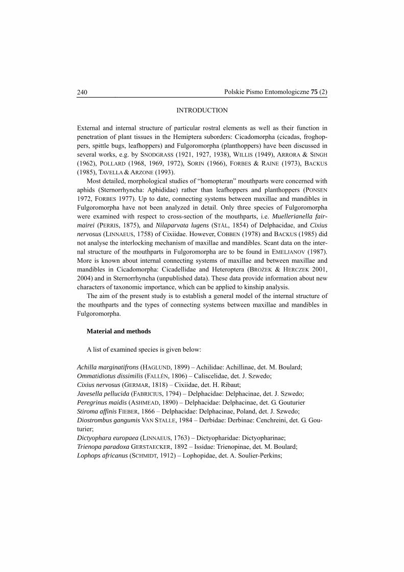
Fig. 1 – Head of the planthopper in lateral view and dorsal view of mouthparts at the point of cross section.
Material for analysis was obtained from the collection of the Museum National d’Historie Naturelle in Paris: Peregrinus maidis, Cixius nervosus, Achilla marginatifrons, Dictyophara europaea, Trienopa paradoxa, Lophops africanus, and Distrombus gangumis; and from the collection of the Department of Zoology, Faculty of Biology and Environ-mental Protection, University of Silesia in Katowice: Stiroma affinis, Javesella pellucida and Ommatidiotus dissimilis. The basal part of head with a part of rostrum was glued verti-cally onto the stage of a scanning microscope. Thus prepared material was dusted with gold and photographs were taken in SEM, Jeol SM III. The obtained documentation helped to reconstruct the rostrum from the point of cross-section. The structure of mouthparts was analysed in cross-section through the subapical rostral segment of adult specimens. This segment is usually well-developed and present in all adult planthoppers. According to other researchers, the internal structure of rostrum is irregular, so it was essential that comparison of the same structural elements in the studied groups be conducted with reference to the same level of cross-section (FORBES 1976).
Figure 1 presents dorsal view of the mouthparts of planthopper and the point of cross-
section through the subapical segment of mouthparts. Figure 2 present a general model of connecting systems between maxillae and mandi-
bles in Fulgoroidea.
242 Polskie Pismo Entomologiczne 75 (2)
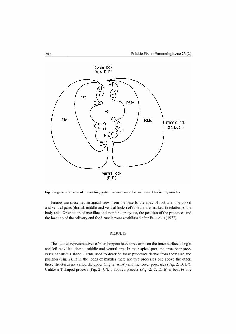
Fig. 2 – general scheme of connecting system between maxillae and mandibles in Fulgoroidea.
Figures are presented in apical view from the base to the apex of rostrum. The dorsal and ventral parts (dorsal, middle and ventral locks) of rostrum are marked in relation to the body axis. Orientation of maxillae and mandibular stylets, the position of the processes and the location of the salivary and food canals were established after POLLARD (1972).
RESULTS The studied representatives of planthoppers have three arms on the inner surface of right
and left maxillae: dorsal, middle and ventral arm. In their apical part, the arms bear proc-esses of various shape. Terms used to describe these processes derive from their size and position (Fig. 2). If in the locks of maxilla there are two processes one above the other, these structures are called the upper (Fig. 2: A, A’) and the lower processes (Fig. 2: B, B’). Unlike a T-shaped process (Fig. 2: C’), a hooked process (Fig. 2: C, D, E) is bent to one
BROŻEK J. et al.: The interlocking mechanism of Fulgoroidea mouthparts 243 side, while a straight process (Fig. 2: D’) is not deflected.
The number of processes on the arms of right and left maxillae is different. On the right maxilla there are five processes, while on the left maxilla there are four. The relative placement of processes on the arms of right and left maxilla forms complex connecting structures, the locks. Representatives of Fulgoromorpha have three locks: dorsal, middle and ventral.
The dorsal lock comprises four processes (Fig. 2): upper – hooked (A’) and straight (A); and lower – straight (B’) and hooked (B). Two hooked processes (A’) and (B) interlock. Straight processes (A) and (B’) form a cover for the interlocking hooked processes. They prevent the unhooking of hooked processes when maxillae are moving. They may also prevent various elements from penetrating into the lock between the inner faces of hooked processes.
The middle lock is composed of three processes (Fig. 2): a T-shaped process (C’) and two hooked processes (C, D). Hooked processes (C, D) interlock with the T-shaped process (C’). The glide path for the T-shaped process is formed by hooked processes, which prevent the maxillae from opening.
The ventral lock comprises only two processes (Fig. 2): a straight process (D’) and a hooked process (E).
The food canal is formed by both maxillae and is placed centrally in subdorsal portion. It is usually oval in cross-section due to concave inner walls of maxillae.
The salivary canal is formed by both maxillae. Process (E), which belongs to the ventral lock, is short, and the left maxilla forms a lateral wall closing the salivary canal (Fig. 2).
The canals differ in size. The diameter of the food canal was found to be much greater than that of the salivary canal.
Mandibles are placed laterally in relation to maxillae. The concave, smooth surfaces of mandibles are contiguous with the convex walls of maxillae (Fig. 2).
Cixiidae In representatives of the family Cixiidae the dorsal lock is formed by four processes. On
the left maxilla, there are processes: upper hooked and lower straight. On the right maxilla, there are two processes: upper straight and lower hooked (Figs 3 & 13).
The middle lock is formed by three processes. On the left maxilla, there is a T-shaped process. On the right maxilla, there are two hooked processes (Figs 3 & 13).
The ventral lock is formed by only two processes: straight and hooked. On the left max-illa, there is a straight process which interacts with the hooked process of the right maxilla (Figs 3 & 13).
The food canal is formed by both maxillae and is placed centrally in subdorsal portion. The salivary canal is formed by both maxillae. The process on the right maxilla which be-longs to the ventral lock is short, and the left maxilla forms a lateral wall closing the sali-vary canal. Mandibles are placed laterally in relation to maxillae. Mandibles are enlarged
244 Polskie Pismo Entomologiczne 75 (2) laterally and surround the dorsal lock from above and the ventral lock from beneath (Figs 3 & 13).
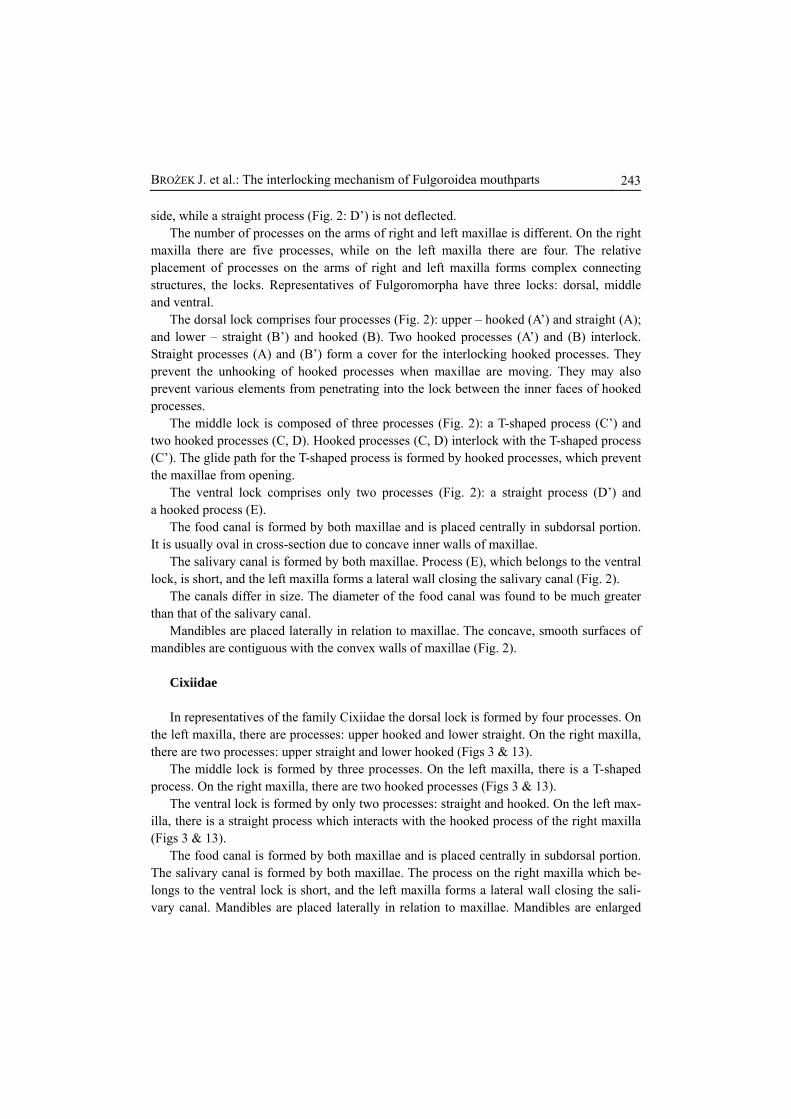
Figs 3–12. Cross section through by subapical segment of the rostrum in Fulgoromorpha. 3 - Cixius nervosus (Cixiidae), 4 - Stiroma affinis (Delphacidae), 5 - Javesella pellucida (Delphacidae), 6 - Peregrinus maidis (Delphacidae), 7 - Achilla marginatifrons (Achilidae), 8 - Diostrombus gangumis (Derbidae), 9 - Dictyophara europea (Dictyopharidae), 10 - Trienopa paradoxa (Issidae), 11 - Om-matidiotus dissmilis (Caliscelidae), 12 - Lophops africanus (Lophopidae).
BROŻEK J. et al.: The interlocking mechanism of Fulgoroidea mouthparts 245
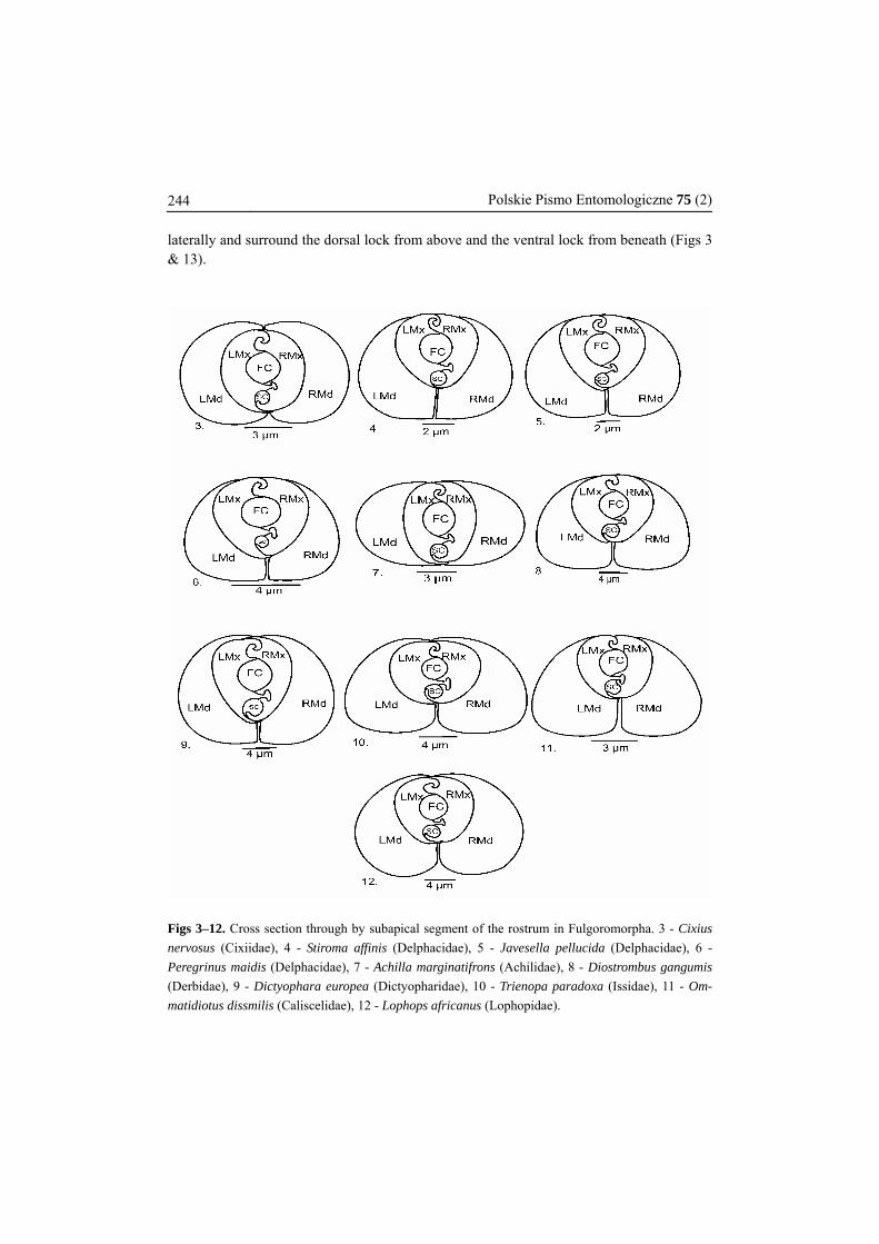
Figs. 13–18. Cross section through by subapical segment of the rostrum in Fulgoromorpha: 13 - Cixius nervosus (Cixiidae), 14 - Stiroma affinis (Delphacidae), 15 - Javesella pellucida (Delphacidae), 16 - Peregrinus maidis (Delphacidae), 17, 18 - Achilla marginatifrons (Achilidae).
246 Polskie Pismo Entomologiczne 75 (2)
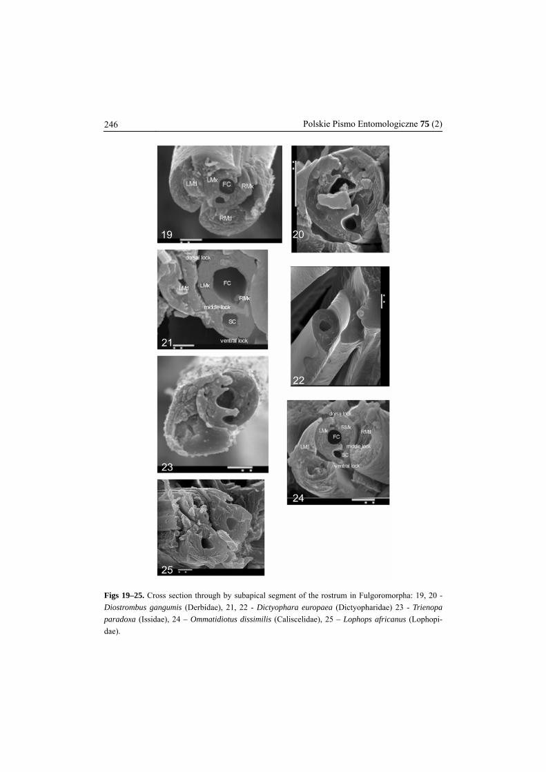
Figs 19–25. Cross section through by subapical segment of the rostrum in Fulgoromorpha: 19, 20 - Diostrombus gangumis (Derbidae), 21, 22 - Dictyophara europaea (Dictyopharidae) 23 - Trienopa paradoxa (Issidae), 24 – Ommatidiotus dissimilis (Caliscelidae), 25 – Lophops africanus (Lophopi-dae).
BROŻEK J. et al.: The interlocking mechanism of Fulgoroidea mouthparts 247
Delphacidae In representatives of the family Delphacidae the dorsal lock is formed by four processes.
On the left maxilla there are processes: upper, hooked and lower straight. On the right max-illa there are two processes: upper straight and lower hooked (Figs 4, 5 & 6).
The middle lock is formed by three processes. On the left maxilla, there is a T-shaped process. On the right maxilla, there are two hooked processes (Figs 4, 5 & 6).
The ventral lock is formed by only two processes: straight and hooked. On the left max-illa, there is a straight process, which interacts with the hooked process of the right maxilla.
The food canal is formed by both maxillae and is placed centrally in subdorsal portion. The salivary canal is formed by both maxillae. The process on the right maxilla which be-longs to the ventral lock is short, and the left maxilla forms a lateral wall closing the sali-vary canal (Figs 4–6, 14–16).
Mandibles are placed laterally in relation to maxillae. Mandibles are better developed in their ventral part and surround the ventral lock from beneath. (Figs 4, 5, 6, 14, 15 & 16).
Achilidae In representatives of the family Achilidae the dorsal lock is formed by four processes.
On the left maxilla, there are processes: upper hooked and lower straight. On the right max-illa, there are two processes: upper straight and lower hooked (Figs 7, 17 & 18).
The middle lock is formed by three processes: straight and hooked. On the left maxilla, there is a T–shaped process. On the right maxilla, there are two hooked processes (Figs 7 & 17).
The ventral lock is formed by only two processes. On the left maxilla, there is a straight process, which interacts with the hooked process of the right maxilla (Figs 7 & 17).
The food canal is formed by both maxillae and is placed centrally in subdorsal portion. The salivary canal is formed by both maxillae. The process on the right maxilla which be-longs to the ventral lock is short, and the left maxilla forms a lateral wall closing the sali-vary canal.
Mandibles are placed laterally in relation to maxillae. Mandibles are enlarged laterally and surround the dorsal lock from above and the ventral lock from beneath (Figs 7 & 17).
Derbidae In representatives of the family Derbidae the dorsal lock is formed by four processes.
On the left maxilla, there are processes: upper hooked and lower straight. On the right max-illa, there are two processes: upper straight and lower hooked (Figs 8, 19 & 20).
The middle lock is formed by three processes. On the left maxilla, there is a T–shaped process. On the right maxilla, there are two hooked processes (Figs 8, 19 & 20).
248 Polskie Pismo Entomologiczne 75 (2)
The ventral lock is formed by only two processes: straight and hooked. On the left max-illa, there is a straight process, which interacts with the hooked process of the right maxilla (Figs 8, 19 & 20).
The food canal is formed by both maxillae and is placed centrally in subdorsal portion. The salivary canal is formed by both maxillae. The process on the right maxilla which be-longs to the ventral lock is short, and the left maxilla forms a lateral wall closing the sali-vary canal.
Mandibles are placed laterally in relation to maxillae. Mandibles are better developed in their ventral part and surround the ventral lock from beneath (Figs 8, 19 & 20).
Dictyopharidae In representatives of the Dictyopharidae four processes form the dorsal lock. On the left
maxilla, there are processes: upper hooked and lower straight. On the right maxilla, there are two processes: upper straight and lower hooked (Figs 9, 21 & 22).
Three processes form the middle lock. On the left maxilla, there is a T-shaped process. On the right maxilla, there are two hooked processes (Figs 9, 21 & 22).
Two processes form the ventral lock: straight and hooked. On the left maxilla, there is a straight process, which interacts with the hooked process of the right maxilla (Figs 9, 21 & 22).
The food canal is formed by both maxillae and is placed centrally in subdorsal portion. The salivary canal is formed by both maxillae. The process on the right maxilla, which belongs to the ventral lock, is short, and the left maxilla forms a lateral wall closing the salivary canal.
Mandibles are placed laterally in relation to maxillae. Mandibles are better developed in their ventral part and surround the ventral lock from beneath (Figs 9, 21 & 22).
Issidae In representatives of the family four processes form the dorsal lock. On the left maxilla,
there are processes: upper hooked and lower straight. On the right maxilla, there are two processes: upper straight and lower hooked (Figs 10 & 23).
Three processes form the middle lock. On the left maxilla, there is a T-shaped process. On the right maxilla, there are two hooked processes (Figs 10 & 23).
Two processes form the ventral lock: straight and hooked. On the left maxilla, there is one straight process, which interacts with the hooked process of the right maxilla (Figs 10 & 23).
The food canal is formed by both maxillae and is placed centrally in subdorsal portion. The salivary canal is formed by both maxillae. The process on the right maxilla that be-longs to the ventral lock is short, and the left maxilla forms a lateral wall closing the sali-vary canal.
BROŻEK J. et al.: The interlocking mechanism of Fulgoroidea mouthparts 249
Mandibles are placed laterally in relation to maxillae. Mandibles are better developed in their ventral part and surround the ventral lock from beneath (Figs 10 & 23).
Caliscelidae In representatives of the family four processes form Caliscelidae the dorsal lock. On the
left maxilla, there are processes: upper hooked and lower straight. On the right maxilla, there are two processes: upper straight and lower hooked (Figs 11 & 24).
Three processes form the middle lock. On the left maxilla, there is a T-shaped process. On the right maxilla, there are two hooked processes (Figs 11 & 24).
Only only two processes form the ventral lock: straight and hooked. On the left maxilla, there is a straight process, which interacts with the hooked process of the right maxilla (Figs 11 & 24).
The food canal is formed by both maxillae and is placed centrally in subdorsal portion. The salivary canal is formed by both maxillae and is placed centrally. The process on the right maxilla that belongs to the ventral lock is short, and the left maxilla forms a lateral wall closing the salivary canal.
Mandibles are placed laterally in relation to maxillae. Mandibles are better developed in their ventral part and surround the ventral lock from beneath (Figs 11 & 24).
Lophopidae
In representatives of the family of Lophopidae four processes form the dorsal lock. On the left maxilla, there are processes: upper hooked and lower straight. On the right maxilla, there are two processes: upper straight and lower hooked (Fig. 12).
Three processes form the middle lock. On the left maxilla, there is a T-shaped process. On the right maxilla, there are two hooked processes (Fig. 12).
Only two processes form the ventral lock: straight and hooked. On the left maxilla, there is a straight process, which interacts with the hooked process of the right maxilla (Fig. 12).
The food canal is formed by both maxillae and is placed centrally in subdorsal portion. The salivary canal is formed by both maxillae. The process on the right maxilla that be-longs to the ventral lock is short, and the left maxilla forms a lateral wall closing the sali-vary canal.
Mandibles are placed laterally in relation to maxillae. Mandibles are best developed in their ventral part and surround the ventral lock from beneath (Figs 12 & 25).
DISCUSSION
An interesting aspect of the inner structure of mouthparts in the studied species is the high structural similarity observed in different families of Fulgoromorpha. One of the characteristic
250 Polskie Pismo Entomologiczne 75 (2) features of planthoppers is the different number of processes constituting each of the three locks (dorsal lock – 4 processes, middle lock – 3 processes and ventral lock – 2 processes). Another model was observed in Cicadomorpha (BROŻEK & HERCZEK 2001), where there are only two locks: dorsal and ventral (and the locks are formed by four processes each). Fulgoro-morpha and Cicadomorpha have long been regarded as sister groups – a claim supported by YOSHIZAWA & SAIGUSA (2001) and BOULARD (2002). It appears, however, that these two groups should perhaps receive a different treatment (BROŻEK et al. 2003; BOURGOIN & CAMPBELL 2002). A close relationship between Fulgoromorpha and Cicadomorpha does not seem to be supported by the characters of the internal connecting systems of the mouthparts. The presence of three locks in the mouthparts has been observed in both Heteroptera (BROŻEK & HERCZEK 2004) and Sternorrhyncha (BROŻEK, in press). The number of locks in Fulgoro-morpha, Heteroptera and Sternorrhyncha offers an interesting insight into the phylogenetic relationship among these groups. Some authors have suggested the possibility of a sister-group relationship between Fulgoromorpha and Heteroptera (GOODCHILD 1966; HAMILTON 1981; BOURGOIN 1986, 1993; BOURGOIN et al. 1997; SWEET 1996; SORENSEN et al. 1995; CAMPBELL et al. 1995).
A position of the food canal is stable, i.e. central in the dorsal part of the rostrum. It has been observed that the shape and size of the food canal are similar in all the analyzed species of Fulgoromorpha (Figs 2–24). The stable central position of the food canal has also been ob-served in other groups of Heteroptera (BROŻEK & HERCZEK 2004) and in Sternorrhyncha.
The salivary canal is placed in both maxillae in all the studied representatives of various families, and the size of the canal is similar. A unique feature of this group is the type of closure of the salivary canal. In representatives of the examined families, the salivary canal is closed by a short process of the right maxilla (Figs 2–8, 10–12), and by the wall of the left maxilla. On the basis of COBBEN’s (1978) photograph it can be established that in Muellerianella fairmairei (PERRIS, 1857) (Delphacidae) the salivary canal is closed in a similar way. A different situation can be observed in Nilaparvata lugens (STÅL, 1854) (Delphacidae), where, according to Fig. 7.7 in BACKUS (1985), the salivary canal is entirely closed by a very long process of the right maxilla. This type of closure of the salivary canal in heteropteran groups (BROŻEK & HERCZEK 2004) is reminiscent of Fulgoromorpha. Another type of closure of the salivary canal is ob-served in Cicadellidae. In this group, the upper process of the ventral lock, which is placed on the left maxilla (BROŻEK & HERCZEK 2001), close the salivary canal.
In the analysed species of Fulgoromorpha, no connection has been found between maxillae and mandibles. Mandibles lie close to maxillae, which is also true of Cicadomorpha. In con-trast to Fulgoromorpha and Cicadomorpha (BROŻEK & HERCZEK 2001), in Heteroptera maxil-lae and mandibles are connected by processes (COBBEN 1978; BROŻEK & HERCZEK 2004).
In the examined representatives of Delphacidae, Derbidae, Dictyopharidae, Issidae, Calis-celidae and Lophopidae, mandibles are better developed in the ventral part. On the basis of Fig. 7.7 of Nilaparvata lugens (Delphacidae) in BACKUS (1985) and on the basis of COBBEN’s (1978) data on Muellerianella fairmairei (Delphacidae), it can be established that also in these two species mandibles are better developed in their ventral part. Only in Cixiidae and Achilidae
BROŻEK J. et al.: The interlocking mechanism of Fulgoroidea mouthparts 251 mandibles are laterally enlarged (Figs 3 & 7). Resemblance between the shape of maxillae and mandibles in Cixiidae and Achillidae may result from their phylogenetic relationships.
According to BOURGOIN (1986), the clades Cixiidae and Achilidae are probably closely re-lated. However, ASCHE (1987) and EMELJANOV (1991), based on morphology, argue that the nearest group to Cixiidae is Delphacidae. It was also supported by female genitalia structures (BOURGOIN 1993) and molecular data (BOURGOIN et al. 1997, YEH et al. 2005). ASCHE (1987), EMELJANOV (1991) BOURGOIN (1993), BOURGOIN et al. (1997) agreed that Meenopliidae is a sister group to Kinnaridae, Fulgoridae is a sister group to Dictyopharidae, Achilidae and Derbidae are closely related. Based on the molecular research the relationship between Meenoplidae and Delphacidae is distant (YEH et al. 1998), which is in disagreement with stud-ies of nymphal characters (CHEN & YANG 1995). YANG & FANG (1993), based on nymphal characters divided Fulgoroidea into 2 groups Delphacidae–Meenopliidae–Cixiidae–Achilidae–Derbidae, with the rest in and individual group. CHEN & YANG (1995) divided them into five family groups (I: Tettigometridae-group – Tettigometridae; II: Ricaniidae-group – Ricaniidae, Lophopidae, Eurybrachidae, Hypochtonellidae, Gengidae; III: Tropiduchidae-group: Tropiduchidae, Flatidae, Nogodinidae, Issidae, Acanaloniidae, IV: Fulgoridae-group: Fulgori-dae, Dictyopharidae; V: Cixiidae-group: Delphacidae, Meenoplidae, Kinnaridae, Cixiidae, Achilidae, Achilixidae, Derbidae). YANG & CHANG (2000) based on nymphal features and male genital structures, proposed sister groups Achilidae and Achilixidae, Cixiidae and Der-bidae, Fulgoridae and Dictyopharidae, Kinnaridae and Meenopliidae as sister group to Delpha-cidae. According to YEH et al. (2005) Delphacidae and Cixiidae are sister groups, Derbidae is sister group to Tropiduchidae, Fulgoridae to Dictyopharidae, and Meenoplidae to Lophopidae. All these families forms basal lineage of Fulgoroidea, where relationships among Meenoplidae, Delphacidae–Cixiidae, Lophopidae and Achilidae cannot be clearly resolved. Several groups among families in the derived lineages are revealed, including Derbidae-Tropiduchidae, Dic-tyopharidae-Fulgoridae, and Eurybrachidae–Ricaniidae, but relationships among the remaining families were poorly resolved (YEH at al. 2005).
Different morphological characters appear to reveal different relative phylogenetic posi-tions of particular families, molecular results are also ambiguous. Thus, the only conclusion generally accepted in Fulgoroidea systematics is that the Fulgoridae and Dictyopharidae are sister groups, the families Cixiidae, Achilidae and Delphacidae – and Derbidae, Kinnaridae and Meenoplidae – are recognized as a cluster of closely related planthopper groups lying near the common stem. The study of the internal structure of the mouthparts allows adding more impor-tant data for studies on Fulgoroidea phylogeny.
REFERENCES ARRORA G.L., SINGH S. 1962. Morphology and musculature of the head and mouth–parts of Idiocerus
atkinsoni LETH. (Jassidae, Homoptera). Journal of Morphology 110: 131–140. ASCHE M. 1987. Preliminary thoughts in the phylogeny of Fulgoromorpha (Homoptera: Auchenor-
rhyncha). Proceedings of the 6th International Auchenorrhyncha Meeting, Turin, Italy, 47–53.
252 Polskie Pismo Entomologiczne 75 (2) BACKUS E.A. 1985. Anatomical and sensory mechanisms of leafhopper and planthopper feeding
behavior. In: NAULT L.R., RODRIGUEZ J.G. (eds.). The Planthoppers and Leafhoppers). John Wiley and Sons: 163–194.
BOULARD M. 2002. Diversité des Auchènorhynques Cicadomorphes formes, couleurs et comporte-ments. Denisia 04, zugleich Katalogue des OÖ. Landesmuseums, Neue Folge Nr. 176: 171–214.
BOURGOIN TH. 1986. Morphologie imaginale du tentorium des Hemiptera, Fulgoromorpha. Interna-tional Journal of Insect Morphology & Embryology 15(4): 237–252.
BOURGOIN TH. 1993. Female genitalia in Hemiptera: Fulgoromorpha, morphological and phylogenetic date. Annales de la Société entomologique de France (N.S.) 29(3): 225–244.
BOURGOIN TH., STEFFEN-CAMPBELL, CAMPBELL B.C. 1997. Molecular phylogeny of Fulgoromorpha (Insecta: Hemiptera, Archaeorrhyncha). The enigmatic Tettigometridae; evolutionary affiliatons and historical biogeography. Cladistics 13: 207–224.
BOURGOIN TH., CAMPBELL B.C. 2002. Inferring a phylogeny for Hemiptera: Falling into the ‘Autapo-morphic Trap’. Denisia 04, zugleich Kataloge des OÖ. Landesmuseums, Neue Folge Nr. 176: 67–82.
BROŻEK J., HERCZEK A. 2001. Modification in the mouthparts structure in selected species of Cicadel-lidae (Hemiptera: Cicadomorpha). Acta entomologica silesiana 7–8: 19–25.
BROŻEK J., SZWEDO J., GAJ D., PILARCZYK S. 2003. Former and current views on the classification of the bugs (Insecta, Hemiptera). Genus. International Journal of Invertebrate Taxonomy, (Supple-ment): 85–110
BROŻEK J., HERCZEK A. 2004. Internal structure of the mouthparts of true bugs (Hemiptera, Heterop-tera). Polskie Pismo Entomologiczne 73(2), 79–106.
CAMPBELL B.C., STEFFEN-CAMPBELL J.D., SORENSEN J.T., GILL R.J. 1995. Paraphyly of Homoptera and Auchenorrhyncha inferred from 18S rDNA nucleotide sequence. Systematic Entomology 20: 175–194.
CHEN S., YANG C-T. 1995. The metatarsi of the Fulgoroidea (Homoptera: Auchenorrhyncha). Chinese Journal of Entomology 15: 257–269.
COBBEN R.H. 1978. Evolutionary trends in Heteroptera. Part II. Mouthpart–structures and feeding strategies. Mededelingen. Landbouwhogeschool Wageningen 78: 1–401.
EMELJANOV A.F. 1987. Filogenia cikadovykh (Homoptera, Cicadina) po sravnitel’no-morfologicheskim dannym. [The phylogeny of cicadoids (Homoptera, Cicadina) based on com-parative morphological data]. Trudy Vsesoyuznogo Entomologicheskogo Obshchestva 69: 19–109. [in Russian]
EMELJANOV A.F. 1990. Opyt postroenya filogeneticheskogo dereva fulgoroidnykh tsikadovykh (Ho-moptera, Cicadina). Entomologicheskoe Obozrenie 69(2): 353–356. [In Russian] Published in English as EMELJANOV A.F. 1991. An attempt to construct a phylogenetic tree for planthoppers (Homoptera, Cicadina). Entomological Revue 70(1): 24–28.
FORBES A.R., RAINE J. 1973. The stylets of the six-spotted leafhopper, Macrosteles fascifrons (Ho-moptera: Cicadellidae). Canadian Entomologist 105: 559–567.
FORBES A.R. 1977. Mouthparts and feeding mechanism of aphids. [In:] HARRIS K.F., MARAMOROSCH K. (Eds.), Aphids as Virus Vectors, New York: 83–103.
GOODCHILD A.J.P. 1966. Evolution of the alimentary canal in the Hemiptera. Biological Review 41: 97–140.
HAMILTON K.G.A. 1981. Morphology and evolution of the Rhynchotan head (Insecta: Hemiptera, Homoptera). Canadian Entomologist 113(11): 953–974.
POLLARD D.G. 1968. Stylet penetration and feeding damage of Eupteryx mellissae CURTIS (Hemiptera: Cicadellidae) on sage. Bulletin of the Entomological Research 58: 55–71.
POLLARD D.G. 1969. Directional control of the stylets in phytophagous Hemiptera. Proceedings of the Royal Entomological Society of London (A), 44 (10-12): 173–185.
BROŻEK J. et al.: The interlocking mechanism of Fulgoroidea mouthparts 253 POLLARD D.G. 1972. The stylet structure of leafhopper (Eupteryx mellissae CURTIS: Homoptera, Ci-
cadellidae). Journal of Natural History 6: 261–271. PONSEN M.B. 1972. The site of potato leafroll virus multiplication in its vectors, Myzus persicae.
Mededelingen. Landbouwhogeschool Wageningen, 1–147. SNODGRASS R.E. 1921. The mouth parts of the cicada. Proceedings of the Entomological Society of
Washington 23: 1–15. SNODGRASS R.E. 1927. The head and mouth parts of the cicada. Proceedings of the Entomological
Society of Washington 29: 1–16. SNODGRASS R.E. 1938. The loral plates and the hypopharynx of Hemiptera. Proceedings of the Ento-
mological Society of Washington 40: 228–236. SORENSEN J.T., CAMPBELLB.C., GILL, R.J., STEFFEN-CAMPBELL J.D. 1995. Non-monophyly of
Auchenorrhyncha (“Homoptera”), based upon 18S rDNA phylogeny: eco-evolutionary and cladistic implications with in pre-heteropterodea Hemiptera (s.l) and a proposal for new mono-phyletic suborders. Pan-Pacific Entomologist 71(1): 31–60.
SORIN M. 1966. Physiological and morphological studies on the suction mechanism of plant juice by aphids. Bulletin of University of Osaka Prefecture B, 18: 95–135.
SWEET M.H. 1996. Comparative external morphology of the pregenital abdomen of the Hemiptera. In: SCHAEFER C.W. (ed.). Studies on Hemiptera Phylogeny. Thomas Say Publications in Entomology 1: 119–158.
TAVELLA L., ARZONE A. 1993. Comparative morphology of mouth parts of Zyginidia pullula, Em-poasca vitis, and Graphocephala fennahi (Homoptera, Auchenorrhyncha). Bolletin de Zoologia 60: 33–39.
YANG C.-T., CHANG T.-Y. 2000. The external male genitalia of Hemiptera (Homoptera - Heteroptera). Shich Way Publishers, Taichung, R.O.C. 1–746.
YANG C.-T., FANG S.-J. 1993. Phylogeny of fulgoromorpha nymphs, first results. In: DROSOPOULOS S., PETRAKIS P.V., CLARIDGE M.F., DE VRIJER P.W.F. (eds.), Proceedings of the 8th Auchenorrhyncha Congress, Delphi, Greece, 9–13 August 1993: 25–26.
YEH W.-B., YANG C.-T., HUI C.-F. 1998. Phylogenetic relationships of the Tropiduchidae–group (Ho-moptera: Fulgoroidea) of planthoppers inferred through nucleotide sequences. Zoological Studies 37(1): 45–55.
YEH W.B., YANG C.-T., HUI C.-F. 2005. A molecular phylogeny of planthoppers (Hemiptera: Fulgor-oidea) inferred from mitochondrial 16S rDNA sequences. Zoological Studies, 44(4): 519–535.
WILLIS D. 1949. The anatomy and histology of the head, gut and associated structures of Typhlocyba ulmi. Proceedings of the Zoological Society of London 118: 984–1001.
YOSHIZAWA K., SAIGUSA T. 2001. Phylogenetic anylysis of paraneopteran orders (Insecta: Neoptera) based on forewing base structure, with comments on monophyly of Auchenorrhyncha (Hemip-tera). Systematic Entomology 26: 1–13.
Received: 20 December 2005 Accepted: 29 March 2006

P O L S K I E P I S M O E N T O M O L O G I C Z N EP O L I S H
J O U R N A L
O F E N T O M O L O G Y
VOL. 75: 239–253 Bydgoszcz 30 June 2006


![PISMO TECHNICZNE - zsdydnia.eu · Pismo techniczne PN-EN ISO 3098 (PN-80/N-01606) Rozróżniamy rodzaje pisma :-PISMO TYPU A [d=(h/14)] -PISMO TYPU B [d=(h/10)] UWAGA !](https://static.fdocuments.pl/doc/165x107/604e38a455fe982c4f3c3745/pismo-techniczne-pismo-techniczne-pn-en-iso-3098-pn-80n-01606-rozrniamy.jpg)