Algorithm in abdominal trauma Algorytm w obrażeniach jamy ...
15
THE JOURNAL OF ORTHOPAEDICS TRAUMA SURGERY AND RELATED RESEARCH Algorithm in abdominal trauma WALDEMAR HŁADKI Klinika Medycyny Ratunkowej i Obrażeń Wielonarządowych II Katedry Chirurgii Ogólnej UJCM w Krakowie Kierownik katedry: prof. dr hab. D. Karcz Address for correspondence/Adres do korespondencji: dr hab. n. med. Waldemar Hładki Klinika Medycyny Ratunkowej i Obrażeń Wielonarządowych UJ CM w Krakowie ul. Kopernika 21, 31-501 Kraków tel. 012-424-82-13, fax 012-421-34-56, e-mail: [email protected] © J ORTHOP TRAUMA SURG REL RES 1 (13) 2009 Review article/Artykuł poglądowy Algorytm w obrażeniach jamy brzusznej Summary Even though abdominal traumas still constitute a small percentage of bodily injuries, their number has been growing considerably over the recent years, and they are one of the main life threats after accidents. A particular increase in the number of patients with abdominal trauma can be observed in patients with multiple bodily injuries. The efficacy of their therapy depends on correct proce- dures, equipment and specialistic treatment at the pre-hospital stage, at the hospital emergency ward, at the operation theatre and at the intensive care unit. Procedures complying to the rules of the Advanced Trauma Live Support at the site of the accident and while transporting the patient, modern diagnostic methods with an application of ultrasonographic examination and computed tomography allow to increase the survival rate of the patients. A quick diagnosis in the case of massive ble- edings into the abdominal cavity following multiorgan injuries offers a chance of survival. The paper presents the rules of proceeding with patients suffering from blunt and penetrating injuries of the abdominal cavity. The main factor, decisive for the diagnostic possibilities and the choice of tre- atment is the patient’s haemodynamic stability. The paper describes the emergency procedure in abbreviated laparotomy and damage control procedures as well as the procedure of staged laparo- tomy. Attention has been paid to a partly different procedure in the case of abdominal trauma coexisting with injuries of other body areas, and to the growing number of indications for prese- rvative treatment, mainly of isolated abdominal traumas, including the use of interventional radio- logy. Key words: algorithm, management, abdominal injuries, multiple injuries Streszczenie Choć obrażenia narządów jamy brzusznej stanowią nadal niewielki odsetek obrażeń ciała to ich liczba w ostatnich latach znacznie wzrasta, a są one jedną z głównych przyczyn zagrożenia życia po urazach. Szczególny wzrost liczby chorych z obrażeniami brzucha nastąpił u chorych z mnogimi obrażenia- mi ciała. Skuteczność leczenia chorych zależy od prawidłowego postępowania, zaopatrzenia i leczenia specjalistycznego na etapie postępowania przedszpitalnego, w szpitalnym oddziale ratun- kowym, oraz na sali operacyjnej i w oddziale intensywnej terapii. Postępowanie zgodnie z usta- leniami Advanced Trauma Live Support na miejscu zdarzenia i w czasie transportu chorego, no- woczesne metody diagnostyczne z zastosowaniem badania ultrasonograficznego, oraz tomografii komputerowej pozwalają obecnie na poprawienie przeżywalności chorych. Szybkie postawienie rozpoznania w przypadku masywnych krwotoków do jamy brzusznej w następstwie uszkodzeń wie- lonarządowych daje szansę przeżycia. W artykule przedstawiono zasady postępowania u chorych z obrażeniami tępymi i przenikającymi jamy brzusznej. Głównym czynnikiem decydującym o moż- liwościach diagnostycznych i wyborze sposobu leczenia jest stan hemodynamicznej stabilności pa- Statistic/Statystyka Word count/Liczba słów 3266/2703 Tables/Tabele 0 Figures/Ryciny 2 References/Piśmiennictwo 55 Received: 22.12.2007 Accepted: 17.05.2008 Published: 01.02.2009
Transcript of Algorithm in abdominal trauma Algorytm w obrażeniach jamy ...
THE JOURNAL OF ORTHOPAEDICS TRAUMA SURGERY
AND RELATED RESEARCH
Algorithm in abdominal trauma
WALDEMAR HŁADKI
Klinika Medycyny Ratunkowej i Obrażeń Wielonarządowych II Katedry ChirurgiiOgólnej UJCM w KrakowieKierownik katedry: prof. dr hab. D. Karcz
Address for correspondence/Adres do korespondencji:dr hab. n. med. Waldemar HładkiKlinika Medycyny Ratunkowej i Obrażeń Wielonarządowych UJ CM w Krakowieul. Kopernika 21, 31-501 Krakówtel. 012-424-82-13, fax 012-421-34-56, e-mail: [email protected]
© J ORTHOP TRAUMA SURG REL RES 1 (13) 2009Review article/Artykuł poglądowy
Algorytm w obrażeniach jamy brzusznej
Summary
Even though abdominal traumas still constitute a small percentage of bodily injuries, their number
has been growing considerably over the recent years, and they are one of the main life threats after
accidents. A particular increase in the number of patients with abdominal trauma can be observed
in patients with multiple bodily injuries. The efficacy of their therapy depends on correct proce-
dures, equipment and specialistic treatment at the pre-hospital stage, at the hospital emergency ward,
at the operation theatre and at the intensive care unit. Procedures complying to the rules of the
Advanced Trauma Live Support at the site of the accident and while transporting the patient, modern
diagnostic methods with an application of ultrasonographic examination and computed tomography
allow to increase the survival rate of the patients. A quick diagnosis in the case of massive ble-
edings into the abdominal cavity following multiorgan injuries offers a chance of survival. The paper
presents the rules of proceeding with patients suffering from blunt and penetrating injuries of the
abdominal cavity. The main factor, decisive for the diagnostic possibilities and the choice of tre-
atment is the patient’s haemodynamic stability. The paper describes the emergency procedure in
abbreviated laparotomy and damage control procedures as well as the procedure of staged laparo-
tomy. Attention has been paid to a partly different procedure in the case of abdominal trauma
coexisting with injuries of other body areas, and to the growing number of indications for prese-
rvative treatment, mainly of isolated abdominal traumas, including the use of interventional radio-
logy.
Key words: algorithm, management, abdominal injuries, multiple injuries
Streszczenie
Choć obrażenia narządów jamy brzusznej stanowią nadal niewielki odsetek obrażeń ciała to ich liczba
w ostatnich latach znacznie wzrasta, a są one jedną z głównych przyczyn zagrożenia życia po urazach.
Szczególny wzrost liczby chorych z obrażeniami brzucha nastąpił u chorych z mnogimi obrażenia-
mi ciała. Skuteczność leczenia chorych zależy od prawidłowego postępowania, zaopatrzenia
i leczenia specjalistycznego na etapie postępowania przedszpitalnego, w szpitalnym oddziale ratun-
kowym, oraz na sali operacyjnej i w oddziale intensywnej terapii. Postępowanie zgodnie z usta-
leniami Advanced Trauma Live Support na miejscu zdarzenia i w czasie transportu chorego, no-
woczesne metody diagnostyczne z zastosowaniem badania ultrasonograficznego, oraz tomografii
komputerowej pozwalają obecnie na poprawienie przeżywalności chorych. Szybkie postawienie
rozpoznania w przypadku masywnych krwotoków do jamy brzusznej w następstwie uszkodzeń wie-
lonarządowych daje szansę przeżycia. W artykule przedstawiono zasady postępowania u chorych
z obrażeniami tępymi i przenikającymi jamy brzusznej. Głównym czynnikiem decydującym o moż-
liwościach diagnostycznych i wyborze sposobu leczenia jest stan hemodynamicznej stabilności pa-
Statistic/Statystyka
Word count/Liczba słów 3266/2703
Tables/Tabele 0
Figures/Ryciny 2
References/Piśmiennictwo 55
Received: 22.12.2007Accepted: 17.05.2008Published: 01.02.2009
63Algorithm in abdominal trauma
1 (13) 2009
WSTĘPObrażenia jamy brzusznej stanowią około 5% wszystkich
obrażeń ciała, a połowie z nich towarzyszą obrażenia
innych okolic ciała (głowy, kończyn, klatki piersiowej).
Urazy jamy brzusznej mogą prowadzić do uszkodzenia
narządów miąższowych (najczęściej śledziona i wątroba)
z towarzyszącym im krwawieniem, lub masywnym krwo-
tokiem wewnętrznym, czy też do uszkodzenia światła
przewodu pokarmowego i zapalenia otrzewnej. Do krwo-
toków do jamy brzusznej może dochodzić także przy
uszkodzeniu naczyń krwionośnych zaopatrujących trzust-
kę i jelita, oraz przy uszkodzeniu nerek, naczyń krwio-
nośnych przestrzeni zaotrzewnowej, oraz narządów mied-
nicy znajdujących się poza jamą otrzewnej (pęcherz
moczowy, macica, przydatki), kiedy krwotok przedosta-
je się do jamy otrzewnej. Konieczność szybkiej interwen-
cji operacyjnej, lub możliwość wykonania niezbędnych
badań diagnostycznych i dopiero potem podjęcia właści-
wego leczenia zależy w głównej mierze od stabilności
hemodynamicznej pacjenta. Obrażenia narządów jamy
brzusznej mogą w bardzo krótkim czasie spowodować
zagrożenie życia i określać właściwy priorytet zaopatry-
wania uszkodzeń innych okolic ciała. Z drugiej strony,
możliwość pojawienia się objawów klinicznych z pew-
nym opóźnieniem, może prowadzić do trudności diagno-
stycznych, szczególnie w początkowym okresie po ura-
zie. Obiektywna ocena narządów jamy brzusznej z uży-
ciem badania ultrasonograficznego (USG), tomografii
komputerowej (KT) czy diagnostycznego płukania
otrzewnej (DPO) jest niezbędna u pacjentów stabilnych
hemodynamicznie. Natomiast dla sposobu zaopatrzenia
ma także znaczenie, czy obrażenie jest izolowane, czy też
nie. Spośród pacjentów z mnogimi obrażeniami ciała
(MOC), istotne dla chorego obrażenia brzucha ma ponad
25% poszkodowanych i odsetek ten powoli, ale systema-
tycznie wzrasta w ciągu ostatnich kilkunastu lat. W tej
sytuacji rozpoznanie obrażeń narządów jamy brzusznej
może być utrudnione z powodu współistnienia innych
obrażeń [25,30,34,38].
cjenta. Omówiono postępowanie ratunkowe w ramach skróconej laparotomii – abbreviated lapa-
rotomy i procedury damage control, oraz postępowania w ramach laparotomii etapowej – staged
laparotomy. Zwrócono uwagę na częściową odmienność postępowania w przypadku obrażeń brzu-
cha współistniejących i obrażeniami innych okolic ciała, oraz na coraz szersze wskazania do le-
czenia zachowawczego głównie izolowanych obrażeń jamy brzusznej, także z wykorzystanie ra-
diologii interwencyjnej.
Słowa kluczowe: algorytm, postępowanie, obrażenia jamy brzusznej, mnogie obrażenia ciała
INTRODUCTIONAbdominal traumas constitute about 5% of all bodily
injuries, half of them being accompanied by injuries of
other body areas (head, limbs, chest). Abdominal trau-
ma may lead to the damage of parenchymatous organs
(usually spleen and liver) with an accompanying bleeding
or a massive internal bleeding, or to the damage of the
lumen of the alimentary tract and to peritonitis. Bleedings
into the abdominal cavity may also occur in the case of
injuring the blood vessels of the pancreas and intestines,
injuring the kidneys, blood vessels of the retroperitoneal
space, and the organs of the pelvis beyond the peritoneal
cavity (urinary bladder, uterus, adnexa), when the ble-
eding enters the peritoneal cavity. The necessity of
a prompt surgical intervention or the possibility to per-
form the necessary diagnostic examinations before star-
ting the treatment itself depend mainly on the patient’s
haemodynamic stability. Abdominal trauma may endan-
ger the patient’s life in a very short time, which deter-
mines the right priority of treating the injuries of other
body areas. On the other hand, the possibility of clinical
symptoms occurring with a certain delay may pose dia-
gnostic difficulties, particularly in the initial period after
the accident. An objective evaluation of abdominal or-
gans with the use of ultrasonographic examination, com-
puted tomography or diagnostic peritoneal lavage is
necessary in haemodynamically stable patients. However,
the procedure depends also on the injury being isolated
or not. Among the patients with multiple bodily injuries,
25% display significant abdominal trauma, the percenta-
ge growing slowly but steadily over the last decade or
two. In these circumstances, diagnosing an abdominal
trauma may prove difficult due to a coexistence of other
injuries [25, 30, 34, 38].
64 W. HŁADKI
THE JOURNAL OF ORTHOPAEDICS TRAUMA SURGERY
AND RELATED RESEARCH
Spośród tych, którzy giną na miejscu wypadku aż
u 50% główną przyczyną bywa intensywne krwawienie
do jamy brzusznej. W Polsce 80% obrażeń jamy brzusz-
nej to obrażenia zamknięte (tępe), które powstają głów-
nie w następstwie wypadków komunikacyjnych i upad-
ków z wysokości, czy bezpośredniego uderzenia, np.
uderzenia pięścią, lub kopnięcia. Tępe obrażenia jamy
brzusznej są trudniejsze do rozpoznania. Warto pamiętać,
że tępe obrażenia dolnej części klatki piersiowej ze zła-
maniami żeber mogą powodować obrażenia śledziony
i wątroby, a złamania miednicy prowadzić do uszkodzeń
w obrębie jelit. W Stanach Zjednoczonych prawie poło-
wę przypadków stanowią uszkodzenia otwarte(przenika-
jące) (rany postrzałowe i kłute). Wyraźnie dominują
mężczyźni (ponad 75% poszkodowanych) i ludzie mło-
dzi (średni wiek niewiele ponad 30 lat) [14,24,41,48].
Obrażenia drążące do jamy brzusznej są głównie powo-
dowane użyciem noży lub broni palnej, jednakże wiel-
kość rany zewnętrznej zwykle nie odpowiada rozległo-
ści obrażeń wewnątrz jamy brzusznej. Prawie zawsze
wymagają leczenia operacyjnego. Ich rozpoznanie jest
łatwe, a zaopatrzenie powinno odbyć się w trybie pilnym
bądź natychmiastowym w zależności od rodzaju uszko-
dzeń i stanu hemodynamicznego chorego [34,38,50].
Umownie przyjęty czas w ramach którego należy wstęp-
nie zaopatrzyć chorego, w tym także poszkodowanego
z obrażeniami jamy brzusznej, zabezpieczyć jego czyn-
ności życiowe i odwrócić procesy metaboliczne mogące
doprowadzić do jego zgonu nazwano „złota godziną”.
Czas ten musi być optymalnie wykorzystany przez ze-
spół lekarzy, ratowników, pielęgniarek i diagnostów
[13,14, 17,25].
POSTĘPOWANIE W WARUNKACH PRZEDSZPITALNYCHU pacjentów z podejrzeniem obrażeń brzucha postępo-
wanie na miejscu wypadku jest uzależnione od stabilno-
ści hemodynamicznej chorego. U chorego stabilnego
hemodynamicznie sprowadza się to do szybkiego stoso-
wania zaleceń ATLS (Advanced Trauma Life Support)
wg reguły „zostań i działaj” – „stay and play” [2].
W pierwszej kolejności, do wstępnego dokładnego bada-
nia rozebranego całkowicie poszkodowanego wg schema-
tu ABC….., rozpoznania zamkniętego lub drążącego ob-
rażenia jamy brzusznej (rany brzucha, otarcia naskórka,
skórne podbiegnięcia krwawe, powiększenie obwodu
brzucha ze stwierdzanym w badaniu fizykalnym płynem
w jamie brzusznej), oraz poszukiwania objawów wstrząsu
krwotocznego, a także rozpoznania lub wykluczenia
obrażeń innych okolic ciała, które również należy wstęp-
nie zaopatrzyć. Obrażenia przewodu pokarmowego bez
następowego intensywnego krwawienia nie powodują
doraźnie bezpośredniego zagrożenia życia i dopiero za-
palenie otrzewnej istotnie pogarsza stan chorego. W przy-
padku chorych niestabilnych hemodynamicznie, kiedy
rozpoczęta resuscytacja płynowa i farmakologiczna oka-
zuje się nieskuteczna, lub kiedy czas dojazdu do najbliż-
szego szpitala potrwa najwyżej do 5 minut należy postę-
pować wg reguły „bierz i pędź” – „scoop and run” [4,28],
In the victims who die at the site of the accident, in
50% of cases it is a massive abdominal bleeding that
constitutes the main cause of death. In Poland, 80% of
abdominal injuries are closed (blunt) injuries, occurring
mostly due to traffic accidents and falls from a height or
a direct blow e.g. a fist blow or a kick. Blunt abdominal
traumas are more difficult to diagnose. It is worth remem-
bering that blunt injuries of the lower chest with broken
ribs may damage spleen and liver, while pelvis breakag-
es may lead to injuries within the area of the intestines.
In the USA almost half of the cases are open (penetrat-
ing) injuries (gunshot and stab wounds). There is
a marked dominance of men (over 75% of the casualties)
and young people (the average age being hardly over 30)
[14, 24, 41, 48]. Injuries penetrating into the abdominal
cavity are usually caused by the use of knives or shot-
guns; however, the size of the external wound does not
usually correspond to the extent of damage inside the ab-
dominal cavity. Almost always, they require surgical
treatment. Their diagnosis is easy and the treatment
should be provided urgently or immediately, according
to the type of injury and the patient’s haemodynamic
condition [34, 38, 50]. A conventional period of time,
within which the patient (including patients with abdom-
inal traumas) should undergo initial treatment, his life
functions should be protected, and potentially lethal
metabolic processes should be reversed, is called „the
golden hour”. That time must be utilized to the utmost
by the team of doctors, rescuers, nurses, and diagnosti-
cians [13, 14, 17, 25].
PRE-HOSPITAL PROCEDURESIn the case of patients with suspected abdominal trauma,
the procedure at the site of the accident depends on the
patient’s haemodynamic stability. If the patient is haemo-
dynamically stable, the procedure is reduced to a quick
application of the ATLS (Advanced Trauma Life Support)
recommendations according to the rule „stay and play”
[2]. First of all, it includes a thorough initial examina-
tion of a fully undressed patient according to the ABC
scheme, diagnosing a closed or penetrating abdominal
injury (abdominal wound, skin abrasion, skin ecchymo-
ses, increased circumference of the abdomen with liquid
in the abdominal cavity found in the course of a physi-
cal examination), looking for symptoms of a haemorrha-
gic shock as well as diagnosing or excluding injuries of
other body areas which should also be initially treated.
Injuries of the alimentary tract without subsequent mas-
sive bleeding do not pose an immediate threat to the
patient’s life and it is only peritonitis that significantly
aggravates the patient’s condition. In the case of haemo-
dynamically unstable patients, when an applied liquid and
pharmacological resuscitation proves ineffective, or when
the transport to the nearest hospital takes not more than
5 minutes, the rule of „scoop and run” should be follo-
wed [4, 28], or rather, as modified by the author of the
paper: „scoop, run, and play”, so that the time of trans-
port is used, as far as the circumstances allow, for pro-
65Algorithm in abdominal trauma
1 (13) 2009
a w rozumieniu autora tego opracowania wg własnego
rozszerzenia „bierz, pędź i działaj” – „scoop, run and
play”, tak aby czas transportu wykorzystać w miarę jego
warunków do krążeniowego, oddechowego i farmakolo-
gicznego zabezpieczenia poszkodowanego, oraz monito-
ringu parametrów życiowych. Należy niezwłocznie po-
wiadomić szpital, o konieczności przygotowania sali
operacyjnej, personelu medycznego, oraz kilku jednostek
krwi uniwersalnej na przyjęcie poszkodowanego. Jedno-
cześnie nadal należy prowadzić intensywne leczenie
przeciwwstrząsowe, stosować tlenoterapię bierną, lub
jeżeli są wskazania to stosować ustną intubację ze wen-
tylacją mechaniczną, wspomaganą lub zastępcza. Zabez-
pieczyć dostęp do żył. Pobrać krew na oznaczenie grupy
krwi i próbę krzyżową [1,5,14,40,42]. Jeżeli uzyskamy
dostęp do żyły, terapia płynowa preparatami koloidowy-
mi i krystaloidami jest w tej sytuacji najistotniejszym
elementem postępowania przeciwwstrząsowego. Dextran
70, Hydroksyetyloskrobia (HES), buforowany mleczanem
roztwór Ringera, oraz hipertoniczny 7,5% roztwór soli
przynoszą oczekiwany efekt [1,14,25,34,38]. Resuscyta-
cję płynową należy uzupełnić resuscytacją inotropową
(dopamina, dobutamina).
Ponieważ zawsze należy się liczyć z możliwością
uszkodzenia przewodu pokarmowego, podanie antybio-
tyku o szerokim spektrum działania już na etapie zaopa-
trzenia przez zespół karetki pogotowia jest jak najbardziej
wskazane. Podawanie na czas transportu krótko działa-
jących środków przeciwbólowych jest również właściwe
i nie powinno ograniczać się tylko do osób przytomnych.
Kolejne istotne procedury ratunkowe to zaopatrzenie ran
jałowymi opatrunkami, którymi także okrywamy wytrze-
wione jelita bez wprowadzania ich z powrotem do jamy
otrzewnej. Chorych przytomnych, zgłaszających dolegli-
wości bólowe brzucha, na czas transportu należy ułożyć
z kończynami zgiętymi w stawach biodrowych i kolano-
wych dla zmniejszenia napięcia mięśni brzucha i osłabie-
nia bodźców bólowych z drażnionej otrzewnej. W celu
ograniczenia utraty ciepła i zapobiegania hipotermii,
szczególnie u chorych niestabilnych hemodynamicznie
okrycie kocem, czy folią termoizolacyjną jest niezbędne.
Transport chorego powinien odbywać się zawsze „na
sygnale”. W tym czasie należy przygotować dokumen-
tację medyczną dotyczącą chorego [2,13,30,50].
POSTĘPOWANIE W WARUNKACH SZPITALNYCHSzpitalny oddział ratunkowy
Przekazanie chorego do oddziału ratunkowego powinno
odbyć się sprawnie. Zespół karetki pogotowia informuje
personel w formie ustnej i dokładnej dokumentacji pisem-
nej o stanie pacjenta, zebranym wywiadzie i rozpozna-
nych wstępnie obrażeniach ciała, ewentualnie przekazu-
je pobraną wcześniej krew poszkodowanego do oznacze-
nia grupy krwi i innych badań laboratoryjnych. Szpital-
ny oddział ratunkowy jest miejscem dalszej diagnostyki
i wstępnego leczenia chorych z obrażeniami jamy brzusz-
nej, a możliwy zakres wykonywanych badań diagnostycz-
nych, czy procedur ratunkowych zależy od stanu ogól-
tecting the patient in the circulatory, respiratory, and
pharmacological aspect as well as for monitoring his life
parameters. The hospital must be immediately notified of
the need to prepare an operating theatre, medical staff,
and several universal blood units for the patient’s admis-
sion. Simultaneously, an intensive anti-shock treatment
should be continued, passive oxygen therapy should be
administered, and – if there are indications – oral intu-
bation with mechanical ventilation (assisted or substitu-
tive) should be applied. An access to veins should be
provided. Blood should be drawn for blood typing and
for a cross-matching test [1, 5, 14, 40, 42]. If an access
to a vein is obtained, liquid therapy with colloidal pre-
parations and crystalloids proves to be the most impor-
tant part of anti-shock treatment in these circumstances.
The desired effect is achieved with the use of Dextran
70, hydroxyethyl starch (HES), lactated Ringer’s solution,
and hypertonic 7.5% salt solution. [1, 14, 25, 34, 38].
Liquid resuscitation should be supplemented with inotro-
pic resuscitation (dopamine, dobutamine).
The possibility of an alimentary tract injury must
always be taken into account, therefore it is by all means
advisable to administer a broad-spectrum antibiotic as
early as the initial procedures are undertaken by the
ambulance team. It is also advisable to administer short-
action analgesics for the time of transport, not only to
conscious patients. Other important rescue procedure is
applying sterile dressings to the wounds. Exenterated
intestines should also be covered with sterile dressing,
without being placed back in the peritoneal cavity. Con-
scious patients, complaining of abdominal pain, should
be put in transport with their legs bent in the hip and knee
joints so that the tension of the abdominal muscles is
released and pain stimuli from the irritated peritoneum
are reduced. In order to prevent heat loss and hypother-
mia, the patients, particularly those haemodynamically
unstable, must be covered with a blanket or a foil hypo-
thermia blanket. The transporting ambulance should al-
ways have the warning signal turned on. Meanwhile, the
patient’s medical documentation should be prepared [2,
13, 30, 50].
HOSPITAL PROCEDURESHospital emergency ward
The patient should be transferred swiftly to the emergency
ward. The ambulance team informs the medical staff,
orally and with precise written documentation, of the
patient’s condition, his collected history and initially
diagnosed bodily injuries; if needed, the formerly drawn
blood is left for blood typing and other laboratory assays.
In the hospital emergency ward further diagnosis and
initial treatment of patients with abdominal traumas ta-
kes place, and the possible extent of diagnostic exami-
nations or life-saving procedures depends on the patient’s
general condition at the moment of his being transferred
by the ambulance doctor. The emergency ward doctor ex-
amines the patient again and evaluates his condition ac-
cording to the ATLS standards, paying attention first to
66 W. HŁADKI
THE JOURNAL OF ORTHOPAEDICS TRAUMA SURGERY
AND RELATED RESEARCH
nego chorego w chwili przekazania go przez lekarza po-
gotowia ratunkowego. Lekarz oddziału ratunkowego
dokonuje powtórnego badania i oceny chorego wg stan-
dardów ATLS, zwracając najpierw uwagę na wydolność
układu oddechowego i stabilność krążenia. Jeżeli chory
jest stabilny hemodynamicznie powstaje różnie długa
rezerwa czasowa w ramach której (okres złotej godziny)
zespół oddziału ratunkowego stara się wykluczyć, lub
rozpoznać obrażenia innych okolic ciała, szczególnie
tych, które mogą razem z obrażeniami brzucha, lub same
przez się być przyczyną złego stanu pacjenta, np. odmę,
czy krwawienie do jamy opłucnej. Powinien sprawdzić
czy nie ma objawów stłuczenia powłok jamy brzusznej
i klatki piersiowej, np. skórnych odcisków pasów bezpie-
czeństwa. Badanie tułowia powinno dotyczyć zarówno
przodu jak i jego tyłu. Pomiar obwodu brzucha może być
pomocny również przy dalszej obserwacji krwawienia do
jamy otrzewnej, a badanie palpacyjne pozwoli ocenić
napięcie mięśniowe powłok i obecność objawów otrzew-
nowych. Zbadanie krocza i badanie przez odbytnicę jest
niezbędne dla oceny czucia w obrębie krocza, napięcia
kanału odbytu, obecności krwi w odbytnicy i stanu gru-
czołu krokowego. W tym czasie jest kontynuowana
dalsza resuscytacja. W razie konieczności lekarz szpital-
nego oddziału ratunkowego, a obecnie jest to często
specjalista medycyny ratunkowej korzysta z pomocy
lekarzy konsultantów: chirurga ogólnego, traumatologa,
urologa neurochirurga i innych, którzy powinni być
dostępni w szpitalu. Choremu pobiera się krew na bada-
nia rutynowe, w tym na grupę krwi, jeżeli nie została
pobrana przez zespół ambulansu ratunkowego i między
innymi na morfologię, układ krzepnięcia, gazometrię,
elektrolity, amylazy, czy enzymy wątrobowe. Wykorzy-
stanie analizatorów parametrów krytycznych pozwala
prawie natychmiast uzyskać znaczną część istotnych dla
chorego wyników badań. Jako pierwsze badanie diagno-
styczne u chorych z obrażeniami brzucha wykonuje się
ultrasonograficzne badanie sposobem FAST (Focused
Assessment with Sonography for Trauma). Należy wy-
konać je zarówno u chorych stabilnych jak i niestabilnych
hemodynamicznie z tępymi i przenikającymi obrażenia-
mi brzucha. Daje ono w ciągu kilkudziesięciu sekund
odpowiedź na pytanie czy w obrębie jamy brzusznej
znajduje się płyn. Jest on możliwy do zobrazowania przy
ilości już 100-200 ml. Należy go poszukiwać w zachył-
ku wątrobowo-nerkowym (Morrisona), w bezpośredniej
bliskości śledziony i w zachyłkach otrzewnej dołu brzu-
cha. Dodatkowe przyłożenia głowicy pozwalają ocenić
obecność płynu w worku osierdziowym, ale także akcję
serca, szerokość żyły próżnej dolnej (hipowolemia, nie-
wydolność krążenia pochodzenia sercowego), płyn
w zachyłkach żebrowo-przeponowych, lub odmę (przy
zmianie głowicy na liniową). W sprzyjających warunkach
czasowych wynikających z lepszego stanu pacjenta
i technicznych (brak otyłości, niewielka ilości gazów je-
litowych) istnieje także możliwość oceny przestrzeni
zaotrzewnowej i nerek. Podanie soli fizjologicznej do
pęcherza moczowego przez cewnik Foley’a daje możli-
the efficiency of his respiratory system and circulatory
stability. If the patient is haemodynamically stable, a time
reserve of varying length is available, when (within the
golden hour) the emergency staff tries to exclude or
diagnose injuries of other areas, particularly those which
could aggravate the patients condition by themselves or
in combination with the abdominal trauma, e.g. pneumo-
thorax or bleeding into the pleural cavity. It should be
checked whether there are no symptoms of contusion of
abdominal and pectoral integuments, e.g. skin imprints
from safety belts. The torso should be examined from its
front as well as its back. A measurement of the circum-
ference of the abdomen may be helpful in further moni-
toring of bleeding into the peritoneal cavity, while pal-
pation examination allows to assess the muscle tension
of integuments and the presence of peritoneal symptoms.
Perineum and rectal examinations are necessary to assess
sensibility in the perineal area, tension of the anal canal,
presence of blood in the rectum and the condition of the
prostata. Meanwhile, further resuscitation is carried on.
If necessary, the emergency ward doctor, nowadays often
a specialist in emergency medicine, consults other do-
ctors: a general surgeon, traumatologist, urologist, neu-
rosurgeon and other specialists that should be available
in hospital. The patient’s blood sample is drawn (if it was
not drawn by the ambulance team) for routine examina-
tions, including blood typing, and, among others, mor-
phology, blood clotting, gasometry, electrolytes, amyla-
ses, or liver enzymes. Thanks to the use of analysers of
critical parameters, a large portion of significant exami-
nation results may be obtained almost immediately. The
first diagnostic examination performed in patients with
abdominal trauma is an ultrasonographic examination
with the FAST method (Focused Assessment with Sono-
graphy for Trauma). It should be performed in both
haemodynamically stable and unstable patients, with
blunt and penetrating abdominal injuries. Within a minute
or two, the examination reveals whether there is liquid
inside the abdominal cavity.Even an amount as small as
100-200 ml can be revealed by the imaging. The liquid
should be looked for in the hepatorenal (Morrison’s)
recess, in the immediate vicinity of the spleen, and in
peritoneal recesses of lower abdomen. Additional appli-
cations of the probe allow to recognize the presence of
liquid in the pericardial sac, but also the heart action, the
diameter of the inferior vena cava (hypovolaemia, circu-
latory failure of heart origin), liquid in the costodiaph-
ragmatic recesses, or pneumothorax (when exchanging
the probe for a linear one). Under favourable time cir-
cumstances, when the patient’s health and technical
condition is better (no obesity, little intestinal gas), there
is also a possibility to assess the retroperitoneal space and
the kidneys. Administering physiological saline into the
bladder through a Foley catheter allows to assess the
bladder in search for its injuries. The examination does
not allow, however, to evaluate unmistakably injuries of
parenchymatous organs, intestines, or diaphragm. The
67Algorithm in abdominal trauma
1 (13) 2009
wość oceny pęcherza w poszukiwaniu jego uszkodzeń.
Badanie to nie pozwala jednoznaczni ocenić uszkodzeń
samych narządów miąższowych, jelit, czy przepony.
Badanie FAST można wykonać również w warunkach
sali operacyjnej tuż przed nagłą laparotomią u niestabil-
nego hemodynamicznie pacjenta, który trafia na salę
operacyjną tzw. „krótką ścieżką” z ominięciem oddziału
ratunkowego. Ostatnio coraz większe nadzieje pokłada się
w badaniu FAST z wcześniejszym podaniem kontrastu
dla uwidocznienia uszkodzeń narządów miąższowych
i zbliżeniu wydolności diagnostycznej badania do bada-
nia KT [9,20,29,35,45,54,55]. Przy braku możliwości wy-
konania badania sposobem FAST wykonuje się diagno-
styczne nakłucie otrzewnej z płukaniem (DPO). Pojawie-
nie się krwi zaraz po wykonaniu nakłucia, lub jej obec-
ność w założonym drenie po wpuszczeniu do brzucha
1000 ml. soli fizjologicznej jest wynikiem dodatnim.
Obiektywny wynik otrzymuje się podczas badania mor-
fologii płynu płuczącego (erytrocyty ponad 100 tyś.
w 1 mm3, hematokryt powyżej 1%, leukocyty ponad 500
w 1 mm3) [9,13,20,30,34]. Rtg przeglądowe jamy brzusz-
nej jest wykonywane rzadko i tylko u chorych stabilnych
hemodynamicznie w poszukiwaniu objawów uszkodze-
nia przewodu pokarmowego (obecność gazu w jamie
otrzewnej), oraz współistnienia złamań dolnych żeber lub
miednicy. Powinno być wykonane w pozycji wyprostnej,
a jeżeli stan chorego na to nie pozwala, to na leżąco
z ułożeniem na lewym boku. Badanie to powinno być
także wykonane u chorych stabilnych z ranami penetru-
jącymi do jamy brzusznej w poszukiwaniu ciał obcych,
w tym pocisków. Badanie z użyciem tomografu kompu-
terowego jest coraz częściej wykonywanym badaniem
w urazach brzucha. Nowe generacje aparatów do wyko-
nywania spiralnej tomografii znacznie skróciły czas jej
wykonania umożliwiając jednocześnie szybką diagnosty-
kę także głowy, kręgosłupa szyjnego, czy klatki piersio-
wej, co w przypadku obrażeń mnogich, współistniejących
z obrażeniami brzucha ma kapitalne znaczenie. Umiej-
scowienie zakładu diagnostyki obrazowej w bezpośred-
niej bliskości oddziału ratunkowego jeszcze bardziej
ułatwia sytuację. Rozległa tomografia komputerowa uzu-
pełniona badaniem angio-KT dla identyfikacji źródła
krwotoku powinna być wykonywana u wszystkich sta-
bilnych hemodynamicznie poszkodowanych w wyniku
wypadków. Powinno się ją także wykonać u chorych nie-
stabilnych z mnogimi obrażeniami, w tym brzucha, po
uzyskaniu wcześniejszej stabilizacji krążenia, likwidując
hipowolemię bolusem soli fizjologicznej dla poszukiwa-
nia wszystkich źródeł krwawienia. W ciągu kilkunastu
minut można wykonać rozległe badanie diagnostyczne.
Drugie tyle w wykonaniu sprawnego lekarza diagnosty-
ki obrazowej trwa analiza otrzymanych informacji
[6,12,19,36,43,46]. Przy utrzymującym się mimo lecze-
nia krwawieniu do przestrzeni zaotrzewnowej bywa
konieczne wykonanie angiografii dla odszukania miejsca
uszkodzenia naczynia krwionośnego i jego embolizacji
[39]. Tylko nieliczni chorzy z izolowanymi, tępymi
obrażeniami jamy brzusznej, ranami powłok jamy brzusz-
FAST examination may also be performed in operating
theatre conditions before a sudden laparotomy in a ha-
emodynamically unstable patient who is directed to the
operating theatre by the so-called „short path”, omitting
the emergency ward. Recently, increasing hopes have
been associated with the FAST examination after a pre-
vious contrast medium administration for visualizing
injuries of parenchymatous organs and approaching the
diagnostic efficiency of computed tomography [9, 20, 29,
35, 45, 54, 55]. When the FAST examination is unava-
ilable, a diagnostic puncture and lavage of the peritoneum
is performed. Blood appearing just after the puncture or
present in a drain after 1000 ml of physiological saline
has been introduced into the abdomen gives a positive
result. An objective result is obtained in the course of
a morphological examination of the rinsing liquid (ery-
throcytes: over 100,000 in 1 mm3, haematocrit over 1%,
leucocytes: over 500 in 1 mm3) [9, 13, 20, 30, 34].
A roentgenological overview of the abdominal cavity is
performed rarely and only in haemodynamically stable
patients in order to find symptoms of alimentary tract
injuries (presence of gas in the peritoneal cavity) or
coexisting fractures of lower ribs or pelvis. The exami-
nation should be performed in a straightened position, and
if the patient’s condition does not allow it, then – lying
on the left side. The examination should also be perfor-
med in stable patients with wounds penetrating into the
abdominal cavity, to find foreign bodies, including bul-
lets. An increasingly popular examination in abdominal
trauma is computed tomography. New generations of
spiral tomographs have shortened radically the duration
of the examination, allowing as well to diagnose quickly
the head, cervical spine, and chest, which is of cardinal
importance in the case of multiple injuries coexisting with
abdominal trauma. The task is easier when the unit of
imaging diagnostics is located in the direct vicinity of the
emergency ward. Extensive computed tomography, com-
plemented with computed tomography angiography to
identify the bleeding source, should be performed in all
haemodynamically stable accident victims. It should be
performed as well in unstable patients with multiple
injuries, including abdominal trauma, after circulation had
been stabilized and hypovolaemia had been alleviated
with a bolus of physiological saline, in order to discover
all sources of bleeding. Within a quarter of an hour
extensive diagnostic examinations may be completed.
Another quarter of an hour is needed for a proficient
imaging diagnostician to analyse collected information [6,
12, 19, 36, 43, 46]. If bleeding into the retroperitoneal
space continues in spite of the treatment, angiography
may prove necessary to determine the place of blood
vessel injury and to perform its embolization [39]. Only
certain patients with isolated blunt abdominal traumas,
wounds of abdominal integuments not penetrating into the
peritoneal cavity, haemodynamically stable, with a con-
tusion of abdominal integuments without visible bleeding
and injuries of parenchymatous organs may be observed
in a hospital emergency ward for 48 hours. During that
68 W. HŁADKI
THE JOURNAL OF ORTHOPAEDICS TRAUMA SURGERY
AND RELATED RESEARCH
nej, nie penetrującymi do jamy otrzewnej, stabilni hemo-
dynamicznie, ze stłuczeniem powłok jamy brzusznej bez
uwidocznionego krwawienia i obrażeń narządów miąż-
szowych mogą być obserwowani w szpitalnym oddziale
ratunkowym przez okres 48 godzin. W tym czasie prze-
chodzą ponowne badanie USG, maja kontrolne badania
morfologii i są monitorowani. Pozostałych należy skie-
rować do dalszego leczenia w oddziale chirurgicznym.
Stabilność hemodynamiczna pacjenta i rodzaj uszkodzeń
decydują o rodzaju i czasie zaopatrzenia obrażeń.
U umierających, skrajnie ciężkich chorych, po urazach
tępych, z objawami narastającego masywnego krwotoku
do jamy brzusznej, lub po przenikających obrażeniach
jamy brzusznej z intensywnym krwotokiem na zewnątrz
należy rozważyć wykonanie już w warunkach oddziału
ratunkowego torakotomii ratunkowej (emergency thora-
cotomy) dla zatrzymania krwotoku poprzez założenie
zacisku na aortę piersiową. U chorych niestabilnych
krążeniowo z tępymi, lub otwartymi obrażeniami jamy
brzusznej należy niekiedy wykonać już w warunkach sali
operacyjnej oddziału ratunkowego procedurę ratunkową
skróconej laparotomii (abbreviated laparotomy), a w jej
trakcie procedurę damage control” [13,25,38,41,48,49].
Dalsze leczenie szpitalne
Sposób dalszego postępowania z chorymi po obrażeniach
narządów jamy brzusznej zależy nadal od ich stanu
i stwierdzanych uszkodzeń. Część chorych z izolowany-
mi obrażeniami brzucha, tych stabilnych krążeniowo i bez
objawów otrzewnowych trafia na oddział chirurgiczny
gdzie jest monitorowana, nadal diagnozowana i dopiero
w razie pogorszenia ich stanu w razie konieczności
operowana. Coraz częściej są oni leczeni zachowawczo
po dokładnej ocenie stopnia uszkodzeń w badaniach
tomograficznych a swoje nieocenione usługi oddaje tu
nowoczesna tomografia komputerowa i badanie USG.
Dotyczy to nie tylko chorych z tępymi obrażeniami jamy
brzusznej I,II, czy III stopnia wg klasyfikacji Amerykań-
skiego Towarzystwa Chirurgii Urazowej (AAST), ale
także chorych z urazami przenikającymi jamy brzusznej
po zaopatrzeniu chirurgicznym rany, których można
uważnie obserwować i w razie konieczności zoperować
[16,44]. Taki sposób postępowania okazuje się w wybra-
nych przypadkach skuteczny także w klinice w której
pracuje autor. Leczenie zachowawcze można prowadzić
tylko przy zabezpieczeniu stałego dostępu do zespołu
operującego, sali operacyjnej i oddziału intensywnej
terapii. Zaopatrzenie rany powłok brzucha powinno
odbyć się w warunkach sali operacyjnej. Polega ono na
wykonaniu hemostazy w kanale rany i sprawdzenia czy
rana nie drąży do jamy otrzewnej, a następnie na jej
warstwowym zamknięciu z ewentualnym pozostawie-
niem drenu. Alternatywnym postępowaniem może być
laparoskopia mająca nie tylko znaczenie diagnostyczne,
szczególnie w przypadkach wątpliwych, ale pozwalają-
ca także na zaopatrzenia niewielkich uszkodzeń i wybra-
nie w razie potrzeby odpowiedniego dostępu operacyj-
nego [3,7,10,30,31]. W przypadku obrażeń mnogich
time, they are monitored and undergo again ultrasonogra-
phic examination and morphological assay. Other patients
should be transferred for further treatment to the surgi-
cal unit. It is the patient’s haemodynamic stability and the
kind of injuries that decide of the method and time of
treatment. In the case of moribund patients in extremely
poor condition after blunt injuries, with the symptoms of
increasingly massive bleeding into the abdominal cavity,
or after penetrating abdominal injuries with a massive
external bleeding, emergency thoracotomy performed at
the emergency ward should be considered in order to stop
the bleeding by means of clamping the thoracic aorta.
Patients with unstable circulation and blunt or open
abdominal injuries may sometimes require the emergen-
cy procedure of abbreviated laparotomy performed in the
operating theatre of the emergency ward, with damage
control procedure applied throughout the operation [13,
25, 38, 41, 48, 49].
Further hospital treatment
The way of further treatment of patients with abdominal
trauma still depends on their condition and kind of in-
juries. Some patients with isolated abdominal trauma
– those with stable circulation and without peritoneal
symptoms – are transferred to the surgical unit where they
are monitored, diagnosed, and only in case of aggrava-
tion of their condition they undergo surgical treatment if
necessary. Increasingly, they undergo preservative treat-
ment after a detailed assessment of the degree of injury
by means of tomography. Computed tomography and ul-
trasonographic examination offer invaluable help here.
This refers not only to patients with blunt abdominal
injuries of the I, II, or III degree according the the AAST
classification, but also to patients with penetrating abdom-
inal injuries after a surgical treatment of the wound, who
can be monitored and operated on, if a necessity arises
[16, 44]. This procedure has proved effective in selected
cases in the author’s clinic, as well. Preservative treat-
ment may be applied only if constant access to a surgi-
cal team, operating theatre, and intensive care unit is
provided. The wound of abdominal integuments should
be treated and dressed in operating theatre conditions. The
treatment includes haemostasis in the wound’s canal,
checking whether the wound does not penetrate the
peritoneum, and then closing it layer by layer, leaving
a drain if necessary. An alternative procedure may be lap-
aroscopy which has not only diagnostic importance,
particularly in doubtful cases, but also allows to treat
smaller injuries and to choose adequate surgical access
in case of necessity [3, 7, 10, 30, 31]. In multiple inju-
ries, preservative treatment of blunt and penetrating
abdominal traumas should not be applied due to a dif-
ferent character of the patients’ requirements [14, 23, 24,
27]. The patient’s condition is determined according to
the Revised Trauma Score (RTS). Patients with stable
circulation or stabilized after preliminary resuscitation, in
whom diagnostic examinations revealed an increasing
amount of liquid in the abdominal cavity and/or perito-
69Algorithm in abdominal trauma
1 (13) 2009
zachowawcze leczenie obrażeń tępych i przenikających
jamy brzusznej nie powinno być stosowane ze względu
na odmienną specyfikę tych chorych [14,23,24,27]. Stan
chorych ocenia się wg skali ciężkości obrażeń RTS
(Revised Trauma Score). Chorzy stabilni krążeniowo, lub
ustabilizowani po wstępnym leczeniu resuscytacyjnym,
u których w badaniach diagnostycznych (USG), stwier-
dza się narastanie płynu w jamie brzusznej i/lub objawy
otrzewnowe są kwalifikowani do zabiegu operacyjnego,
a decyzja o rozległości zabiegu z wykorzystaniem ogól-
nie uznanych procedur chirurgicznych zależy od operu-
jącego chirurga i rodzaju zastanych obrażeń. Dokonuje
się szerokiego otwarcia jamy brzusznej najczęściej
z cięcia środkowego górnego, przedłużając go, jeśli to
konieczne, w zależności od miejsca krwotoku (ustalane-
go nierzadko śródoperacyjnie) w kierunku lewego bądź
prawego łuku żebrowego, w dół z obejściem pępka
z lewej strony. Chorzy niestabilni hemodynamicznie,
w stanie krytycznym, z objawami zapalenia otrzewnej
wymagają pilnej laparotomii i trafiają na salę operacyj-
ną bezpośrednio z oddziału ratunkowego, lub po krótkim
przygotowaniu w oddziale intensywnej terapii. Elemen-
ty triady śmierci, tj. hipotermia poniżej 34 st.C, kwasica
z pH poniżej 7,2 , oraz zaburzenia krzepnięcia są wska-
zaniami do szybkiej i krótkiej interwencji chirurgicznej.
Postępowanie w tej sytuacji sprowadza się do skróconej
laparotomii (abbreviated laparotomy) z wykorzystaniem
procedur mających na celu zatrzymanie masywnego
krwotoku i zapobieganiu kontaminacji jamy brzusznej
w ramach damage control – doraźnego opanowania bez-
pośrednich zagrożeń. Można zastosować ręczny ucisk
krwawiących narządów, czy naczyń, np. manewr Pringla,
usuniecie krwawiącej śledziony, packing wątroby, czy
przestrzeni zaotrzewnowej, tymczasowe założenie zaci-
sków na krwawiące naczynia krwionośne. Uszkodzenia
przewodu pokarmowego z jego otwarciem należy zabez-
pieczyć czasowo szwami mechanicznymi, czy kapciucho-
wymi, lub poprzez założenie zacisków. Odcinkowe
uszkodzenia przewodu pokarmowego należy wyciąć bez
zespalania. Powłoki brzucha zamyka się szwami przez
wszystkie warstwy, lub tylko skórę, a niekiedy z koniecz-
nością wszycia folii polietylenowej (torba Bogoty), dla
uniknięcia zespołu nadciśnienia śródbrzusznego (abdomi-
nal compartment syndrome). Zabieg nie powinien trwać
dłużej niż kilkanaście minut [4,11,15,17,18,25,48,49]. Jest
on początkowym elementem etapowej laparotomii (sta-
ged laparotomy). Po zabiegu pacjent pozostaje w oddziale
intensywnej terapii dla wyrównania zaburzeń metabolicz-
nych i homeostazy, wykonania dodatkowych badań. Po
określeniu strategii ostatecznego postępowania chory jest
ponownie operowany po upływie ok. 24 godzin. Jego
celem jest usunięcie chust laparotomijnych założonych
w ramach packingu i ostateczne zaopatrzenie uszkodzeń
wątroby, przewodu pokarmowego i naczyń krwionośnych
po usunięciu czasowo pozostawionych zacisków. Także
zaopatrzenie tych obrażeń, które nie zostały rozpoznane
podczas poprzedniego zabiegu [13,14,34,37]. Każdy
zabieg operacyjny wykonany zarówno w warunkach sta-
neal symptoms, are qualified for surgical treatment, and
the decision on the surgery’s extent with the use of
generally accepted surgical procedures depends on the
operating surgeon and on the kind of injuries. A broad
opening of the abdominal cavity is made, usually from
a median upper incision, and lengthened, if necessary,
depending on the site of the bleeding (often determined
in the course of surgery) towards the left or right costal
arch, downwards with by-passing the navel on the left.
Haemodynamically unstable patients, in critical condition,
with the symptoms of peritonitis, require urgent laparo-
tomy and are transferred to the operating theatre directly
from the emergency ward or after a short preparation at
the intensive care unit. The elements of the trauma triad
of death, i.e. hypothermia below 34°C, acidosis with pH
below 7.2, and coagulation disorders, constitute indica-
tions for a prompt and short surgical intervention. The
procedure is reduced to abbreviated laparotomy with an
employment of measures designed to stop the massive
bleeding and to prevent abdominal cavity contamination
within the damage control proceeding – immediate elim-
ination of direct dangers. The measures may include:
pressing the bleeding organs or vessels with hands, e.g.
Pringle maneuver, removal of the bleeding spleen, liver
packing, retroperitoneal space packing, temporary clamp-
ing of bleeding vessels. Injuries opening the alimentary
tract should be temporarily protected with mechanical or
purse-string sutures, or with clamps. Segmental injuries
of the alimentary tract should be cut out without connect-
ing. The abdominal integuments are closed with sutures
throughout all the layers or only through the skin; some-
times it is necessary to sew in polythene foil (Bogota bag)
to avoid the abdominal compartment syndrome. The
operation should not take longer than approximately
a quarter of an hour [4, 11, 15, 17, 18, 25, 48, 49]. It is
an initial element of staged laparotomy. After the oper-
ation, the patient is retained at the intensive care unit to
have metabolic disorders and homeostasis compensated
as well as additional examinations performed. After the
final strategy of treatment has been determined, the
patient is operated on again after approximately 24 ho-
urs. The aim of the operation is to remove laparotomy
sheets used for packing and to treat finally the injuries
of liver, alimentary tract and blood vessels after the tem-
porary clamps have been removed. The injuries that
were not discovered during the previous operation are
treated as well [13, 14, 34, 37]. Each operation perfor-
med while the patient’s condition is stable and while
time comfort is available as well as another operation
on a stable patient within the procedure of staged lapa-
rotomy is completed with renewed control of all the
abdominal organs, thorough washing of the peritoneal
cavity with physiological saline, and draining. In cer-
tain cases, when closing the abdomen in one step is
impossible, another operation performed several days
later may be necessary.
70 W. HŁADKI
THE JOURNAL OF ORTHOPAEDICS TRAUMA SURGERY
AND RELATED RESEARCH
bilności chorego i komforcie czasowym, jak i kolejny
zabieg u stabilnego chorego w ramach etapowej laparo-
tomii (staged laparotomy) kończy ponowna kontrola
wszystkich narządów jamy brzusznej, dokładne płukanie
jamy otrzewnej płynem fizjologicznym i drenaż. W nie-
których przypadkach przy braku możliwości jednoczaso-
wego zamknięcia brzucha bywa konieczny kolejny zabieg
wykonany kilka, czy kilkanaście dni później.
Swoje miejsce w leczeniu krwawień z powodu uszko-
dzeń narządów jamy brzusznej i naczyń krwionośnych,
także w przestrzeni zaotrzewnowej znalazła kardiologia
interwencyjna. Możliwość skorzystania z pracowni radio-
logicznej znajdującej się w bezpośredniej bliskości sali
operacyjnej, lub wręcz wykonanie angiografii na sali
operacyjnej i embolizacja lub założenie stentu do uszko-
dzonego naczynia poprawiły przeżywalność w tej grupie
chorych, którzy mimo zabiegu operacyjnego wymagają
dodatkowo takiego sposobu leczenia. Jednocześnie jest
ona coraz szerzej stosowana do zachowawczego lecze-
nia wybranych uszkodzeń śledziony i wątroby [8,21,22,
32,39,47,51,52,53].
W sytuacji braku poprawy po leczeniu operacyjnym,
czy embolizacji większych naczyń i utrzymującego się
krwawego „siąpienia” z rozległych powierzchni uszko-
dzonych narządów z pomocą przychodzi stosowany od
kilku lat rekombinowany aktywny VII czynnik krzepnię-
cia krwi [26,33]. Opisane powyżej postępowanie w ob-
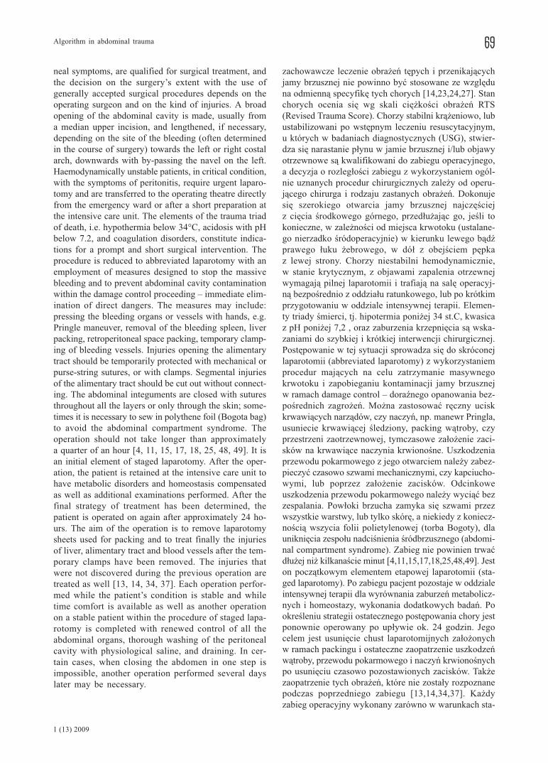
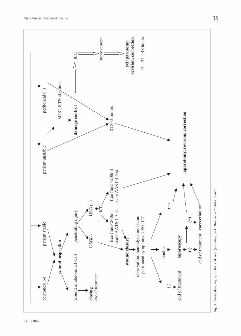
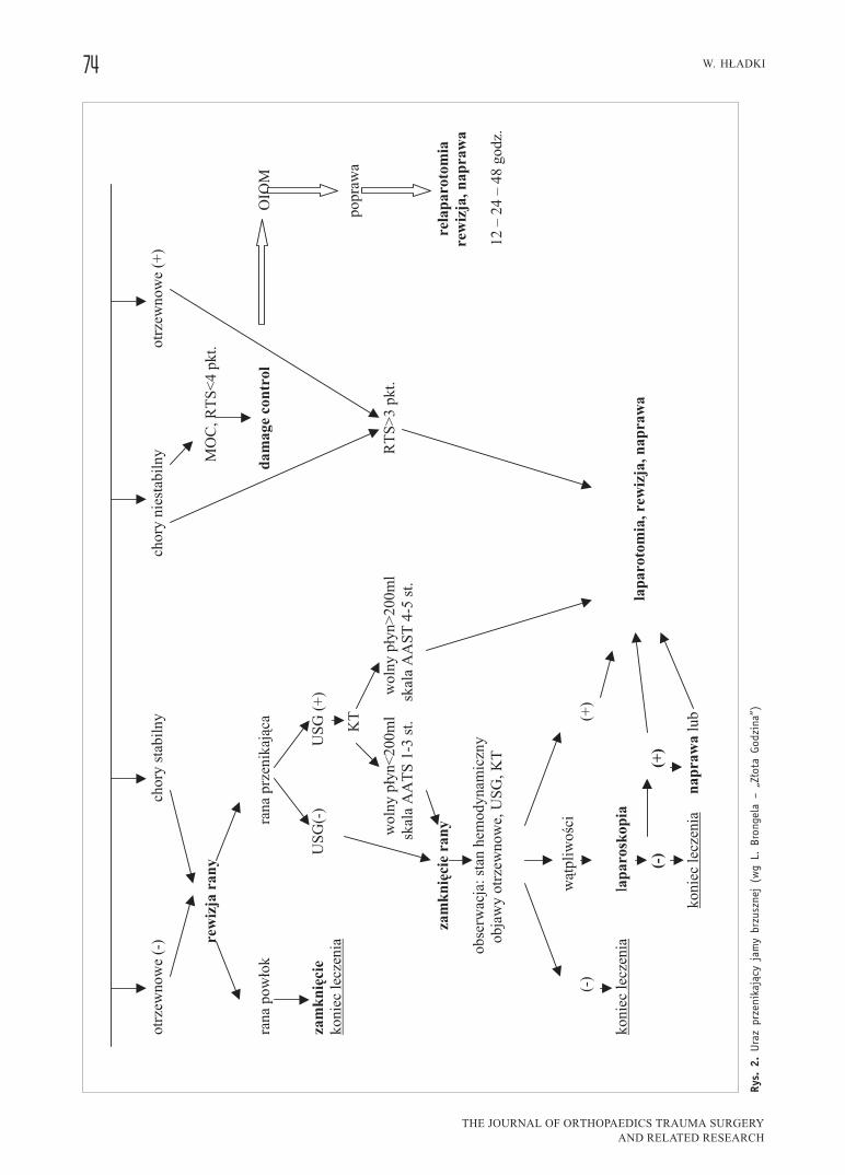
rażeniach jamy brzusznej przedstawiono schematycznie
na rysunkach nr 1 i nr 2.
Interventional cardiology has also found its place in
the treatment of bleedings caused by abdominal trauma
or blood vessel injuries, in the retroperitoneal space as
well. The possibility of using a radiological laboratory
located near the operating theatre, or even of performing
angiography at the operating theatre, of embolization and
inserting a stent into an injured blood vessel, has in-
creased the survival rate among the patients who require,
apart from an operation, this way of treatment as well.
It is also used increasingly often for preservative treat-
ment of selected injuries of spleen and liver [8, 21, 22,
32, 39, 47, 51, 52, 53].
If there is no improvement after surgical treatment or
embolization of larger vessels and blood is still oozing
from extensive surfaces of injured organs, the recombi-
nant activated coagulation factor VII, in use for several
years now, proves very helpful [26, 33]. The procedures
applied in the case of abdominal trauma, described above,
have been presented schematically in fig.1 and fig.2.
71Algorithm in abdominal trauma
1 (13) 2009
Fig
. 1
. C
lose
d i
nju
ry t
o t
he
abd
om
en (
acco
rdin
g t
o L
. B
ron
gel
– “
Go
lden
Ho
ur”
)
72 W. HŁADKI
THE JOURNAL OF ORTHOPAEDICS TRAUMA SURGERY
AND RELATED RESEARCH
Rys.
1.
Ura
z za
mkn
ięty
jam
y br
zusz
nej
(wg
L. B
rong
ela
– „Z
łota
God
zina
”)
73Algorithm in abdominal trauma
1 (13) 2009
Fig.
2.
Pene
trat
ing
inju
ry t
o th
e ab
dom
en (
acco
rdin
g to
L.
Bron
gel
– “G
olde
n H
our”
)
74 W. HŁADKI
THE JOURNAL OF ORTHOPAEDICS TRAUMA SURGERY
AND RELATED RESEARCH
Rys.
2.
Ura
z pr
zeni
kają
cy j
amy
brzu
szne
j (w
g L.
Bro
ngel
a –
„Zło
ta G
odzi
na”)
75Algorithm in abdominal trauma
1 (13) 2009
References/Piśmiennictwo:
1. Abu-Hatoum O., Bashenko Y., Hirsh M., Krausz M.: Continuous
fluid resuscitation and splenectomy for treatment of incontrolled
hemorrhagic shock after massive splenic injury. J Trauma, 2002,
52, 253 – 258.
2. American College of Surgeons, Committee on Trauma. Advan-
ced Trauma Life Support Manual. Chicago; American College
of Surgeons. 1997.
3. Alimov A.N., Isaev A.F, Safronov E.P., Otlygin I.V., Useinov E.B.,
Muradov I.U. et al.: Surgical policy and future of endo-surgery
of closed abdominal injuries in severe combined trauma.[in
Russian] Khirurgiia, 2006, 1, 34-37.
4. Andeweg C.S., Vingerhoedt N.M., van Vugt A.B., Haerkens M.H.:
Damage control surgery in polytraumatized patients. Ned Tijd-
schr Geneeskd, 2006, 150, 1503-1507.
5. Beal S.L.: Fatal hepatic hemorrhage: an unresolved problem in
the management of complex liver injuries. J Trauma, 1990, 30,
163-169.
6. Becker C.D., Poletti P.A.: The trauma concept: the role of MDCT
in the diagnosis and management of visceral injuries. Eur Ra-
diol, 2005, 15 Suppl. 4, 105-109.
7. Becker H.P., Willms A., Schwab R.: Laparoscopy in abdominal
trauma [in German]. Chirurg,. 2006, 77, 1007- 1013.
8. Bessoud B., Denys A., Calmes J.M., Madoff D., Qanadli S.,
Schnyder P. et al.: Nonoperative management of traumatic sple-
nic injuries: is there a role for proximal splenic artery emboli-
zation? Am J Roentgenol, 2006, 186, 779-785.
9. Blaivas M.: Emergency diagnostic paracentesis to determine in-
traperitoneal fluid identity discovered bedside ultrasound of
unstable patients. J Emerg Med, 2005, 29, 461-465.
10. Bobrzyński A., Brongel L., Kuliś M., Budzyński A., Hładki W.:
The place of laparoscopy at the diagnostics and treatment of ab-
dominal trauma consequences [in Polish]. Gdańska Wiosna Trau-
matologiczna, II Sympozjum Sekcji Chirurgii Urazowej TCHP, 2
– 3 czerwca 2005, 19.
11. Bose D., Tejwani N.C.: Evolving trends in the care of polytrau-
ma patients. Injury, 2006, 37, 20-28.
12. Brofman N., Atri M., Hanson J.M., Grinblat L., Chughtai T.,
Brenneman F.: Evaluation of bowel and mesenteric blunt trau-
ma with multidetector CT. Radiographics, 2006, 26, 1119-1131.
13. Brongel L. – Golden Hour [in Polish]. Wydawnnictwo Medycz-
ne, Kraków 2007.
14. Brongel L.: Injuries of abdominal cavity. In: Multiple and Mul-
tiorgan body injuries, Brongel L., Duda K. [in Polish]. Wydaw-
nictwo Lekarskie PZWL, Warszawa 2001, 228–243.
15. Burch J.M., Ortiz V.B., Richardson R.J., Martin R.R., Mattox K.L.,
Fordan G.L.Jr.: Abbreviated laparotomy and planned reopera-
tion for critically injuried patients. Ann Surg, 1992, 215, 476 –
483.
16. Chiu W.C., Wong-You-Cheong J.J., Rodriguez A., Shanmugana-
than K., Mirvis S.E., Scalea T.M.: Ultrasonography for interval
assessment in the nonoperative management of hepatic trauma.
Am Surg, 2005, 71, 841-846.
17. Clarce J.R., Trooskin S.Z., Doshi P., Grenwald L., Charles J.M.:
Time of laparotomy for intra-abdominal bleeding from trauma
does affect survival for delays up to 90 minutes. J Trauma, 2002,
52, 420 – 425.
18. Denton J.R., Moore E.E., Coldwell D.M.: Multimodality treatment
for grade V hepatic injuries: perihepatic packing, arterial em-
bolization, and venous stenting. J Trauma, 1997, 42, 964 – 967.
19. Fang J.F., Wong Y.C., Lin B.C., Hsu Y.P., Chen M.F.: Usefulness of
multidetector computed tomography for the initial assessment of
blunt abdominal trauma patients. World J Surg. 2006, 30, 176-182.
20. Feliciano D.V. Diagnostic modalities in abdominal trauma: pe-
ritoneal lavage, ulrasonogaphy, computed tomography scaning
and arteriography. Surg Clin North Am, 1991, 71, 241 – 256.
21. Haan J.M., Biffl W., Knudson M.M., Davis K.A., Oka T., Majer-
cik S. et al.: Western Trauma Association Multi-Institutional
Trials Committee.: Splenic embolization revisited: a multicenter
review. J Trauma 2004, 56, 542-547.
22. Hagiwara A., Murata A., Matsuda T., Matsuda H., Shimazaki S.:
The efficacy and limitation of transarterial embolization for
severe hepatic injury. J Trauma, 2002, 52, 1091 –1996.
23. Hirshberg A., Wall M.J.Jr., Allen M.K., Mattox K.L.: Double je-
opardy: thoracoabdominal injuries requiring surgery in both
chest and abdomen. J Trauma, 1995, 39, 225-229.
24. Hładki W., Anielski R., Cichoń S., Pałka G.: Thoraco-abdomi-
nal injuries in multiple trauma. [in Polish] Wybrane zagadnie-
nia z chirurgii. Biblioteka Polskiego Przeglądu Chirurgicznego.
1999, t.1, 54 – 59.
25. Hładki W., Brongel L.: Posttraumatic massive hemorrhage to the
abdominal cavity [in Polish]. Pol Przeg Chir, 2003, 75, 1237-
1242.
26. Jarzynowski W., Brongel L., Hładki W., Budzyński P., Magiera
J., Guzik P.: RFVIIA (Novoseven) in hemorrhagic shock control
in multitraumatized patients. International Proceedings, Medi-
mond, 2006. 115-117.
27. Jarzynowski W., Brongel L., Bobrzyński A., Kuliś M., Hładki W.:
Algorithm of abdominal injuries [in Polish]. Gdańska Wiosna
Traumatologiczna, II Sympozjum Sekcji Chirurgii Urazowej
TCHP, 2 – 3 czerwca 2005, 47.
28. Johnson W., Gracias V., Gupta R., Guillamondegui O., Reilly M.,
Shapiro M. et al.: Hepatic angiography in patients undergoing
damage control laparotomy. J Trauma, 2002, 52, 1102 – 1106.
29. Kirkpatric A.W., Sirois M., Laupland K.B., Goldstein L., Brown
D.R. Simons R.K. et al.: Prospctive evaluation of hand-held
focused abdominal sonography for trauma (FAST) in blunt ab-
dominal trauma. Can J Surg, 2005, 48, 453-460.
30. Kuliś M., Brongel L., Budzyński P., Hładki W., Lorkowski J.,
Nazimek R. et al.: Selected aspects in abdominal trauma mana-
gement [in Polish]. Pol Przegl Chir, 2007, 79, 253-361.
31. Lazzara S., Palmeri R., Melita G., Trovato M., Iapichino G.,
Cucinotta E. et al.: Laparoscopy in traumatic abdominal emer-
gencies. Chir Ital, 2006, 58, 485-491.
32. Liu P.P., Lee W.Ch., Cheng Y.F., Hsieh P.M., Hsieh Y.M., Tan B.L.
et al.: Use of splenic artery embolization as an adjunct to non-
surgical management of blunt splenic injury. J Trauma, 2004, 56,
768-773.
33. Martinowitz U., Keneth G., Segal E., Luboshitz J., Lubetsky A.,
Ingerslev J. et al.: Recombinant activated factor VII for adjunc-
tive hemorrhage control in trauma. J Trauma, 2001, 51, 431 –
438.
34. Marx J.A., Isenhour J., Abdominal Trauma. In: Marx I.A. (ed.),
Rosen’s Emergency Medicine,6th Ed. Mosby Elsevier, Philadel-
phia 2006, 489-514.
35. McGahan J.P., Horton S., Gerscovich E.O., Gillen M., Ri-
chards J.R., Cronan M.S. et al.: Appearance of solid organ
injury with contrast-enhanced sonography in blunt abdominal
trauma: preliminary experience. AJR Am J Roentgeno, 2006,
187, 658-666.
36. Menegaux F., Tresallet C., Gosgnach M., Nguyen-Thanh Q,
Langeron O, Riou B.: Diagnosis of bowel and mesenteric inju-
ries in blunt abdominal trauma: a prospective study. Am J Emerg
Med, 2006, 24, 19-24.
37. Moore E.E.: Staged laparotomy for the hypothermia, acidosis,
and coagulopathy Syndrome. Am J Surg, 1996, 172, 405 - 410.
38. Nelson J., Barr M., Witucki P., Abdominal Trauma. In: Roppolo
L.P. (ed.), Emergency Medicine, Clinical Concepts for Clinical
Practice. Mosby Elsevier, Philadelphia 2007, 155-163.
39. Nicholson A.: Vascular radiology in trauma. Cardiovasc Intervent
Radiol, 2004, 27, 105-120.
76 W. HŁADKI
THE JOURNAL OF ORTHOPAEDICS TRAUMA SURGERY
AND RELATED RESEARCH
40. Omert L., Yeaney W., Mizikowski S., Protetch J.: Role of emer-
gency medicine physician in airway management of the trauma
patient. J Trauma, 2001, 51, 1065 – 1068.
41. Pachter H.L., Spencer F.C., Hofstetter S.R., Liang H.G., Coppa
G.F.: Significant trends in the treatment of hepatic trauma:
Experience with 411 injuries. Ann. Surg., 1992, 215, 492. –
500.
42. Platz S.H., Adler J.N.: Emergency Medicine [in Polish]. Wyda-
nie I polskie pod redakcją J. Jakubaszki. Urban & Partner. 2000,
Wrocław.
43. Prokop A., Hotte H., Kruger K., Rhem K.E., Isenberg J., Schiffer
G..: Multislice CT in diagnostic work-up of polytrauma. Unfal-
lchirurg 2006, 109, 545-550.
44. Richardson J.D. Changes in the management of injuries to the
liver and spleen. J Am Coll Surg. 2005, 200, 648-699.
45. Salen P.N., Melanson S.W., Heller M.B.: The focused abdominal
sonography for trauma (FAST) examination: considerations and
recommendations for training physicians in the use of a new
clinical tool. Acad Emerg Med, 2000, 7, 162 – 168.
46. Salim A., Sangthong B., Martin M., Brown C., Plurad D., Deme-
triades D.: Whole body imaging in blunt multisystem trauma
patients without obvious signs of injury: results of a prospective
study. Arch Surg 2006, 141, 468-473.
47. Scalfani S.J.A.: The use of angiographic haemostasis in salvage
of the injured spleen. Radiology 1981; 141: 645-651.
48. Shapiro M.B., Jenkins D.H., Schwab C.W., Rotondo M.F.: Damage
control: collective review. J Trauma, 2000, 49, 969 – 978.
49. Sharp K., Locicero R.: Abdominal packing for surgically uncon-
trollable hemorrhage. Ann Surg, 1992, 215, 467-474.
50. Todd S.R.: Critical concepts in abdominal injury. Crit Care Clin.
2004. 20, 119-134
51. Thompson C.S., Rodriguez J.A., Ramaiah V.G., DiMungo L.,
Shafigue S., Olsen D. et al.: Acute traumatic rupture of the
thoracic aorta treated with endoluminal stent grafts. J Trauma,
2002, 52, 1173 – 1177.
52. Vargo D., Sorensen J., Barton R.: Repair of a grade VI hepatic injury:
case report and literature review. J Trauma, 2002, 53, 823 – 824.
53. Wahl W., Ahrns K., Brandt M., Franklin G., Taheri F.: The need
for early angiografic embolization in blunt liver injuries. J Trau-
ma, 2002, 52, 1097 – 1104.
54. Walcher F., Weinlich M., Conrad G., Schweigkofler U., Breitkrentz
R., Kirsching T. et al.: Prehospital ultrasound imaging improves
management of abdominal trauma. Br J Surg, 2006, 93, 238-242.
55. Zhang M., Liu Z.H., Yang J.X., Gan J.X., Xu S.W., You X.D. et
al.: Rapid detection of pneumothorax by ultrasonography in
patients with multiple trauma. Crit Care, 2006, 10, R112.