The effecTs of 2-MeThoxyeThanol and 2-eThoxyeThanol on...
13
Medycyna Pracy 2015;66(3):303–315 http://medpr.imp.lodz.pl/en ORIGINAL PAPER Beata Starek-Świechowicz 1 Katarzyna Miranowicz-Dzierżawska 2 Bogusława Budziszewska 1,3 Andrzej Starek 1 THE EFFECTS OF 2-METHOXYETHANOL AND 2-ETHOXYETHANOL ON HEMATOLOGICAL CHANGES INDUCED BY 2-BUTOXYETHANOL WPłYW 2-METOKSYETANOLU I 2-ETOKSYETANOLU NA ZMIANY HEMATOLOGICZNE INDUKOWANE PRZEZ 2-BUTOKSYETANOL 1 Jagiellonian University / Uniwersytet Jagielloński, Kraków, Poland Medical College, Faculty of Pharmacy, Department of Biochemical Toxicology / Collegium Medicum, Wydział Farmaceutyczny, Zakład Biochemii Toksykologicznej 2 Central Institute for Labour Protection – National Research Institute / Centralny Instytut Ochrony Pracy – Państwowy Instytut Badawczy, Warszawa, Poland Department of Chemical, Aerosol and Biological Hazards, Laboratory of Toxicology / Zakład Zagrożeń Chemicznych, Aerozolowych i Biologicznych, Laboratorium Toksykologiczne 3 Polish Academy of Sciences / Polska Akademia Nauk, Kraków, Poland Institute of Pharmacology, Department of Experimental Neuroendocrinology / Instytut Farmakologii, Zakład Neuroendokrynologii Doświadczalnej Abstract Background: Alkoxyethanols (ethylene glycol alkyl ethers) are used as mixtures in a variety of industrial and household prod- ucts. e aim of this study has been to evaluate the effects of 2-methoxyethanol (ME) and 2-ethoxyethanol (EE) on hematologi- cal changes induced by 2-butoxyethanol (BE) in rats. Material and Methods: Experiments were performed on male Wistar rats treated subcutaneously with BE, ME, and EE alone (in the dose of 0.75 mM/kg/day and 1.25 mM/kg/day) and their mixtures with the molar ratio 1:1, for 4 weeks. Hematological analyses were performed on the day 0, 4, 11, 18, and 29. Hemoglobin (HGB) con- centration in the urine was also determined in the rats treated with BE alone and co-exposed to BE and ME and also BE and EE. Results: e rats co-exposed to BE and ME or BE and EE demonstrated significantly less pronounced hematological changes in comparison with animals treated with BE alone at the beginning of exposure. At the later period the hematological alterations in the same animals were markedly pronounced and progressing with exposure time. e rats co-exposed to BE and ME or BE and EE did not demonstrate hemoglobinuria. Conclusions: ME or EE co-administered to rats with BE lead to the amelioration in the majority of the hematological parameters at the beginning of the exposure. e hematological changes at the end of the co-exposure to BE and ME or BE and EE were markedly pronounced. e effects observed in this study appear to be related with metabolic interactions of the examined ether. Med Pr 2015;66(3):303–315 Key words: rats, 2-butoksyethanol, 2-methoxyethanol, 2-ethoxyethanol, interactions, combined exposure Streszczenie Wstęp: Alkoksyetanole (etery alkilowe glikolu etylowego) są stosowane jako mieszaniny w różnych produktach przemysłowych i domowych. Celem badania była ocena wpływu 2-metoksyetanolu (ME) i 2-etoksyetanolu (EE) na zmiany hematologiczne indu- kowane przez 2-butoksyetanol (BE) u szczurów. Materiał i metody: Samce szczurów rasy Wistar narażano drogą podskórną na BE, ME lub EE (w dawce 0,75 mM/kg/dzień i 1,25 mM/kg/dzień) oraz na ich mieszaniny w stosunku molowym (1:1) przez 4 tygod- nie. Badania hematologiczne krwi obwodowej przeprowadzono w 0., 4., 11., 18. i 29. dniu doświadczenia. Ponadto na początku narażenia oznaczono stężenie hemoglobiny w moczu szczurów otrzymujących tylko BE oraz tylko jego mieszaninę z ME lub EE. Wyniki: Szczury narażone łącznie na badane związki wykazywały na początku ekspozycji słabiej zaznaczone zmiany hematologicz- ne niż zwierzęta otrzymujące tylko BE. W późniejszym okresie zmiany te były wyraźne i narastały wraz z czasem trwania naraże- nia. U szczurów eksponowanych na mieszaniny alkoksyetanoli nie zaobserwowano hemoglobinurii charakterystycznej dla działa- nia samego BE. Wnioski: Poprawa parametrów hematologicznych na początku narażenia może być spowodowana hamującym dzia- łaniem ME i EE na metabolizm BE. Z kolei akumulacja kwasów metoksyoctowego i etoksyoctowego, czyli metabolitów odpowied- nio ME i EE, może być przyczyną wyraźnych zmian hematologicznych na końcu ekspozycji. Med. Pr. 2015;66(3):303–315 Słowa kluczowe: szczury, 2-butoksyetanol, 2-metoksyetanol, 2-etoksyetanol, interakcje, narażenie łączne Corresponding author / Autorka do korespondencji: Beata Starek-Świechowicz, Jagiellonian University, Medical College, Faculty of Pharmacy, Department of Biochemical Toxicology, Medyczna 9, 30-688 Kraków, Poland, e-mail: [email protected] Received: October 14, 2014, accepted: January 20, 2015 http://dx.doi.org/10.13075/mp.5893.00126
-
Upload
hoanghuong -
Category
Documents
-
view
213 -
download
0
Transcript of The effecTs of 2-MeThoxyeThanol and 2-eThoxyeThanol on...
Medycyna Pracy 2015;66(3):303–315http://medpr.imp.lodz.pl/en
ORIGINAL PAPERBeata Starek-Świechowicz1
Katarzyna Miranowicz-Dzierżawska2
Bogusława Budziszewska1,3
Andrzej Starek1
The effecTs of 2-MeThoxyeThanol and 2-eThoxyeThanol on heMaTological changes induced by 2-buToxyeThanolWPłyW 2-mEtOksyEtANOLu I 2-EtOksyEtANOLu NA zmIANy hEmAtOLOGIczNE INdukOWANE PRzEz 2-butOksyEtANOL
1 Jagiellonian University / Uniwersytet Jagielloński, Kraków, Poland Medical College, Faculty of Pharmacy, Department of Biochemical Toxicology / Collegium Medicum, Wydział Farmaceutyczny, Zakład Biochemii Toksykologicznej2 Central Institute for Labour Protection – National Research Institute / Centralny Instytut Ochrony Pracy – Państwowy Instytut Badawczy, Warszawa, Poland Department of Chemical, Aerosol and Biological Hazards, Laboratory of Toxicology / Zakład Zagrożeń Chemicznych, Aerozolowych i Biologicznych, Laboratorium Toksykologiczne 3 Polish Academy of Sciences / Polska Akademia Nauk, Kraków, PolandInstitute of Pharmacology, Department of Experimental Neuroendocrinology / Instytut Farmakologii, Zakład Neuroendokrynologii Doświadczalnej
AbstractBackground: Alkoxyethanols (ethylene glycol alkyl ethers) are used as mixtures in a variety of industrial and household prod-ucts. The aim of this study has been to evaluate the effects of 2-methoxyethanol (ME) and 2-ethoxyethanol (EE) on hematologi-cal changes induced by 2-butoxyethanol (BE) in rats. Material and Methods: Experiments were performed on male Wistar rats treated subcutaneously with BE, ME, and EE alone (in the dose of 0.75 mM/kg/day and 1.25 mM/kg/day) and their mixtures with the molar ratio 1:1, for 4 weeks. Hematological analyses were performed on the day 0, 4, 11, 18, and 29. Hemoglobin (HGB) con-centration in the urine was also determined in the rats treated with BE alone and co-exposed to BE and ME and also BE and EE. Results: The rats co-exposed to BE and ME or BE and EE demonstrated significantly less pronounced hematological changes in comparison with animals treated with BE alone at the beginning of exposure. At the later period the hematological alterations in the same animals were markedly pronounced and progressing with exposure time. The rats co-exposed to BE and ME or BE and EE did not demonstrate hemoglobinuria. Conclusions: ME or EE co-administered to rats with BE lead to the amelioration in the majority of the hematological parameters at the beginning of the exposure. The hematological changes at the end of the co-exposure to BE and ME or BE and EE were markedly pronounced. The effects observed in this study appear to be related with metabolic interactions of the examined ether. Med Pr 2015;66(3):303–315Key words: rats, 2-butoksyethanol, 2-methoxyethanol, 2-ethoxyethanol, interactions, combined exposure
StreszczenieWstęp: Alkoksyetanole (etery alkilowe glikolu etylowego) są stosowane jako mieszaniny w różnych produktach przemysłowych i domowych. Celem badania była ocena wpływu 2-metoksyetanolu (ME) i 2-etoksyetanolu (EE) na zmiany hematologiczne indu-kowane przez 2-butoksyetanol (BE) u szczurów. Materiał i metody: Samce szczurów rasy Wistar narażano drogą podskórną na BE, ME lub EE (w dawce 0,75 mM/kg/dzień i 1,25 mM/kg/dzień) oraz na ich mieszaniny w stosunku molowym (1:1) przez 4 tygod-nie. Badania hematologiczne krwi obwodowej przeprowadzono w 0., 4., 11., 18. i 29. dniu doświadczenia. Ponadto na początku narażenia oznaczono stężenie hemoglobiny w moczu szczurów otrzymujących tylko BE oraz tylko jego mieszaninę z ME lub EE. Wyniki: Szczury narażone łącznie na badane związki wykazywały na początku ekspozycji słabiej zaznaczone zmiany hematologicz-ne niż zwierzęta otrzymujące tylko BE. W późniejszym okresie zmiany te były wyraźne i narastały wraz z czasem trwania naraże-nia. U szczurów eksponowanych na mieszaniny alkoksyetanoli nie zaobserwowano hemoglobinurii charakterystycznej dla działa-nia samego BE. Wnioski: Poprawa parametrów hematologicznych na początku narażenia może być spowodowana hamującym dzia-łaniem ME i EE na metabolizm BE. Z kolei akumulacja kwasów metoksyoctowego i etoksyoctowego, czyli metabolitów odpowied-nio ME i EE, może być przyczyną wyraźnych zmian hematologicznych na końcu ekspozycji. Med. Pr. 2015;66(3):303–315Słowa kluczowe: szczury, 2-butoksyetanol, 2-metoksyetanol, 2-etoksyetanol, interakcje, narażenie łączne
Corresponding author / Autorka do korespondencji: Beata Starek-Świechowicz, Jagiellonian University, Medical College, Faculty of Pharmacy, Department of Biochemical Toxicology, Medyczna 9, 30-688 Kraków, Poland, e-mail: [email protected]: October 14, 2014, accepted: January 20, 2015
http://dx.doi.org/10.13075/mp.5893.00126
B. Starek-Świechowicz et al. Nr 3304
parison with the control group (laminate workers) [13]. The frequency of anemia in the group exposed to ME was significantly higher as compared to the correspond-ing control group. While, RBC was significantly nega-tively associated with air concentrations of ME, HGB, PCV, and RBC were negatively correlated with uri-nary levels of methoxyacetic acid (MAA), a metabolite of ME. Similarly, in another survey of male workers chronically exposed to ME, similar hematological dis-orders in peripheral blood were observed [14].
In shipyard painters exposed to mixed solvents con-taining 2-ethoxyethanol acetate it was observed that this chemical might be toxic to the bone marrow [15].
In the workers exposed to a low level of BE, a sta-tistically significant decrease in PCV value and an in-crease in mean cell hemoglobin concentration (MCHC) value were seen [16].
Ethylene glycol ether is often used as mixtures in industrial or domestic solvent compositions [17]. To our knowledge, there are no studies describing the hematological effects of repeated exposure to any mixtures of these chemicals in experimental animals. The objective of this study has been to evaluate the ef-fects of ME and EE on hematological changes induced by BE in rats.
MaTeRial and MeThods
ChemicalsThe substances: ME, EE, and BE were purchased from Sigma-Aldrich Ltd, Poland. Other chemicals were ob-tained from POCH (Poland). Methoxyethanol, EE, and BE solutions and their mixtures were made up in sa-line, immediately before dosing.
AnimalsAdult male Wistar rats (12 weeks old, with an initial body weight about 290 g) were purchased from the Jagiellonian University, Pharmacy Breeding Labo-ratory (Kraków, Poland). The animals were kept in clean polypropylene cages under standard laboratory conditions (temperature at 22±2°C, relative humidity of 50±10% and a 12/12 h light/dark cycle, the light on at 8:00), with food (Murigram, Motycz, Poland) and water available ad libitum. All the treatments were started after at least 1 week of acclimatization to labo-ratory conditions.
All experiments were carried out according to the National Institutes of Health Guide for the Care and Use of Laboratory Animals, and were approved by
inTRoducTion
A number of natural and synthetic chemicals are now clearly recognized to be both hemopoietic and hemo-lytic xenobiotics. For example, benzene is a classical he-mopoietic xenobiotic, for which toxic and leukemogenic effects in humans and animal species are well-docu-mented in the literature [1,2]. Lead causes inhibition of hemoglobin (HGB) biosynthesis and is a causal fac-tor of hemolytic anemia induced by a decrease in Na+, K+-ATPase activity in erythrocytic membrane [3]. Exposure to ethylene glycol ether (EGEs) and related acetates may lead to peripheral hematological disorders and bone marrow injuries [4], sometimes complicated by aplastic anemia or immunodeficiency. Ethylene gly-col ether was found to be potent hemopoietic xenobi-otics in vitro, which acts directly on the hemopoietic cells [5].
Ethylene glycol ether, especially 2-methoxyetha-nol (ME) (CAS No. 109-86-4), 2-ethoxyethanol (EE) (CAS No. 110-80-5), and 2-butoxyethanol (BE) (CAS No. 111-76-2), has the unique solvent characteristics, i.e., solubility in water and organic compounds such as oils. The amphiphilic structure, the hydrophilic part binds to water, whereas lypophilic part binds to oils. The ether is applied to dissolve, extract, suspend, dry, and synthesize. Ethylene glycol ether is widely used in industry as an ingredient of paints, inks, water-based cleaners, adhesives, detergents, cosmetics, cut- ting fluids, agrochemicals, jet fuels, and hydraulic brake fluids [6].
Ethylene glycol ether may cause adverse hematolog-ical effects through inhalation, dermal absorption, and ingestion. It was suggested that skin absorption of BE is a predominant factor in occupational exposure [7]. Such ether is oxidized by NAD-dependent dehydro-genases in the liver, testicles, and skin [8,9] to corre-sponding alkoxyacetic acids (AAs), which have been identified as primary urinary metabolites in humans and animals. Alkoxyacetic acids are responsible for toxic effects of EGEs [10]. These metabolites exert in-hibitory effect on Na+-, K+- and Ca2+-ATPase in eryth-rocyte membrane in vitro [11].
Data on human exposure to EGEs indicates that ME in occupational conditions lead to leucopenia, pancy-topenia, bone marrow depression, and decline in the number of red blood cells (RBC), and hemoglobin concentration [12]. In male impregnation workers chronically exposed to ME, the HGB, packed cell vol-ume (PCV), and RBC were significantly lower in com-
Hematological effects of exposure to ME, EE, BENr 3 305
the Local Ethics Committee, Kraków, Poland (the cer-tificate No. 33/2008).
Animal treatmentThe rats were randomly assigned to 11 groups of 5 ani-mals each. The rats (6 groups) were treated with BE, ME, and EE alone in doses of 0.75 and 1.25 mM/kg/day by subcutaneous injections at a fixed volume of 2 ml/kg of body weight (b.w.), regardless of a dose, once daily, 5 days per week, for 4 weeks. Other 4 groups of rats were exposed to 2-component mixtures of the examined compounds, i.e., BE and ME or BE and EE, in the same manner and in the same doses with the molar ratio 1:1. Control rats (1 group) were given the equivalent volume of saline. The applied doses and route of administration were chosen based on the previous work output [18].
Blood samples from the tail vein of rats were col-lected for hematological analyses directly before the experiment, during exposure and after its termination, i.e., on the day 0, 4, 11, 18, and 29.
Hematological analysesData on the hematological analyses has been de-scribed elsewhere [18]. Hematological analyses were systematically checked. To this end a standard hu-man blood CBC-3D Hematology Control (R&D Sys-tem Inc., Minneapolis, MN, USA) was used. A day-to-day precision of RBC, PCV, and mean corpuscular volume (MCV) measurements (N = 25) in blood stood at 4.4%, 4.8%, and 4.6%, respectively.
Urine examinationRats treated with BE in a dose of 1.25 mM/kg/day or ex-posed to BE and ME or BE and EE mixtures in the same doses were placed in individual glass metabolic cages for urine collection. Urine was collected every day for the first 3 days of exposure. Hemoglobin concentration was determined by means of Drabkin’s method and creatinine level – by means of Jaffe’s reaction. The HGB concentration in urine was calculated for 1 g creatinine after its determination by Jaffe’s reaction.
Statistical analysesAll data is expressed as mean ± standard deviation. Data was analyzed by means of a 2-way analysis of vari-ance with repeated measures for 1 factor (time or dose) and evaluation of simple effects. The analysis was per-formed with the SPSS 14.0 statistical packet (SPSS Inc., Chicago, IL, USA). Results were considered statistically significant when p ≤ 0.05.
ResulTs
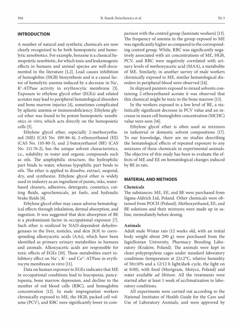
Hematological effects2-Butoxyethanol alone, administered in the lower dose of 0.75 mM/kg/day caused a significant decrease in RBC on each test day, PCV – only on the day 4, and HGB – on the days 4, 11, and 29 of the experiment. Moreover, an increase in both MCV on each test day, and reticulocyte (RET) on the days 4, and 11 was ob-served (Figure 1).
2-Methoxyethanol alone in the same dose as BE (0.75 mM/kg/day) led to only a weakly pronounced but statistically significant decrease in RBC on the day 18 of exposure. In the rats co-exposed to BE and ME in the lower doses (0.75 mM + 0.75 mM), significantly less pronounced hematological changes vs. BE alone group were seen on the days 4 or 4 and 11, mainly in PCV, HGB, and RBC, respectively. In these rats significantly less pronounced changes in MCV in comparison with animals exposed to BE alone on each test day were observed. In a later period, i.e., mainly on the days 18 and 29 of exposure, the hematological alterations (RBC, PCV, HGB, and RET) in rats simultaneously treated with BE and ME were similar or more pronounced than after BE alone treatment (Figure 1).
2-Butoxyethanol administration in a dose of 1.2 mM/kg/day resulted in a decrease in RBC and also in an increase in both MCV, and RET on each test day. The decline in PCV on the days 4 and 18, and HGB on the days 4, 18, and 29 was also observed. In addi-tion, BE in the administered dose caused secondary hemoglobinuria on the day 1 of exposure. Hemoglob-in concentrations in urine of treated and control rats were 345±6.1 mg/g of creatinine and 43±7.9 mg/g of creatinine, respectively.
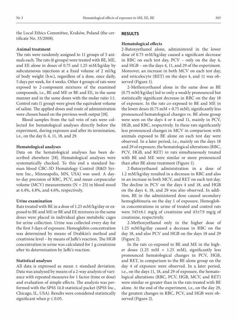
2-Methoxyethanol only in the higher dose of 1.25 mM/kg/day caused a decrease in RBC on the day 18, and also PCV and HGB on the days 18 and 29 (Figure 2).
In the rats co-exposed to BE and ME in the high-er doses (1.25 mM + 1.25 mM), significantly less pronounced hematological changes in PCV, HGB, and RET, in comparison to the BE alone group on the day 4 of exposure were observed. In a later period, i.e., on the days 11, 18, and 29 of exposure, the hemato-logical alterations (RBC, PCV, HGB, MCV, and RET) were similar or greater than in the rats treated with BE alone. At the end of the experiment, i.e., on the day 29, the greatest changes in RBC, PCV, and HGB were ob-served (Figure 2).
306
RBC
[×10
6 /µl]
Day / Dzień
15
10
5
00 4 11 18 29
PCV
[%]
Day / Dzień
100
50
00 4 11 18 29
a)
b)
HGB
[g/d
l]
Day / Dzień
20
15
10
5
00 4 11 18 29
MCV
[fl]
Day / Dzień
100
50
00 4 11 18 29
c)
d)
RET/
103 R
BC
Day / Dzień
100
50
00 4 11 18 29
e)
control rats BE ME BE+ME
control rats BE ME BE+ME
control rats BE ME BE+ME
control rats BE ME BE+ME
control rats BE ME BE+ME
***
****
***
*****
*****
**
*****
***
****
***
****
*****
***
***
***
****
*****
***
****
****
***
*****
*****
* ***
**** *
*****
*
*****
***** *
*****
***
***
RBC – red blood cells / czerwone krwinki, PCV – packed cell volume / hematokryt, HGB – hemoglobin / hemoglobina, MCV – mean corpuscular volume of red blood cell / średnia objętość krwinki czerwonej, RET – reticulocytes / retikulocyty.p ≤ 0.05 significantly different / istotnie różny:* from control rats / w stosunku do grupy kontrolnej,** from rats treated with BE alone / w stosunku do grupy narażonej tylko na BE,*** from rats treated with ME alone / w stosunku do grupy narażonej tylko na ME.The values are the means ± standard deviation of 5 rats / Wartości przedstawiono jako średnią arytmetyczną ± odchylenie standardowe dla 5 szczurów.
Fig. 1. The effects of combined exposure to 2-butoxyethanol (BE) and 2-methoxyethanol (ME) in the doses of 0.75 mM/kg b.w. on: a) RBC, b) PCV, c) HGB, d) MCV, e) RET values in peripheral blood in male rats Ryc. 1. Wpływ łącznego narażenia na 2-butoksyetanol (BE) i 2-metoksyetanol (ME) w dawkach 0,75 mM/kg m.c. na: a) RBC, b) PCV, c) HGB, d) MCV, e) RET we krwi obwodowej samców szczurów
307
Abbreviations as in Figure 1 / Objaśnienia jak na rycinie 1.
Fig. 2. The effects of combined exposure to BE and ME in the doses of 1.25 mM/kg b.w. on: a) RBC, b) PCV, c) HGB, d) MCV, e) RET values in peripheral blood in male rats Ryc. 2. Wpływ łącznego narażenia na BE i ME w dawkach 1,25 mM/kg m.c. na: a) RBC, b) PCV, c) HGB, d) MCV, e) RET we krwi obwodowej samców szczurów
RBC
[×10
6 /µl]
Day / Dzień
15
10
5
00 4 11 18 29
PCV
[%]
Day / Dzień
60
40
20
00 4 11 18 29
a)
b)
HGB
[g/d
l]
Day / Dzień
20
10
00 4 11 18 29
MCV
[fl]
Day / Dzień
100
50
00 4 11 18 29
c)
d)
RET/
103 R
BC
Day / Dzień
300250
200
150
100
50
00 4 11 18 29
e)
control rats BE ME BE+ME
control rats BE ME BE+ME
control rats BE ME BE+ME
control rats BE ME BE+ME
control rats BE ME BE+ME
****
*
*****
****
*
**
****
***
***
*
***
***
***** *
*****
*
***
***
*
****
***
****
**
*****
**
**
*****
**
*** *****
**
**
*****
*
****
* *****
308
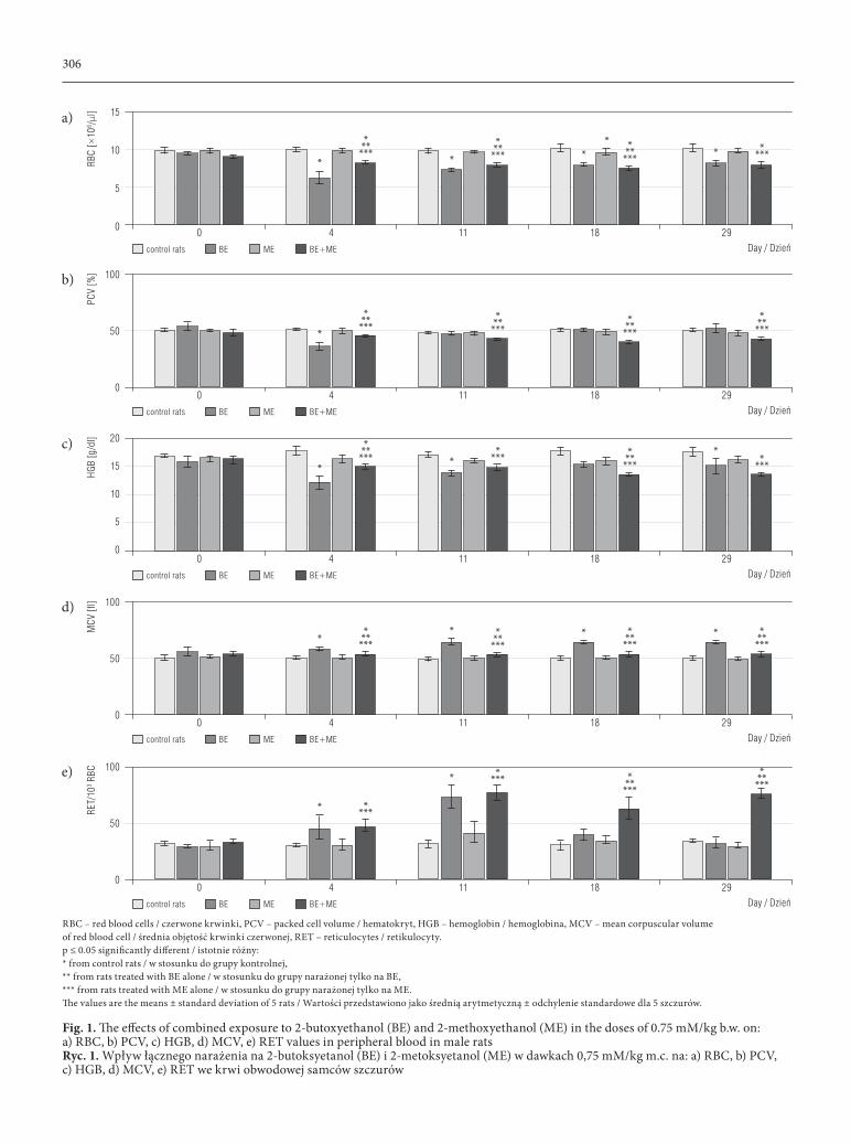
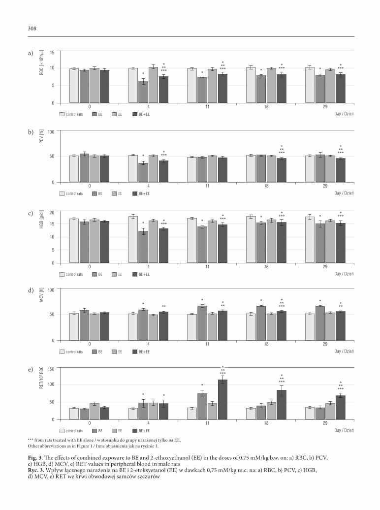
*** from rats treated with EE alone / w stosunku do grupy narażonej tylko na EE.Other abbreviations as in Figure 1 / Inne objaśnienia jak na rycinie 1.
Fig. 3. The effects of combined exposure to BE and 2-ethoxyethanol (EE) in the doses of 0.75 mM/kg b.w. on: a) RBC, b) PCV, c) HGB, d) MCV, e) RET values in peripheral blood in male rats Ryc. 3. Wpływ łącznego narażenia na BE i 2-etoksyetanol (EE) w dawkach 0,75 mM/kg m.c. na: a) RBC, b) PCV, c) HGB, d) MCV, e) RET we krwi obwodowej samców szczurów
RBC
[×10
6 /µl]
Day / Dzień
15
10
5
00 4 11 18 29
PCV
[%]
Day / Dzień
100
50
00 4 11 18 29
a)
b)
HGB
[g/d
l]
Day / Dzień
20
15
10
5
00 4 11 18 29
MCV
[fl]
Day / Dzień
100
50
00 4 11 18 29
c)
d)
RET/
103 R
BC
Day / Dzień
150
100
50
00 4 11 18 29
e)
control rats BE EE BE+EE
control rats BE EE BE+EE
control rats BE EE BE+EE
control rats BE EE BE+EE
control rats BE EE BE+EE
***
****
***
****
*****
*****
*****
***
****
*****
*****
*****
*****
*****
*** ***
* ***
****
***
*
**
***
***
*
***
*** ***
***
309
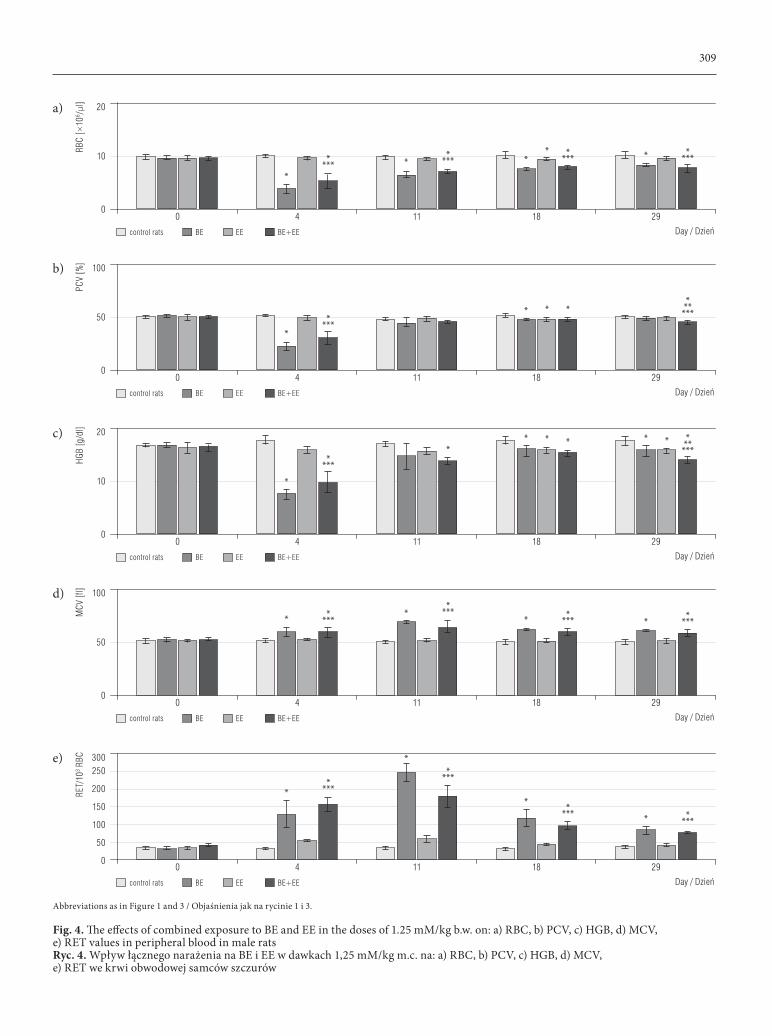
Abbreviations as in Figure 1 and 3 / Objaśnienia jak na rycinie 1 i 3.
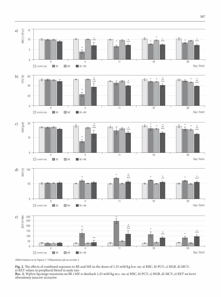
Fig. 4. The effects of combined exposure to BE and EE in the doses of 1.25 mM/kg b.w. on: a) RBC, b) PCV, c) HGB, d) MCV, e) RET values in peripheral blood in male ratsRyc. 4. Wpływ łącznego narażenia na BE i EE w dawkach 1,25 mM/kg m.c. na: a) RBC, b) PCV, c) HGB, d) MCV, e) RET we krwi obwodowej samców szczurów
RBC
[×10
6 /µl]
Day / Dzień
20
10
00 4 11 18 29
PCV
[%]
Day / Dzień
100
50
00 4 11 18 29
a)
b)
HGB
[g/d
l]
Day / Dzień
20
10
00 4 11 18 29
MCV
[fl]
Day / Dzień
100
50
00 4 11 18 29
c)
d)
RET/
103 R
BC
Day / Dzień
300250
200
150
100
50
00 4 11 18 29
e)
control rats BE EE BE+EE
control rats BE EE BE+EE
control rats BE EE BE+EE
control rats BE EE BE+EE
control rats BE EE BE+EE
****
*
*****
****
**
*****
****
*
****
*****
****
*
**** *
*****
**
*****
***** *
**** *****
*****
****
*
****
**
****
B. Starek-Świechowicz et al. Nr 3310
WBC
[×10
3 /µl]
Day / Dzień
20
10
00 4 11 18 29
a)
LYM
[×10
3 /µl]
Day / Dzień
20
15
10
5
00 4 11 18 29
b)
control rats BE ME BE+ME
control rats BE ME BE+ME
**
*
**
*
***
*
In contrast, EE produced less pronounced effects on circulating RBC than BE or ME alone. The lower dose of EE (0.75 mM/kg/day) did not induce hema-tological alterations (Figure 3). Only the higher dose of this compound (1.25 mM/kg) resulted in a de-crease in RBC and PCV on day 18, and HGB on the days 18 and 29 (Figure 4).
In the rats concurrently treated with BE and EE in the lower doses (0.75 mM/kg + 0.75 mM/kg), a sig-nificantly less pronounced decrease in RBC than in BE alone group was found on the days 4 and 11. In a later period (on the days 18, and 29) the same values of RBC and HGB, and a greater decrease in PCV in comparison to BE group were observed. While the in-crease in MCV was significantly lower than in the rats treated with BE alone on each test day, RET were dis-tinctly elevated on the days 11, 18 and 29 (Figure 3). In the rats co-exposed to the higher doses of BE and EE (1.25 mM/kg + 1.25 mM/kg) the hematological chang-es were similar (RBC, MCV, and RET on the days from 4 to 29) or more pronounced (PCV and HGB on the day 29) than in the rats treated with BE (1.25 mM/kg) alone (Figure 4).
The rats concurrently exposed to BE and ME or BE and EE did not demonstrate hemoglobinuria on the 1st day of exposure. Hemoglobin concentrations in urine
of these rats were similar to the control group (66±23.6 and 62±13.4 mg/g of creatinine, respectively).
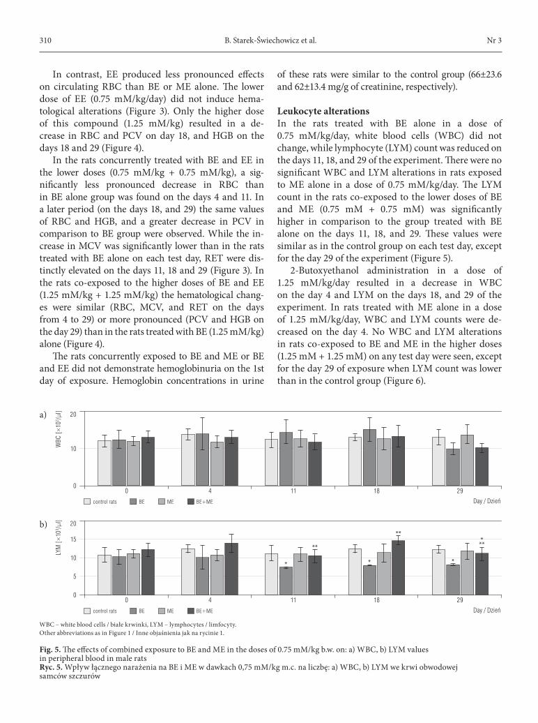
Leukocyte alterationsIn the rats treated with BE alone in a dose of 0.75 mM/kg/day, white blood cells (WBC) did not change, while lymphocyte (LYM) count was reduced on the days 11, 18, and 29 of the experiment. There were no significant WBC and LYM alterations in rats exposed to ME alone in a dose of 0.75 mM/kg/day. The LYM count in the rats co-exposed to the lower doses of BE and ME (0.75 mM + 0.75 mM) was significantly higher in comparison to the group treated with BE alone on the days 11, 18, and 29. These values were similar as in the control group on each test day, except for the day 29 of the experiment (Figure 5).
2-Butoxyethanol administration in a dose of 1.25 mM/kg/day resulted in a decrease in WBC on the day 4 and LYM on the days 18, and 29 of the experiment. In rats treated with ME alone in a dose of 1.25 mM/kg/day, WBC and LYM counts were de-creased on the day 4. No WBC and LYM alterations in rats co-exposed to BE and ME in the higher doses (1.25 mM + 1.25 mM) on any test day were seen, except for the day 29 of exposure when LYM count was lower than in the control group (Figure 6).
WBC – white blood cells / białe krwinki, LYM – lymphocytes / limfocyty.Other abbreviations as in Figure 1 / Inne objaśnienia jak na rycinie 1.
Fig. 5. The effects of combined exposure to BE and ME in the doses of 0.75 mM/kg b.w. on: a) WBC, b) LYM values in peripheral blood in male ratsRyc. 5. Wpływ łącznego narażenia na BE i ME w dawkach 0,75 mM/kg m.c. na liczbę: a) WBC, b) LYM we krwi obwodowej samców szczurów
Hematological effects of exposure to ME, EE, BENr 3 311
WBC
[×10
3 /µl]
Day / Dzień
20
10
00 4 11 18 29
a)
LYM
[×10
3 /µl]
Day / Dzień
20
15
10
5
00 4 11 18 29
b)
control rats BE EE BE+EE
control rats BE EE BE+EE
*
**
*
*
*
WBC
[×10
3 /µl]
Day / Dzień
30
20
10
00 4 11 18 29
a)
LYM
[×10
3 /µl]
Day / Dzień
20
15
10
5
00 4 11 18 29
b)
control rats BE ME BE+ME
control rats BE ME BE+ME
*
* * **
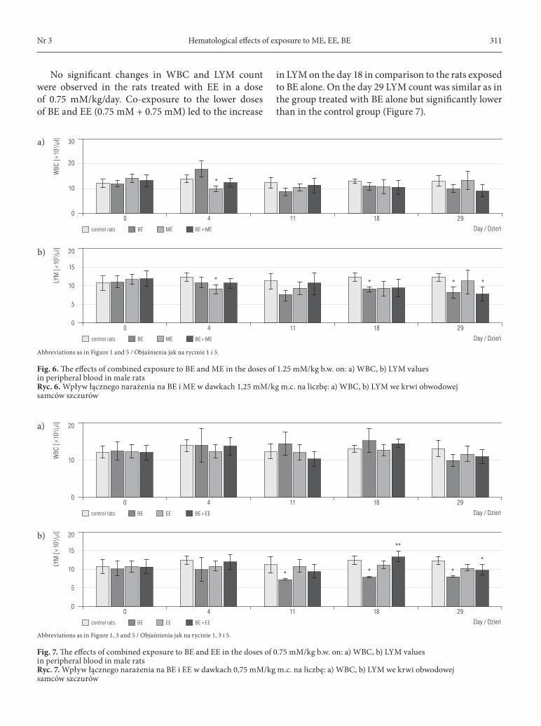
No significant changes in WBC and LYM count were observed in the rats treated with EE in a dose of 0.75 mM/kg/day. Co-exposure to the lower doses of BE and EE (0.75 mM + 0.75 mM) led to the increase
in LYM on the day 18 in comparison to the rats exposed to BE alone. On the day 29 LYM count was similar as in the group treated with BE alone but significantly lower than in the control group (Figure 7).
Abbreviations as in Figure 1, 3 and 5 / Objaśnienia jak na rycinie 1, 3 i 5.
Fig. 7. The effects of combined exposure to BE and EE in the doses of 0.75 mM/kg b.w. on: a) WBC, b) LYM values in peripheral blood in male ratsRyc. 7. Wpływ łącznego narażenia na BE i EE w dawkach 0,75 mM/kg m.c. na liczbę: a) WBC, b) LYM we krwi obwodowej samców szczurów
Abbreviations as in Figure 1 and 5 / Objaśnienia jak na rycinie 1 i 5.
Fig. 6. The effects of combined exposure to BE and ME in the doses of 1.25 mM/kg b.w. on: a) WBC, b) LYM values in peripheral blood in male rats Ryc. 6. Wpływ łącznego narażenia na BE i ME w dawkach 1,25 mM/kg m.c. na liczbę: a) WBC, b) LYM we krwi obwodowej samców szczurów
B. Starek-Świechowicz et al. Nr 3312
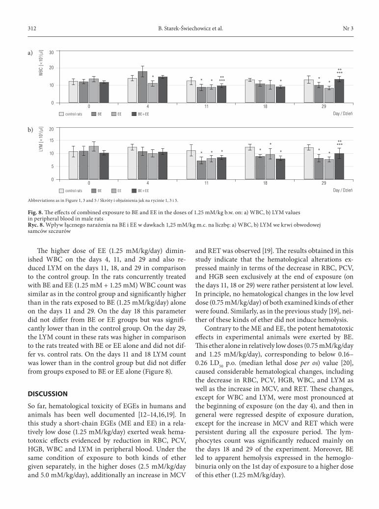
The higher dose of EE (1.25 mM/kg/day) dimin-ished WBC on the days 4, 11, and 29 and also re-duced LYM on the days 11, 18, and 29 in comparison to the control group. In the rats concurrently treated with BE and EE (1.25 mM + 1.25 mM) WBC count was similar as in the control group and significantly higher than in the rats exposed to BE (1.25 mM/kg/day) alone on the days 11 and 29. On the day 18 this parameter did not differ from BE or EE groups but was signifi-cantly lower than in the control group. On the day 29, the LYM count in these rats was higher in comparison to the rats treated with BE or EE alone and did not dif-fer vs. control rats. On the days 11 and 18 LYM count was lower than in the control group but did not differ from groups exposed to BE or EE alone (Figure 8).
discussion
So far, hematological toxicity of EGEs in humans and animals has been well documented [12–14,16,19]. In this study a short-chain EGEs (ME and EE) in a rela-tively low dose (1.25 mM/kg/day) exerted weak hema-totoxic effects evidenced by reduction in RBC, PCV, HGB, WBC and LYM in peripheral blood. Under the same condition of exposure to both kinds of ether given separately, in the higher doses (2.5 mM/kg/day and 5.0 mM/kg/day), additionally an increase in MCV
and RET was observed [19]. The results obtained in this study indicate that the hematological alterations ex-pressed mainly in terms of the decrease in RBC, PCV, and HGB seen exclusively at the end of exposure (on the days 11, 18 or 29) were rather persistent at low level. In principle, no hematological changes in the low level dose (0.75 mM/kg/day) of both examined kinds of ether were found. Similarly, as in the previous study [19], nei-ther of these kinds of ether did not induce hemolysis.
Contrary to the ME and EE, the potent hematotoxic effects in experimental animals were exerted by BE. This ether alone in relatively low doses (0.75 mM/kg/day and 1.25 mM/kg/day), corresponding to below 0.16–0.26 LD50 p.o. (median lethal dose per os) value [20], caused considerable hematological changes, including the decrease in RBC, PCV, HGB, WBC, and LYM as well as the increase in MCV, and RET. These changes, except for WBC and LYM, were most pronounced at the beginning of exposure (on the day 4), and then in general were regressed despite of exposure duration, except for the increase in MCV and RET which were persistent during all the exposure period. The lym-phocytes count was significantly reduced mainly on the days 18 and 29 of the experiment. Moreover, BE led to apparent hemolysis expressed in the hemoglo-binuria only on the 1st day of exposure to a higher dose of this ether (1.25 mM/kg/day).
Abbreviations as in Figure 1, 3 and 5 / Skróty i objaśnienia jak na rycinie 1, 3 i 5.
Fig. 8. The effects of combined exposure to BE and EE in the doses of 1.25 mM/kg b.w. on: a) WBC, b) LYM values in peripheral blood in male rats Ryc. 8. Wpływ łącznego narażenia na BE i EE w dawkach 1,25 mM/kg m.c. na liczbę: a) WBC, b) LYM we krwi obwodowej samców szczurów
WBC
[×10
3 /µl]
Day / Dzień
30
20
10
00 4 11 18 29
a)
LYM
[×10
3 /µl]
Day / Dzień
20
15
10
5
00 4 11 18 29
b)
control rats BE EE BE+EE
control rats BE EE BE+EE
* ******* *
*****
**
*** *
**
*****
**
Hematological effects of exposure to ME, EE, BENr 3 313
It was suggested that hematological disturbances induced by BE in this experiment and the previous study [19] are probably the result of the selective hemol-ysis of the aged erythrocytes. There are many evidences that the aging process of RBC leads to several biochem-ical alterations and to the loss cell deformability and the increased MCV value and osmotic fragility [21,22]. It seems that selective hemolysis of older erythrocytes together with the process of active blood regeneration may lead to tolerance development to the hemolytic action of BE [19,23,24]. On the other hand, the lack of tolerance to ME- and EE-induced hemolysis in rats treated with these kinds of ether was observed.
The main aim of this study has been the evaluation of the effects of ME and EE on the hematological chang-es induced by BE in rats. The obtained results show that short-chain ether could modify the hematological im-pact of BE. This modification, especially by ME, was characterized by 2-step hematological alterations in a different manner than those induced by BE alone. At the beginning of the exposure (mainly on the day 4), the attenuation of the hematotoxic effects of BE expressed in terms of higher values of RBC, PCV, and HGB, and lower values of MCV and RET as well as the lack of he-moglobinuria was seen. On the other hand, at the end of exposure (from the days 11 to 29) these alterations were more pronounced, except for a decrease in MCV for all the exposure period and RET on the days 18 and 29 in the rats co-exposed to BE and ME in the higher doses (1.25 mM/kg/day + 1.25 mM/kg/day). The effects of EE on hematological changes induced by BE were less pro-nounced than ME.
The obtained data demonstrates that in rats con-currently treated with BE and ME or BE and EE the hematological changes have the opposite direction of the course than those in animals exposed to BE alone. Thus, the short-chain ether disturbs the tolerance de-velopment to the hemolytic action of BE.
In contrast with the above described chang-es, in the leukocyte system both kinds of ether (ME in the dose of 0.75 mM/kg/day and EE in doses of 0.75 mM/kg/day and 1.25 mM/kg/day) co-adminis-tered with BE resulted in an increase of the number of lymphocytes at the end of exposure. These results are insufficient for the suggestion that co-exposure to BE and ME or BE and EE may counteract lymphocyto-penia and immunosuppression induced by each ether alone, especially at higher exposure level [19].
In this study, the action of a single compound and 2-component mixtures: the 1st one with lipophilic
properties (BE) and the 2nd hydrophilic one (ME and EE), on the hematological parameters, was ex-amined. In fact, industry workers are exposed mainly to mixtures of EGEs [17]. Also, in household prepara-tions these kinds of ether occur as mixtures.
Few studies, in which the mixtures of EGEs or com-position of these kinds of ether with other chemicals were administered, indicate that examined compounds could modify each other’s toxic effects. For example, ME attenuated hematotoxic action of isopropoxyetha-nol (IPE) in rats at the beginning of the exposure in-creased its harmful effects at the end of the treatment [18]. Aliphatic alcohols, i.e., ethanol, n-propanol, and n-bu-tanol, almost completely inhibited the hemolytic effect of BE in rats, and significantly reduced the urinary excre-tion of butoxyacetic acid (BAA), a metabolite of BE.
In contrast, co-administration of these alcohols with ME did not change the 24 h urinary excretion of MAA. It was suggested that competitive inhibition of alcohol dehydrogenase (ADH) by alcohols results in the diversion of BE metabolism but not ME [25]. In the other study performed on the rats, the intake of ethanol along with short-term repeated exposure to ME, EE (2.5 mM/kg/day and 5.0 mM/kg/day) or BE (0.75 mM/kg/day and 1.25 mM/kg/day) led to marked-ly ameliorated hematological parameters, as compared to those which were altered by these ethers alone [26].
The data cited above indicates that the interactions between EGEs exclusively or these kinds of ether and other compounds may be related to the metabolism of these substances. An increasing body of evidence shows that metabolic activation of EGEs to the appro-priate AAs is prerequisite for hematotoxic and other harmful effects induced by these chemicals [27].
The basal layer of the epidermis contains enzymes that have the capacity to metabolize EGEs [28]. How-ever, it considers that these kinds of ether are mainly metabolized by the liver.
Common metabolic pathway of EGEs is via alcohol and aldehyde dehydrogenases which lead to the forma-tion of AAs. The other 2 pathways of BE biotransfor-mation involve conjugation with glucuronic acid and sulfate [27]. Whereas MAA is conjugated with glicine to methoxyacetylglicine. No glucuronide of sulphate of ME has been observed in urine [25].
It was found that BE metabolism to BAA in vivo is more efficient than biotransformation of ME to MAA and EE to EAA due to the differences in the substrate constant (Km) values which decreased with increas-ing chain length from ME to BE [8]. Thus, at low and
B. Starek-Świechowicz et al. Nr 3314
similar EGEs levels, the metabolism of BE would be more efficient than that of the other 2 kinds of glycol ether. Moreover, the metabolism of BE is saturated at lower concentrations than those of ME and EE. 2-Bu-toxyacetic acid is not accumulated in the organism whereas both MAA and EAA are accumulated [29].
It seems that the effects of ME and EE on the he-matological alterations induced by BE are caused by metabolic interactions of examined kinds of ether. 2-Methoxyethanol and EE may inhibit metabolic acti-vation of BE to BAA and may increase the formation of glucuronide and sulphate of BE excreted with urine, especially at the beginning of exposure. In the later pe-riod, at the end of the exposure there is a possibility of an increase in AAs levels as a result of their accumula-tion. These suggestions should be confirmed by subse-quent studies.
Finally, it should be taken into account that because of the gonadotoxic activity of ME and EE, their pro-duction has been limited whereas the production of BE which is regarded as safer from a toxicological point of view increased. Currently, above a 1/2 of household preparations contains BE [6]. In this study, the exposure to both BE with ME and BE with EE exerted hematotox-ic effects in rats, especially at the end of the short-term repeated exposure. The adverse effects of EGEs indicate the need to limit their use, e.g., by larger substitution of propylene glycol ether [30]. Occupational exposure to mixtures of ether and excessive use of the preparations containing EGEs may lead to adverse consequences in the hematological system and other organs.
conclusions
1. The short-term repeated exposure to BE in rats in relatively low doses leads to well-marked hemato-logical changes which are characteristic of hemo-lytic anemia.
2. The treatment of rats with ME or EE alone in the same manner caused relatively less pronounced he-matological alterations.
3. 2-Methoxyethanol administered to rats exposed to BE alone leads to the amelioration in the major-ity of the hematological parameters and counter- acts hemolysis exclusively at the beginning of the exposure.
4. 2-Ethoxyethanol in the same conditions of the ex-periment exerts qualitatively similar protective ef-fect but quantitatively less pronounced than ME on the hemolytic action of BE.
5. The hematological changes markedly pronounced and progressively increased with exposure time at the end of the co-exposure to BE and ME or BE and EE as compared with BE alone appear to be related with metabolic interactions of these com-pounds.
REFERENCES
1. Cronkite EP. Benzene hematotoxicity and leukemogene-sis. Blood Cells. 1986;12:129–37, http://dx.doi.org/10.1289/ehp.8982109.
2. Maltoni C, Ciliberti A, Cotti G, Conti B, Belpoggi F. Ben-zene, an experimental multipotential carcinogen: Results of the long-term bioassays performed at the Bologne In-stitute of Oncology. Environ Health Perspect. 1989;82: 109–24, http://dx.doi.org/10.1289/ehp.8982109.
3. Grabowska M, Gumińska M. [The effect of lead and cadmium ions on the activity of ATPases from human erythrocyte membranes]. Folia Med Cracov. 1987;28(1–2): 123–30. Polish.
4. Cullen M, Rado T, Waldron JA, Welch LS. Bone mar-row injury in lithographers exposed to glycol ethers and organic solventsused in multicolor offset and ul-traviolet curing printing processes. Arch Environ Health. 1984;38:347–54, http://dx.doi.org/10.1080/00039896.1983.10545819.
5. Ruchaud S, Boiron O, Cicolella A, Lanotte M. Ethylene glycol ethers as hemopoietic toxins – In vitro studies of acute exposure. Leukemia. 1992;6(4):328–34.
6. De Ketttenis P. The historic and current use of glycol ethers: A picture of change. Toxicol Lett. 2005;156:5–11, http://dx.doi.org/10.1016/j.toxlet.2003.12.076.
7. Vincent R, Cicolella A, Subra I, Rieger B, Poirot P, Pierre F. Occupational exposure to 2-butoxyethanol for workers using window cleaning agents. Appl Occup En-viron Hyg. 1993;8:580–6, http://dx.doi.org/10.1080/1047322X.1993.10388162.
8. Aasmoe L, Winberg JO, Aarbakke J. The role of liver alcohol dehydrogenase isoenzymes in the oxidation of glycolethers in male and female rats. Toxicol Appl Pharmacol. 1998;150:86–90, http://dx.doi.org/10.1006/taap.1998.8410.
9. Lockley DJ, Howes D, Williams FM. Cutaneous meta-bolism of glycol ethers. Arch Toxicol. 2005;79:160–8, http://dx.doi.org/10.1007/s00204-004-0619-3.
10. Miller RR, Hermann EA, Young JT, Landry TD, Cal-houn LL. Ethylene glycol monomethyl ether and pro-pylene glycol monomethyl ether: Metabolism, disposi-tion, and subchronic inhalation toxicity studies. Environ
Hematological effects of exposure to ME, EE, BENr 3 315
This work is available in Open Access model and licensed under a Creative Commons Attribution-NonCommercial 3.0 Poland License / Ten utwór jest dostępny w modelu open access na licencji Creative Commons Uznanie autorstwa – Użycie niekomercyjne 3.0 Polska – http://creativecommons.org/licenses/by-nc/3.0/pl/deed.en.
Publisher / Wydawca: Nofer Institute of Occupational Medicine, Łódź, Poland
Health Perspect. 1984;57:233–9, http://dx.doi.org/10.1289/ehp.8457233.
11. Szabla J. [Hemolytic effect of ethylene glycol alkyl ethers – The role of ATPases] [dissertation]. Kraków: Jagiellonian University, Phaculty of Pharmacy; 2007. Polish.
12. Larese F, Fiorito A, Zotti RD. The possible haematologi-cal effects of glycol monomethyl ether in a frame factory. Br J Ind Med. 1992;49:131–3, http://dx.doi.org/10.1136/oem.49.2.131.
13. Shih T-S, Hsieh A-T, Liao G-D, Chen Y-H, Liou S-H. Haematological and spermatotoxic effects of ethylene glycol monomethyl ether in copper clad laminate facto-ries. Occup Environ Med. 2000;57:348–52, http://dx.doi.org/10.1136/oem.57.5.348.
14. Shih T-S, Hsieh A-T, Chen Y-H, Liao G-D, Chen C-Y, Chou J-S, et al. Follow up study of haematological ef-fects in workers exposed to 2-methoxyethanol. Occup Environ Med. 2003;60:130–5, http://dx.doi.org/10.1136/oem.60.2.130.
15. Kim Y, Lee N, Sakai T, Kim KS, Yang JS, Park S, et al. Evaluation of exposure to ethylene glycol monoethyl ether acetates and their possible haematological effects on shipyard painters. Occup Environ Med. 1999;56: 378–82, http://dx.doi.org/10.1136/oem.56.6.378.
16. Haufroid V, Thirion F, Mertens P, Buchet JP, Li-son D. Biological monitoring of workers exposed to low levels of 2-butoxyethanol. Int Arch Occup Envi-ron Health. 1997;70:232–6, http://dx.doi.org/10.1007/s004200050212.
17. Wesołowski W, Gromiec JP. Occupational exposure in Polish paint and lacquer industry. Int J Occup Med En-viron Health. 1997;10:79–88.
18. Starek-Świechowicz B, Miranowicz-Dzierżawska K, Szymczak W, Budziszewska B, Starek A. Hematological effects of exposure to mixtures of selected ethylene gly-col alkyl ethers in rats. Pharmacol Rep. 2012;64:166–78, http://dx.doi.org/10.1016/S1734-1140(12)70743-0.
19. Starek A, Szymczak W, Zapor L. Hematological effects of four ethylene glycol monoalkyl ethers in short-term repeated exposure in rats. Arch Toxicol. 2008;82:125–36, http://dx.doi.org/10.1007/s00204-007-0236-z.
20. Gingell R, Boatman RJ, Bus JS, Cawley TJ, Knaak JB, Krasavage WJ, et al. Glycol ethers and other selected glycol derivatives. In: Clayton GD, Clayton FE, editors.
Patty’s Industrial Hygiene and Toxicology. 4th ed. Vol 2. Part D. New York: John Wiley & Sons, Inc; 1994. p. 2796.
21. Yargicoğlu P, Gümüslüoriob S, Ağa A, Kipmen Kor-gun D, Kücükatay V. Effect of sulfur dioxide inhalation on erythrocyte antioxidant status, food intake, and lipid peroxidation during aging. Arch Environ Health. 2001; 56:53–7, http://dx.doi.org/10.1080/00039890109604054.
22. Udden MM. In vitro sub-hemolytic effects of butoxy-acetic acid on human and rat erythrocytes. Toxicol Sci. 2002;69:258–64, http://dx.doi.org/10.1093/toxsci/69.1.258.
23. Sivarao DV, Mehendale HM. 2-Butoxyethanol autopro-tection is due to resilience of newly formed erythro-cytes to hemolysis. Arch Toxicol. 1995;69:526–32, http://dx.doi.org/10.1007/s002040050207.
24. Ghanayem BI, Sanchez IM, Matthews HB. Develop-ment of tolerance to 2-butoxyethanol-induced hemolytic anemia and studies to elucidate the under lying mech-anisms. Toxicol Appl Pharmacol. 1992;112:198–206, http://dx.doi.org/10.1016/0041-008X(92)90188-X.
25. Morel G, Lambert AM, Rieger B, Subra I. Interactive effect of combined exposure to glycol ethers and alco-hols on toxicodynamic and toxicokinetic parameters. Arch Toxicol. 1996;70:519–25, http://dx.doi.org/10.1007/s002040050309.
26. Starek A, Miranowicz-Dzierżawska K, Starek-Świe-chowicz B. Interactive effect of combined exposure to ethylene glycol ethers and ethanol on hematological parameters in rats. Health. 2010;2(9):1054–64, http://dx.doi.org/10.4236/health.2010.29155.
27. Ghanayem BI, Burka LT, Matthews HB. Metabolic basis of ethylene glycol monobutyl ether (2-butoxyethanol) toxicity: Role of alcohol and aldehyde dehydrogenases. J Pharmacol Exp Ther. 1987;242:222–31.
28. Coomes MW, Norling A, Pohl RJ, Muller D, Fouts IR. For-eign compound metabolism by isolated skin cells from the hairless mouse. J Pharmacol Exp Ther. 1983;225:770–7.
29. Aasmoe L, Mathiesen M, Sager G. Elimination of meth-oxyacetic acid and ethoxyacetic acid in rat. Xenobio-tica. 1999;29:417–24, http://dx.doi.org/10.1080/004982599238597.
30. Spencer PJ. New toxicity data for the propylene glycol ethers – A commitment to public health and safety. Toxi-col Lett. 2005;156:181–8, http://dx.doi.org/10.1016/j.tox-let.2003.09.023.