Case Report Erythema Annulare Centrifugum (Deep Type): A ...
3
DERMATOLOGY Open Journal http://dx.doi.org/10.17140/DRMTOJ-2-119 ISSN 2473-4799 Dermatol Open J Khalid Al Hawsawi, MD 1* ; Hanadi Alzanbagi, MD 2 ; Sara Almatrafi, MD 3 ; Samaher Refae, MD 2 ; Fatmah Al-Shahrani, MD 4 ; Hatem Alsulimani, MD 3 ; Randa Almatrafi, MD 5 1 Dermatology Consultant, Head of Dermatology Department, King Abdul Aziz Hospital, Makkah, Saudi Arabia 2 Dermatology Resident, King Abdul Aziz Hospital, Makkah, Saudi Arabia 3 Medical Intern, Umm Al-Qura University, King Abdul Aziz Hospital, Makkah, Saudi Arabia 4 Medical Intern, King Khalid University, Abha, Saudi Arabia; King Abdul Aziz Hospital, Makkah, Saudi Arabia 5 General Practitioner, Alnoor Hospital, Makkah, Saudi Arabia * Corresponding author Khalid Al Hawsawi, MD Dermatology Consultant King Abdul Aziz Hospital House#4148, Al-Takassosi District Branch#6134, Unit#1 Makkah 24323, Saudi Arabia Tel. 00966-555756499 Fax: 00966-25424449 E-mail: [email protected] Article History Received: February 5 th , 2017 Accepted: February 10 th , 2017 Published: February 10 th , 2017 Copyright ©2017 Al Hawsawi K. This is an open access article distributed un- der the Creative Commons Attribu- tion 4.0 International License (CC BY 4.0), which permits unrestricted use, distribution, and reproduction in any medium, provided the original work is properly cited. Volume 2 : Issue 1 Article Ref. #: 1000DRMTOJ2119 Erythema Annulare Centrifugum (Deep Type): A Rare Case Report Page 4 Case Report Citation Al Hawsawi K, Alzanbagi H, Almatrafi S, et al. Erythema annulare centrifu- gum (deep type): A rare case report. Dermatol Open J. 2017; 2(1): 4-6. doi: 10.17140/DRMTOJ-2-119 ABSTRACT Erythema annulare centrifugum (EAC) is one of the figurate erythemas. It is uncommon in- flammatory condition characterized by annular or arcuate erythematous eruptions that slowly enlarge centrifugally. It persists from few days to several months. It can recurrent. Here in we present a 40-year-old-male otherwise healthy, is working as a nurse in leprosy hospital. He was concerned from having leprosy. He developed recurrent asymptomatic skin lesions on his face for the last 5 months. The lesions start as small pimples that are getting bigger every day. The lesions last for few weeks and then disappear but recur again after weeks to months. Sensation examination of the facial skin was normal. On palpation of periauricular nerves, there was nerve hypertrophy. His older brother had similar skin lesions 10 years ago that lasted for few months and then healed without treatment. Skin examination revealed multiple non-scaly an- nular erythematous plaques, with variable sizes ranging from 2 cm to 4 cm on his face. Skin biopsy showed normal epidermis. The dermis showed moderately dense perivascular and peri- adnexal mononuclear cellular infiltrate in coat sleeve pattern in both upper and lower dermis. Fite stain was negative. The patient was reassured. KEYWORDS: Erythema annulare centrifugum; Figurate erythema; Erythema gyratum perstans. ABBREVIATIONS: EAC: Erythema Annulare Centrifugum; GA: Granuloma Annulare. INTRODUCTION Erythema annulare centrifugum (EAC) is uncommon inflammatory skin disorder character- ized by figurate erythematous eruptions that slowly enlarge centrifugally. 1-3 EAC is self-limited disease with variable course that lasts as little as few weeks to as long as three decades. The exact cause of EAC is not known. However, various agents have been implicated including hypersensitivity reaction to drugs (penicillin, salicylates, hydrochlorothiazide), arthropod bites, infections (bacterial, mycobacterial, viral, fungal, filarial), food allergy (blue cheese Penicilli- um), malignancy as (lymphoma, multiple myeloma, breast cancer), autoimmune and endocrine disease (Hashimoto Thyroiditis , Sjogren syndrome). 4-8 EAC is classified into two types. The superficial type is characterized clinically by presence of fine collarette of scales on the trailing edge of the annular plaques which histo- pathologically show pronounced epidermal changes as well as perivascular cellular infiltrate in superficial dermis. The deep type is characterized clinically by non-scaly annular plaques with infiltrated borders which histopathologically show perivascular cellular infiltrate in both
Transcript of Case Report Erythema Annulare Centrifugum (Deep Type): A ...
DERMATOLOGY
Open Journalhttp://dx.doi.org/10.17140/DRMTOJ-2-119ISSN 2473-4799
Dermatol Open J
Khalid Al Hawsawi, MD1*; Hanadi Alzanbagi, MD2; Sara Almatrafi, MD3; Samaher Refae, MD2; Fatmah Al-Shahrani, MD4; Hatem Alsulimani, MD3; Randa Almatrafi, MD5
1Dermatology Consultant, Head of Dermatology Department, King Abdul Aziz Hospital, Makkah, Saudi Arabia2Dermatology Resident, King Abdul Aziz Hospital, Makkah, Saudi Arabia3Medical Intern, Umm Al-Qura University, King Abdul Aziz Hospital, Makkah, Saudi Arabia4Medical Intern, King Khalid University, Abha, Saudi Arabia; King Abdul Aziz Hospital, Makkah, Saudi Arabia5General Practitioner, Alnoor Hospital, Makkah, Saudi Arabia
*Corresponding authorKhalid Al Hawsawi, MD Dermatology ConsultantKing Abdul Aziz HospitalHouse#4148, Al-Takassosi DistrictBranch#6134, Unit#1Makkah 24323, Saudi ArabiaTel. 00966-555756499Fax: 00966-25424449 E-mail: [email protected]
Article HistoryReceived: February 5th, 2017Accepted: February 10th, 2017Published: February 10th, 2017
Copyright©2017 Al Hawsawi K. This is an open access article distributed un-der the Creative Commons Attribu-tion 4.0 International License (CC BY 4.0), which permits unrestricted use, distribution, and reproduction in any medium, provided the original work is properly cited.
Volume 2 : Issue 1Article Ref. #: 1000DRMTOJ2119
Erythema Annulare Centrifugum (Deep Type): A Rare Case Report
Page 4
Case Report
Citation Al Hawsawi K, Alzanbagi H, Almatrafi S, et al. Erythema annulare centrifu-gum (deep type): A rare case report. Dermatol Open J. 2017; 2(1): 4-6. doi: 10.17140/DRMTOJ-2-119
ABSTRACT
Erythema annulare centrifugum (EAC) is one of the figurate erythemas. It is uncommon in-flammatory condition characterized by annular or arcuate erythematous eruptions that slowly enlarge centrifugally. It persists from few days to several months. It can recurrent. Here in we present a 40-year-old-male otherwise healthy, is working as a nurse in leprosy hospital. He was concerned from having leprosy. He developed recurrent asymptomatic skin lesions on his face for the last 5 months. The lesions start as small pimples that are getting bigger every day. The lesions last for few weeks and then disappear but recur again after weeks to months. Sensation examination of the facial skin was normal. On palpation of periauricular nerves, there was nerve hypertrophy. His older brother had similar skin lesions 10 years ago that lasted for few months and then healed without treatment. Skin examination revealed multiple non-scaly an-nular erythematous plaques, with variable sizes ranging from 2 cm to 4 cm on his face. Skin biopsy showed normal epidermis. The dermis showed moderately dense perivascular and peri-adnexal mononuclear cellular infiltrate in coat sleeve pattern in both upper and lower dermis. Fite stain was negative. The patient was reassured.
KEYWORDS: Erythema annulare centrifugum; Figurate erythema; Erythema gyratum perstans.
ABBREVIATIONS: EAC: Erythema Annulare Centrifugum; GA: Granuloma Annulare.
INTRODUCTION
Erythema annulare centrifugum (EAC) is uncommon inflammatory skin disorder character-ized by figurate erythematous eruptions that slowly enlarge centrifugally.1-3 EAC is self-limited disease with variable course that lasts as little as few weeks to as long as three decades. The exact cause of EAC is not known. However, various agents have been implicated including hypersensitivity reaction to drugs (penicillin, salicylates, hydrochlorothiazide), arthropod bites, infections (bacterial, mycobacterial, viral, fungal, filarial), food allergy (blue cheese Penicilli-um), malignancy as (lymphoma, multiple myeloma, breast cancer), autoimmune and endocrine disease (Hashimoto Thyroiditis , Sjogren syndrome). 4-8
EAC is classified into two types. The superficial type is characterized clinically by presence of fine collarette of scales on the trailing edge of the annular plaques which histo-pathologically show pronounced epidermal changes as well as perivascular cellular infiltrate in superficial dermis. The deep type is characterized clinically by non-scaly annular plaques with infiltrated borders which histopathologically show perivascular cellular infiltrate in both
DERMATOLOGYOpen Journal
http://dx.doi.org/10.17140/DRMTOJ-2-119
Dermatol Open J
ISSN 2473-4799
Page 5
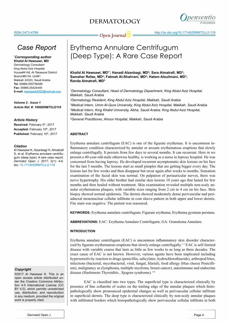
Figure 1: Multiple Non-scaly Annular Erythema-tus Plaques with Infiltrated Border on Peri-auric-ular Area.
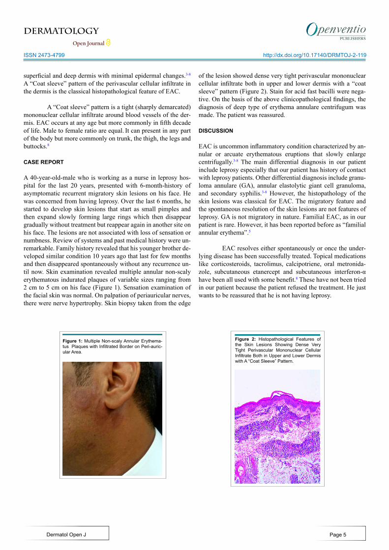
Figure 2: Histopathological Features of the Skin Lesions Showing Dense Very Tight Perivascular Mononuclear Cellular Infiltrate Both in Upper and Lower Dermis with A “Coat Sleeve” Pattern.
superficial and deep dermis with minimal epidermal changes.3-8 A “Coat sleeve” pattern of the perivascular cellular infiltrate in the dermis is the classical histopathological feature of EAC.
A “Coat sleeve” pattern is a tight (sharply demarcated) mononuclear cellular infiltrate around blood vessels of the der-mis. EAC occurs at any age but more commonly in fifth decade of life. Male to female ratio are equal. It can present in any part of the body but more commonly on trunk, the thigh, the legs and buttocks.8
CASE REPORT
A 40-year-old-male who is working as a nurse in leprosy hos-pital for the last 20 years, presented with 6-month-history of asymptomatic recurrent migratory skin lesions on his face. He was concerned from having leprosy. Over the last 6 months, he started to develop skin lesions that start as small pimples and then expand slowly forming large rings which then disappear gradually without treatment but reappear again in another site on his face. The lesions are not associated with loss of sensation or numbness. Review of systems and past medical history were un-remarkable. Family history revealed that his younger brother de-veloped similar condition 10 years ago that last for few months and then disappeared spontaneously without any recurrence un-til now. Skin examination revealed multiple annular non-scaly erythematous indurated plaques of variable sizes ranging from 2 cm to 5 cm on his face (Figure 1). Sensation examination of the facial skin was normal. On palpation of periauricular nerves, there were nerve hypertrophy. Skin biopsy taken from the edge
of the lesion showed dense very tight perivascular mononuclear cellular infiltrate both in upper and lower dermis with a “coat sleeve” pattern (Figure 2). Stain for acid fast bacilli were nega-tive. On the basis of the above clinicopathological findings, the diagnosis of deep type of erythema annulare centrifugum was made. The patient was reassured.
DISCUSSION
EAC is uncommon inflammatory condition characterized by an-nular or arcuate erythematous eruptions that slowly enlarge centrifugally.3-8 The main differential diagnosis in our patient include leprosy especially that our patient has history of contact with leprosy patients. Other differential diagnosis include granu-loma annulare (GA), annular elastolytic giant cell granuloma, and secondary syphilis.3-8 However, the histopathology of the skin lesions was classical for EAC. The migratory feature and the spontaneous resolution of the skin lesions are not features of leprosy. GA is not migratory in nature. Familial EAC, as in our patient is rare. However, it has been reported before as “familial annular erythema”.5
EAC resolves either spontaneously or once the under-lying disease has been successfully treated. Topical medications like corticosteroids, tacrolimus, calcipotriene, oral metronida-zole, subcutaneous etanercept and subcutaneous interferon-α have been all used with some benefit.8 These have not been tried in our patient because the patient refused the treatment. He just wants to be reassured that he is not having leprosy.
DERMATOLOGYOpen Journal
http://dx.doi.org/10.17140/DRMTOJ-2-119
Dermatol Open J
ISSN 2473-4799
Page 6
ACKNOWLEDGMENTS
No sources of funding were used to assist in preparation of this manuscript.
CONFLICTS OF INTEREST
The authors have no conflicts of interest that are directly relevant to the content of this review.
CONSENT STATEMENT
Consent has been taken from the patient for purpose of using patient’s photographs for publication in print or on the internet.
REFERENCES
1. Kavurt S, Aydemir O, Celik U, et al. Erythema annulare cen-trifugum as the presenting sign of pseudomonas aeruginosa sep-sis in a newborn. Eur J Pediatr. 2013; 172(6): 847-849. doi: 10.1007/s00431-012-1848-8
2. Kim DH, Lee JH, Lee JY, Park YM. Erythema annulare cen-trifugum: Analysis of associated diseases and clinical outcomes according to histopathologic classification. Ann Dermatol. 2016; 28(2): 257-259. doi: 10.5021/ad.2016.28.2.257
3. Weyers W, Diaz-Cascajo C, Weyers I. Erythema annulare
centrifugum: Results of a clinicopathologic study of 73 pa-tients. Am Dermatol. 2003; 25(6): 451-462. Web site. http://journals.lww.com/amjdermatopathology/pages/articleviewer.aspx?year=2003&issue=12000&article=00001&type=abstract. Accessed February 4, 2017.
4. Ohmori S, Sugita K, Ikenouchi-Sugita A, et al. Erythema an-nulare centrifugum associated with herpes zoster. J UOEH. 2012; 34(3): 225-229. doi: 10.7888/juoeh.34.225
5. Bressler GS, Jones RE Jr. Erythema annulare centrifugum. J Am Acad Dermatol. 1981; 4(5): 597-602. doi: 10.1016/S0190-9622(81)70063-X
6. Ziemer M, Eisendle K, Zelger B. New concepts on erythema annulare centrifugum: A clinical reaction pattern that does not represent a specific clinicopathological entity. Br J Dermatol. 2009; 160(1): 119-126. doi: 10.1111/j.1365-2133.2008.08803.x
7. Watsky KL, Hansen T. Annular erythema in identical twins. Cutis. 1989; 44(2): 139-140. Web site. http://europepmc.org/abstract/med/2758863. Accessed February 4, 2017.
8. Mshrai H, Fallatah B, Alwafi D, Babkoor D, Al Sufyani H, Al Hawsawi K. Erythema annulare centrifugum (EAC): A case report of annually recurring EAC. J Health Sci. 2016; 6(5): 74-76. doi: 10.5923/j.health.20160605.02