Thoracic endometriosis syndrome – case 1,3 study 1 ... · Endometrioza opłucnej – studium...
3
436 M. Klimek et al. Mateusz KLIMEK 1 Maciej KAJOR 2 Katarzyna WOREK 1,3 Wojciech SZANECKI 1 1 Department of Gynaecology and Obstetrics of the Medical University of Silesia, Katowice, Poland Head: Prof. dr hab. n. med. Andrzej Witek 2 Department of Pathomorphology of the Medical University of Silesia, Katowice, Poland Head: Dr hab. n. med. Maciej Kajor 3 Students’ Scientific Association in Department of Gynaecology and Obsterics, Medical University of Silesia, Katowice, Poland Additional key words: catamenial pneumothorax thoracic endometriosis syndrome videothoracoscopy Dodatkowe słowa kluczowe: zespół endometriozy opłucnej odma katamenalna wideotorakoskopia Address for correspondence: Wojciech Szanecki 40-001 Katowice ul. Medyków 14 tel: +48 697 138 524 e-mail: [email protected] CASE REPORTS Thoracic endometriosis syndrome – case study Endometrioza opłucnej – studium przypadku Conflicts of interest The authors have no conflicts of interest relevant to this article TES (-thoracic endometriosis syn- drome) is a rare disease characteri- zed by the presence of functioning endometrial tissue in pleura, lung pa- renchyma, airways and/or diaphragm. TES may present with catamenial pneumothorax which occur within 72 hours before or after menstruation [1,2]. The authors present a case of a 51 year old female who was presen- ting cyclical recurrent episodes of chest pain, shortness of breath and right side pneumothorax. Histopatho- logical examination after resection of the top of the upper lobe of the right lung revealed clusters of endometrial cells staining positive for estrogen re- ceptor ES (+). TES (ang. thoracic endometrio- sis syndrome) to rzadka choroba charakteryzująca się występowaniem czynnej tkanki endometrialnej w opłucnej, miąższu płuca, drogach oddechowych i/lub w przeponie. Choroba może obejmować nawrotową odmę opłucnową pojawiającą się w ciągu 72 godzin przed lub po rozpoczęciu menstruacji (odma kata- menalna) [1,2]. Autorzy przedstawiają przypadek 51 letniej po dwóch cięciach cesarskich z endometriozą jajnika z nawracającymi cyklicznymi epizodami bólów w klatce piersio- wej, duszności i prawostronnej odmy opłucnowej. Badania histopatolog- iczne wycinków po resekcji szczytu górnego płata płuca prawego ujawniło skupiska komórek endometrialnych z dodatnim barwieniem na receptor estrogenowy ES (+). Introduction Endometriosis affects between 5% and 15% of women in reproductive age, whe- reas no more than 12% of patients have ectopic tissue outside lower pelvis [1,2]. Transposed fragments of active endome- trial tissue may localize in distant areas of the peritoneal cavity, in the umbilical region, lymph nodes, lungs and even bones and heart. Endometriosis is a frequent cause of infertility, painful periods and pelvic pain. The first case of pleural endometriosis was described by Schwarz in 1938 [3]. TES is a constellation of four clinical presenta- tions, such as catamenial pneumothorax, hemoptysis, hemothorax and pulmonary nodules [4]. TES may present with cata- menial pneumothorax which occurs within 72 hours before or after menstruation [5,1]. The first case of catamenial pneumothorax was described in 1958 by Mauer et al. [6], and the term “catamenial pneumothorax” was first introduced into the literature in 1972 by Lillington et al. [7]. Case report 51-yer-old patient presented to her GP for routine health check. She complained of decreased exercisetolerance and effort dyspnea. Physical examination did not show any abnormalities. She had a histo- ry numerous admissions to pulmonology wards and menstrual problems (copious and painful menstruations). She had re- gular periods and was not treated for any chronic condition. At the age of 43 she had an emergen- cy admission to pulmonology ward becau- se of weakness, dyspnea and right-sided chest pain caused by spontaneous pneu- mothorax, diagnosed 5 days earlier on ro- utine plain chest X-ray. On admission, she presented with de- creased oxygen saturation-SpO 2 91% and tachypnea-22 per minute. Laboratory tests and ECG did not show any abnormalities. A series of chest x-ray films in antero-late- ral projection revealed 5 mm air deposit in the right apical region. The 3 consecutive imaging performed every 2 days showed regression of the pneumothorax. Compu- terized tomography (CT) of chest without contrast performed on the day 7 did not show presence of any air in the right pleu- ral cavity, no emphysematous bullae and no focal lesions in the lung parenchyma. Pleural recesses and diaphragm were nor- mal. Ultrasound of abdominal cavity and thyroid were normal. The treatment included oxygen therapy (10 L/min) and estazolam. Patient was di- scharged home in good general condition and referred to outpatient chest clinic for follow-up. Next month, the patient was re-ad- mitted as an emergency to the thoracic surgery ward because of dyspnea, we- akness and pain in the region of the right apex. Standard laboratory tests and ECG results were normal. X-ray examination re- vealed recurrent spontaneous right-sided pneumothorax (Fig. 1). Right pleural cavity
Transcript of Thoracic endometriosis syndrome – case 1,3 study 1 ... · Endometrioza opłucnej – studium...
436 M. Klimek et al.
Mateusz KLIMEK1
Maciej KAJOR2
Katarzyna WOREK1,3
Wojciech SZANECKI1
1Department of Gynaecology and Obstetrics of the Medical University of Silesia, Katowice, PolandHead:Prof. dr hab. n. med. Andrzej Witek
2Department of Pathomorphology of the Medical University of Silesia, Katowice, PolandHead:Dr hab. n. med. Maciej Kajor
3Students’ Scientific Association in Department of Gynaecology and Obsterics, Medical University of Silesia, Katowice, Poland
Additional key words:catamenial pneumothoraxthoracic endometriosis syndrome videothoracoscopy
Dodatkowe słowa kluczowe: zespół endometriozy opłucnejodma katamenalnawideotorakoskopia
Address for correspondence:Wojciech Szanecki40-001 Katowiceul. Medyków 14tel: +48 697 138 524e-mail: [email protected]
CASE REPORTS
Thoracic endometriosis syndrome – case study
Endometrioza opłucnej – studium przypadku
Conflicts of interestThe authors have no conflicts of interest relevant to this article
TES (-thoracic endometriosis syn-drome) is a rare disease characteri-zed by the presence of functioning endometrial tissue in pleura, lung pa-renchyma, airways and/or diaphragm. TES may present with catamenial pneumothorax which occur within 72 hours before or after menstruation [1,2]. The authors present a case of a 51 year old female who was presen-ting cyclical recurrent episodes of chest pain, shortness of breath and right side pneumothorax. Histopatho-logical examination after resection of the top of the upper lobe of the right lung revealed clusters of endometrial cells staining positive for estrogen re-ceptor ES (+).
TES (ang. thoracic endometrio-sis syndrome) to rzadka choroba charakteryzująca się występowaniem czynnej tkanki endometrialnej w opłucnej, miąższu płuca, drogach oddechowych i/lub w przeponie. Choroba może obejmować nawrotową odmę opłucnową pojawiającą się w ciągu 72 godzin przed lub po rozpoczęciu menstruacji (odma kata-menalna) [1,2]. Autorzy przedstawiają przypadek 51 letniej po dwóch cięciach cesarskich z endometriozą jajnika z nawracającymi cyklicznymi epizodami bólów w klatce piersio-wej, duszności i prawostronnej odmy opłucnowej. Badania histopatolog-iczne wycinków po resekcji szczytu górnego płata płuca prawego ujawniło skupiska komórek endometrialnych z dodatnim barwieniem na receptor estrogenowy ES (+).
IntroductionEndometriosis affects between 5% and
15% of women in reproductive age, whe-reas no more than 12% of patients have ectopic tissue outside lower pelvis [1,2]. Transposed fragments of active endome-trial tissue may localize in distant areas of the peritoneal cavity, in the umbilical region, lymph nodes, lungs and even bones and heart. Endometriosis is a frequent cause of infertility, painful periods and pelvic pain. The first case of pleural endometriosis was described by Schwarz in 1938 [3]. TES is a constellation of four clinical presenta-tions, such as catamenial pneumothorax, hemoptysis, hemothorax and pulmonary nodules [4]. TES may present with cata-menial pneumothorax which occurs within 72 hours before or after menstruation [5,1]. The first case of catamenial pneumothorax was described in 1958 by Mauer et al. [6], and the term “catamenial pneumothorax” was first introduced into the literature in 1972 by Lillington et al. [7].
Case report 51-yer-old patient presented to her GP
for routine health check. She complained of decreased exercisetolerance and effort dyspnea. Physical examination did not show any abnormalities. She had a histo-ry numerous admissions to pulmonology wards and menstrual problems (copious and painful menstruations). She had re-gular periods and was not treated for any chronic condition.
At the age of 43 she had an emergen-cy admission to pulmonology ward becau-se of weakness, dyspnea and right-sided chest pain caused by spontaneous pneu-mothorax, diagnosed 5 days earlier on ro-utine plain chest X-ray.
On admission, she presented with de-creased oxygen saturation-SpO2 91% and tachypnea-22 per minute. Laboratory tests and ECG did not show any abnormalities. A series of chest x-ray films in antero-late-ral projection revealed 5 mm air deposit in the right apical region. The 3 consecutive imaging performed every 2 days showed regression of the pneumothorax. Compu-terized tomography (CT) of chest without contrast performed on the day 7 did not show presence of any air in the right pleu-ral cavity, no emphysematous bullae and no focal lesions in the lung parenchyma. Pleural recesses and diaphragm were nor-mal. Ultrasound of abdominal cavity and thyroid were normal.
The treatment included oxygen therapy (10 L/min) and estazolam. Patient was di-scharged home in good general condition and referred to outpatient chest clinic for follow-up.
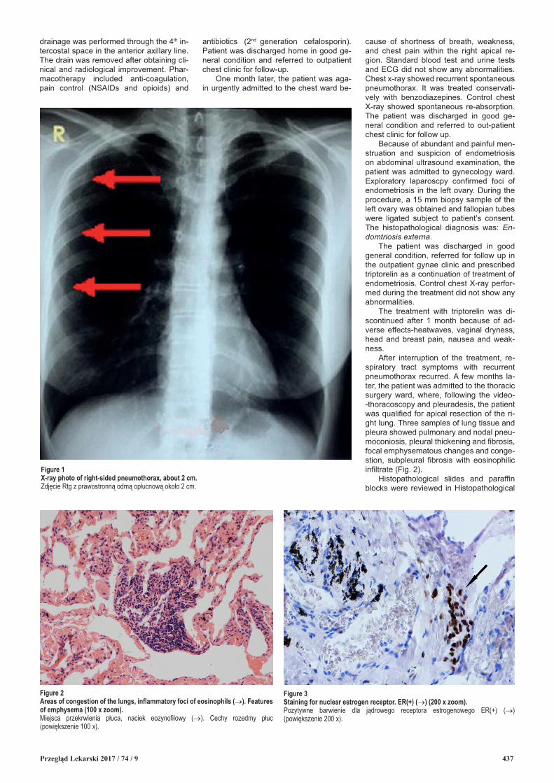
Next month, the patient was re-ad-mitted as an emergency to the thoracic surgery ward because of dyspnea, we-akness and pain in the region of the right apex. Standard laboratory tests and ECG results were normal. X-ray examination re-vealed recurrent spontaneous right-sided pneumothorax (Fig. 1). Right pleural cavity
Przegląd Lekarski 2017 / 74 / 9 437
cause of shortness of breath, weakness, and chest pain within the right apical re-gion. Standard blood test and urine tests and ECG did not show any abnormalities. Chest x-ray showed recurrent spontaneous pneumothorax. It was treated conservati-vely with benzodiazepines. Control chest X-ray showed spontaneous re-absorption. The patient was discharged in good ge-neral condition and referred to out-patient chest clinic for follow up.
Because of abundant and painful men-struation and suspicion of endometriosis on abdominal ultrasound examination, the patient was admitted to gynecology ward. Exploratory laparoscpy confirmed foci of endometriosis in the left ovary. During the procedure, a 15 mm biopsy sample of the left ovary was obtained and fallopian tubes were ligated subject to patient’s consent. The histopathological diagnosis was: En-domtriosis externa.
The patient was discharged in good general condition, referred for follow up in the outpatient gynae clinic and prescribed triptorelin as a continuation of treatment of endometriosis. Control chest X-ray perfor-med during the treatment did not show any abnormalities.
The treatment with triptorelin was di-scontinued after 1 month because of ad-verse effects-heatwaves, vaginal dryness, head and breast pain, nausea and weak-ness.
After interruption of the treatment, re-spiratory tract symptoms with recurrent pneumothorax recurred. A few months la-ter, the patient was admitted to the thoracic surgery ward, where, following the video--thoracoscopy and pleuradesis, the patient was qualified for apical resection of the ri-ght lung. Three samples of lung tissue and pleura showed pulmonary and nodal pneu-moconiosis, pleural thickening and fibrosis, focal emphysematous changes and conge-stion, subpleural fibrosis with eosinophilic infiltrate (Fig. 2).
Histopathological slides and paraffin blocks were reviewed in Histopathological
Figure 1X-ray photo of right-sided pneumothorax, about 2 cm.Zdjęcie Rtg z prawostronną odmą opłucnową około 2 cm.
Figure 2Areas of congestion of the lungs, inflammatory foci of eosinophils (→). Features of emphysema (100 x zoom).Miejsca przekrwienia płuca, naciek eozynofilowy (→). Cechy rozedmy płuc (powiększenie 100 x).
Figure 3Staining for nuclear estrogen receptor. ER(+) (→) (200 x zoom).Pozytywne barwienie dla jądrowego receptora estrogenowego ER(+) (→) (powiększenie 200 x).
drainage was performed through the 4th in-tercostal space in the anterior axillary line. The drain was removed after obtaining cli-nical and radiological improvement. Phar-macotherapy included anti-coagulation, pain control (NSAIDs and opioids) and
antibiotics (2nd generation cefalosporin). Patient was discharged home in good ge-neral condition and referred to outpatient chest clinic for follow-up.
One month later, the patient was aga-in urgently admitted to the chest ward be-
438
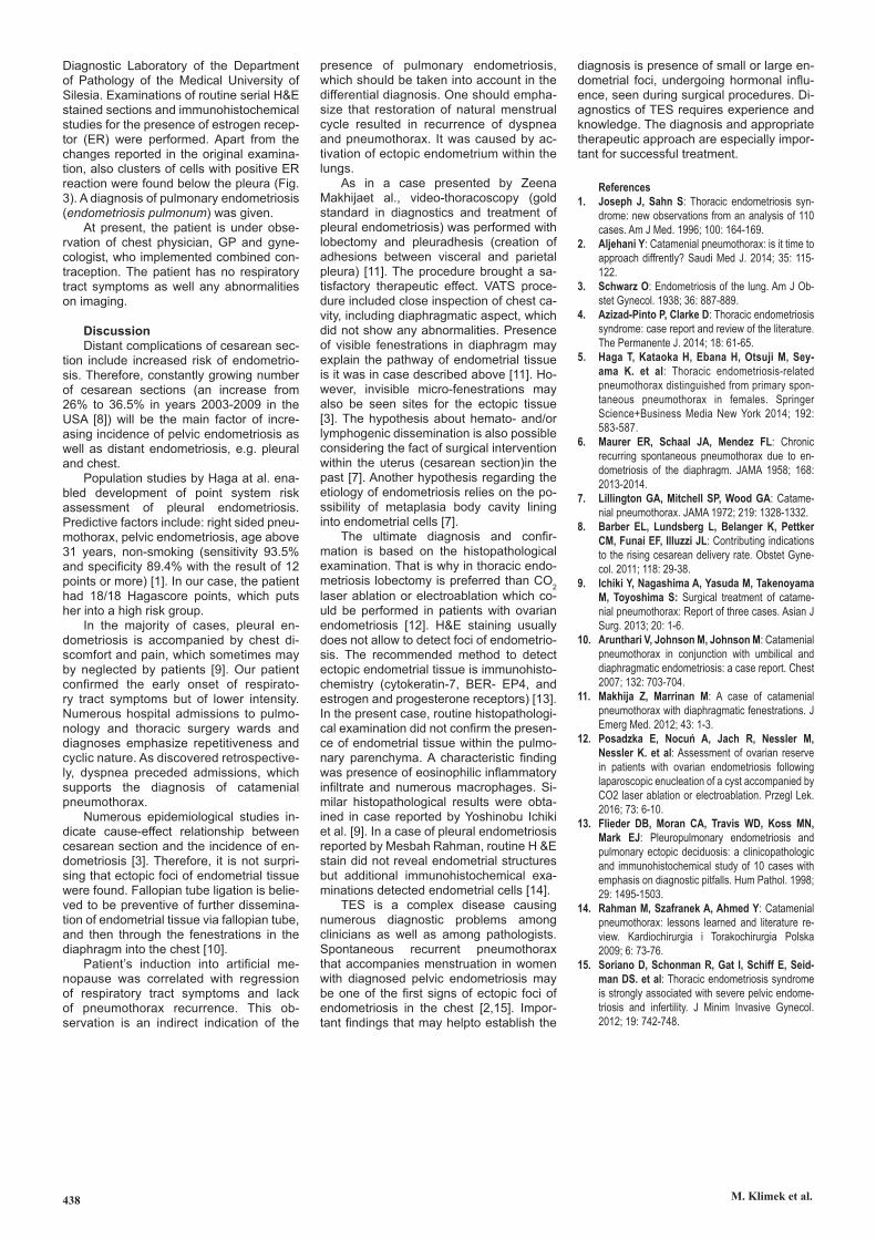
Diagnostic Laboratory of the Department of Pathology of the Medical University of Silesia. Examinations of routine serial H&E stained sections and immunohistochemical studies for the presence of estrogen recep-tor (ER) were performed. Apart from the changes reported in the original examina-tion, also clusters of cells with positive ER reaction were found below the pleura (Fig. 3). A diagnosis of pulmonary endometriosis (endometriosis pulmonum) was given.
At present, the patient is under obse-rvation of chest physician, GP and gyne-cologist, who implemented combined con-traception. The patient has no respiratory tract symptoms as well any abnormalities on imaging.
DiscussionDistant complications of cesarean sec-
tion include increased risk of endometrio-sis. Therefore, constantly growing number of cesarean sections (an increase from 26% to 36.5% in years 2003-2009 in the USA [8]) will be the main factor of incre-asing incidence of pelvic endometriosis as well as distant endometriosis, e.g. pleural and chest.
Population studies by Haga at al. ena-bled development of point system risk assessment of pleural endometriosis. Predictive factors include: right sided pneu-mothorax, pelvic endometriosis, age above 31 years, non-smoking (sensitivity 93.5% and specificity 89.4% with the result of 12 points or more) [1]. In our case, the patient had 18/18 Hagascore points, which puts her into a high risk group.
In the majority of cases, pleural en-dometriosis is accompanied by chest di-scomfort and pain, which sometimes may by neglected by patients [9]. Our patient confirmed the early onset of respirato-ry tract symptoms but of lower intensity. Numerous hospital admissions to pulmo-nology and thoracic surgery wards and diagnoses emphasize repetitiveness and cyclic nature. As discovered retrospective-ly, dyspnea preceded admissions, which supports the diagnosis of catamenial pneumothorax.
Numerous epidemiological studies in-dicate cause-effect relationship between cesarean section and the incidence of en-dometriosis [3]. Therefore, it is not surpri-sing that ectopic foci of endometrial tissue were found. Fallopian tube ligation is belie-ved to be preventive of further dissemina-tion of endometrial tissue via fallopian tube, and then through the fenestrations in the diaphragm into the chest [10].
Patient’s induction into artificial me-nopause was correlated with regression of respiratory tract symptoms and lack of pneumothorax recurrence. This ob-servation is an indirect indication of the
presence of pulmonary endometriosis, which should be taken into account in the differential diagnosis. One should empha-size that restoration of natural menstrual cycle resulted in recurrence of dyspnea and pneumothorax. It was caused by ac-tivation of ectopic endometrium within the lungs.
As in a case presented by Zeena Makhijaet al., video-thoracoscopy (gold standard in diagnostics and treatment of pleural endometriosis) was performed with lobectomy and pleuradhesis (creation of adhesions between visceral and parietal pleura) [11]. The procedure brought a sa-tisfactory therapeutic effect. VATS proce-dure included close inspection of chest ca-vity, including diaphragmatic aspect, which did not show any abnormalities. Presence of visible fenestrations in diaphragm may explain the pathway of endometrial tissue is it was in case described above [11]. Ho-wever, invisible micro-fenestrations may also be seen sites for the ectopic tissue [3]. The hypothesis about hemato- and/or lymphogenic dissemination is also possible considering the fact of surgical intervention within the uterus (cesarean section)in the past [7]. Another hypothesis regarding the etiology of endometriosis relies on the po-ssibility of metaplasia body cavity lining into endometrial cells [7].
The ultimate diagnosis and confir-mation is based on the histopathological examination. That is why in thoracic endo-metriosis lobectomy is preferred than CO2 laser ablation or electroablation which co-uld be performed in patients with ovarian endometriosis [12]. H&E staining usually does not allow to detect foci of endometrio-sis. The recommended method to detect ectopic endometrial tissue is immunohisto-chemistry (cytokeratin-7, BER- EP4, and estrogen and progesterone receptors) [13]. In the present case, routine histopathologi-cal examination did not confirm the presen-ce of endometrial tissue within the pulmo-nary parenchyma. A characteristic finding was presence of eosinophilic inflammatory infiltrate and numerous macrophages. Si-milar histopathological results were obta-ined in case reported by Yoshinobu Ichiki et al. [9]. In a case of pleural endometriosis reported by Mesbah Rahman, routine H &E stain did not reveal endometrial structures but additional immunohistochemical exa-minations detected endometrial cells [14].
TES is a complex disease causing numerous diagnostic problems among clinicians as well as among pathologists. Spontaneous recurrent pneumothorax that accompanies menstruation in women with diagnosed pelvic endometriosis may be one of the first signs of ectopic foci of endometriosis in the chest [2,15]. Impor-tant findings that may helpto establish the
diagnosis is presence of small or large en-dometrial foci, undergoing hormonal influ-ence, seen during surgical procedures. Di-agnostics of TES requires experience and knowledge. The diagnosis and appropriate therapeutic approach are especially impor-tant for successful treatment.
References1. Joseph J, Sahn S: Thoracic endometriosis syn-
drome: new observations from an analysis of 110 cases. Am J Med. 1996; 100: 164-169.
2. Aljehani Y: Catamenial pneumothorax: is it time to approach diffrently? Saudi Med J. 2014; 35: 115-122.
3. Schwarz O: Endometriosis of the lung. Am J Ob-stet Gynecol. 1938; 36: 887-889.
4. Azizad-Pinto P, Clarke D: Thoracic endometriosis syndrome: case report and review of the literature. The Permanente J. 2014; 18: 61-65.
5. Haga T, Kataoka H, Ebana H, Otsuji M, Sey-ama K. et al: Thoracic endometriosis-related pneumothorax distinguished from primary spon-taneous pneumothorax in females. Springer Science+Business Media New York 2014; 192: 583-587.
6. Maurer ER, Schaal JA, Mendez FL: Chronic recurring spontaneous pneumothorax due to en-dometriosis of the diaphragm. JAMA 1958; 168: 2013-2014.
7. Lillington GA, Mitchell SP, Wood GA: Catame-nial pneumothorax. JAMA 1972; 219: 1328-1332.
8. Barber EL, Lundsberg L, Belanger K, Pettker CM, Funai EF, Illuzzi JL: Contributing indications to the rising cesarean delivery rate. Obstet Gyne-col. 2011; 118: 29-38.
9. Ichiki Y, Nagashima A, Yasuda M, Takenoyama M, Toyoshima S: Surgical treatment of catame-nial pneumothorax: Report of three cases. Asian J Surg. 2013; 20: 1-6.
10. Arunthari V, Johnson M, Johnson M: Catamenial pneumothorax in conjunction with umbilical and diaphragmatic endometriosis: a case report. Chest 2007; 132: 703-704.
11. Makhija Z, Marrinan M: A case of catamenial pneumothorax with diaphragmatic fenestrations. J Emerg Med. 2012; 43: 1-3.
12. Posadzka E, Nocuń A, Jach R, Nessler M, Nessler K. et al: Assessment of ovarian reserve in patients with ovarian endometriosis following laparoscopic enucleation of a cyst accompanied by CO2 laser ablation or electroablation. Przegl Lek. 2016; 73: 6-10.
13. Flieder DB, Moran CA, Travis WD, Koss MN, Mark EJ: Pleuropulmonary endometriosis and pulmonary ectopic deciduosis: a clinicopathologic and immunohistochemical study of 10 cases with emphasis on diagnostic pitfalls. Hum Pathol. 1998; 29: 1495-1503.
14. Rahman M, Szafranek A, Ahmed Y: Catamenial pneumothorax: lessons learned and literature re-view. Kardiochirurgia i Torakochirurgia Polska 2009; 6: 73-76.
15. Soriano D, Schonman R, Gat I, Schiff E, Seid-man DS. et al: Thoracic endometriosis syndrome is strongly associated with severe pelvic endome-triosis and infertility. J Minim Invasive Gynecol. 2012; 19: 742-748.
M. Klimek et al.