Results and biomechanical consideration of treatment of ... · Results and biomechanical...
8
Acta of Bioengineering and Biomechanics Original paper Vol. 16, No. 1, 2014 DOI: 10.5277/abb140116 Results and biomechanical consideration of treatment of congenital lower limb shortening and deformity using the Ilizarov method PIOTR MORASIEWICZ* 1 , LESZEK MORASIEWICZ 2 , MAREK STĘPNIEWSKI 1 , WIKTOR ORZECHOWSKI 1 , MAŁGORZATA MORASIEWICZ 2 , ŁUKASZ PAWIK 2 , ZDZISŁAWA WRZOSEK 2 , SZYMON DRAGAN 1 1 Wrocław Medical University, Department and Clinic of Orthopaedic and Traumatologic Surgery, ul. Borowska 213, 50-556 Wrocław, Poland. 2 University of Physical Education in Wrocław, The Chair of Physiotherapy and Occupational Therapy in Motor System Dysfunctions, ul. Paderewskiego 35, 51-612 Wrocław, Poland. One of the applications of the Ilizarov apparatus is the correction of congenital shortening and deformities. Ilizarov external fixator produces biomechanical structure with surrounding tissue, which is the reason why very important is correct stability of fixator. Large distraction in the case of high value of lengthening, and large deformity corection result in shear stresses that occur additionally in the regenerate, which can potentially lead to damage of the regenerating nutritive microcirculation of bone tissue and bone fragment dis- placements. Our objective was to assess the results of the Ilizarov method in the treatment of congenital shortening taking into account treatment strategy and the size of the axis of lengthening and correction. Our research problems include presenting the effects of biome- chanics of musculoskeletal deformations on treatment results, presenting complications and their treatment. Between 1989 and 2009, 62 patients underwent surgery to correct congenital lower limb deficiencies at our Clinic; 33 patients were followed-up. In total, there were 70 surgeries (2.12/patient). Axial correction was performed in 26 patients (78.79%). Average age at the start of the treatment was 15.58 years. Mean follow-up was 8.58 years. Mean lengthening per surgery was 3.17 cm with the lengthening index of 50.7 day/cm. Results were very good for 23 patients, good for 7 patients, satisfactory for 3 patients. Complications appeared in 24 patients, problems occurred in 74.42% of the cases, obstacles in 4.65% of cases, and true complications in 20.93% of the cases. The best results were achieved in the treatment of patients with two-stage and two-segment lengthening with a total elongation of less than 7 cm, and without correction of the axis. Congenital shortening of the lower limb should be treated comprehensively because the shortening applies to all segments, and disturbs biomechanics of all lower limb. In the case of axial correction and large amount of elongation high soft tissue forces counteract the distraction forces. Hybrid construction may help to shorten treatment time, increase fixator stability and decrease rate of complications. We suggest use of hybrid Ilizarov fixator, especially when large elongation and axis corection are planned. Key words: bone elongation, congenital deformity, hybrid external fixator, Ilizarov external fixator 1. Introduction One of the applications of the Ilizarov apparatus is the correction of congenital shortening and deformities. Congenital shortening of the lower limb occurs very rarely, at a rate of 1–3 per 10 000 births [9], [14], [15], [29]. The aetiology is not completely clear, it is known, however, that genetic and environmental factors play a major role [3], [9], [13]–[15], [29]. The clinical picture shows a reduction in the size of the lower ex- tremity, consisting in its shortening and thinning. The shortening applies to the pelvis and all the segments of the lower limb: femur, lower leg, and foot in any ______________________________ * Corresponding author: Piotr Morasiewicz, Department and Clinic of Orthopaedic and Traumatologic Surgery, Wrocław Medical University, ul. Borowska 213, 50-556 Wrocław, Poland. Tel: +48-71-734-32-00, e-mail: [email protected] Received: April 10th, 2013 Accepted for publication: August 7th, 2013
Transcript of Results and biomechanical consideration of treatment of ... · Results and biomechanical...
Acta of Bioengineering and Biomechanics Original paperVol. 16, No. 1, 2014 DOI: 10.5277/abb140116
Results and biomechanical consideration oftreatment of congenital lower limb shortening
and deformity using the Ilizarov method
PIOTR MORASIEWICZ*1, LESZEK MORASIEWICZ2, MAREK STĘPNIEWSKI1, WIKTOR ORZECHOWSKI1,MAŁGORZATA MORASIEWICZ2, ŁUKASZ PAWIK2, ZDZISŁAWA WRZOSEK2, SZYMON DRAGAN1
1 Wrocław Medical University, Department and Clinic of Orthopaedic and Traumatologic Surgery,ul. Borowska 213, 50-556 Wrocław, Poland.
2 University of Physical Education in Wrocław, The Chair of Physiotherapy and Occupational Therapy in Motor System Dysfunctions,ul. Paderewskiego 35, 51-612 Wrocław, Poland.
One of the applications of the Ilizarov apparatus is the correction of congenital shortening and deformities. Ilizarov external fixatorproduces biomechanical structure with surrounding tissue, which is the reason why very important is correct stability of fixator. Largedistraction in the case of high value of lengthening, and large deformity corection result in shear stresses that occur additionally in theregenerate, which can potentially lead to damage of the regenerating nutritive microcirculation of bone tissue and bone fragment dis-placements. Our objective was to assess the results of the Ilizarov method in the treatment of congenital shortening taking into accounttreatment strategy and the size of the axis of lengthening and correction. Our research problems include presenting the effects of biome-chanics of musculoskeletal deformations on treatment results, presenting complications and their treatment.
Between 1989 and 2009, 62 patients underwent surgery to correct congenital lower limb deficiencies at our Clinic; 33 patients werefollowed-up. In total, there were 70 surgeries (2.12/patient). Axial correction was performed in 26 patients (78.79%). Average age at thestart of the treatment was 15.58 years. Mean follow-up was 8.58 years.
Mean lengthening per surgery was 3.17 cm with the lengthening index of 50.7 day/cm. Results were very good for 23 patients, goodfor 7 patients, satisfactory for 3 patients. Complications appeared in 24 patients, problems occurred in 74.42% of the cases, obstacles in4.65% of cases, and true complications in 20.93% of the cases.
The best results were achieved in the treatment of patients with two-stage and two-segment lengthening with a total elongation ofless than 7 cm, and without correction of the axis. Congenital shortening of the lower limb should be treated comprehensively becausethe shortening applies to all segments, and disturbs biomechanics of all lower limb. In the case of axial correction and large amount ofelongation high soft tissue forces counteract the distraction forces. Hybrid construction may help to shorten treatment time, increasefixator stability and decrease rate of complications. We suggest use of hybrid Ilizarov fixator, especially when large elongation and axiscorection are planned.
Key words: bone elongation, congenital deformity, hybrid external fixator, Ilizarov external fixator
1. Introduction
One of the applications of the Ilizarov apparatus isthe correction of congenital shortening and deformities.Congenital shortening of the lower limb occurs veryrarely, at a rate of 1–3 per 10 000 births [9], [14], [15],
[29]. The aetiology is not completely clear, it is known,however, that genetic and environmental factors playa major role [3], [9], [13]–[15], [29]. The clinicalpicture shows a reduction in the size of the lower ex-tremity, consisting in its shortening and thinning. Theshortening applies to the pelvis and all the segmentsof the lower limb: femur, lower leg, and foot in any
______________________________
* Corresponding author: Piotr Morasiewicz, Department and Clinic of Orthopaedic and Traumatologic Surgery, Wrocław MedicalUniversity, ul. Borowska 213, 50-556 Wrocław, Poland. Tel: +48-71-734-32-00, e-mail: [email protected]
Received: April 10th, 2013Accepted for publication: August 7th, 2013
P. MORASIEWICZ et al.134
dimension, and in different proportions in particularsegments. In many cases, other abnormalities, such asvalgus or varus knee deformity, the lack of certainmetatarsal bones, talocalcaneal coalition, and otherfoot deformities co-exist [1], [4], [9], [13], [14], [16],[18], [24], [25], [28]. Ankle joint is unstable and set ina crooked position due to the spherical shape of thetrochlea of the talus in the frontal plane (“ball andsocket ankle joint”) [1], [11], [13], [14], [18], [28].Aside from limb lengthening, patients frequently re-quire correction of the associated deformities. Due tolower osteogenic potential of the bone (there are altera-tions in bone structure that originate during the prenatalperiod), treatment is very difficult [6], [9], [15], [24].
Until the 1970s orthopedic footwear was the mostcommon method of treatment, followed by prosthesisfor large shortenings, and amputation if use of prosthe-sis was not possible [1], [4], [7], [9], [10], [14], [15],[25]. Limb lengthening with the Wagner technique orusing Orthofix stabilizer was used in exceptional casesdue to a large number of complications [12].
The introduction of the external circular fixator byIlizarov and introduction of distraction osteogenesisallowed effective treatment of these patients [9], [11],[12], [16], [25]. Ilizarov external fixator producebiomechanical structure with surrounding tissue,which is the reason why very important is correctstability of fixator. Too flexible, or too rigid anaparatus causes increased rate of complications [22],[23]. In the case of congenital deformity correction,internal forces increased due to the bad condition ofthe soft tissues, and poor elastic properties. Theamount and direction of bone fragment displacementsare associated with the biomechanical conditions inthe bone regenerate, which determine whether or notthe treatment process will progress in an optimalmanner [22], [23]. In addition to distraction, Ilizarov’sapparatus makes it possible to correct the deformity inthree planes [11], [24], [25]. Large distraction in thecase of high value of lengthening, and large deformitycorrection results in shear stresses that occur addition-ally in the regenerate, which can potentially lead todamage of the regenerating nutritive microcirculationof bone tissue and bone fragment displacements. Thusfar, there are only a few publications in internationalliterature on the results and treatment of congenitalshortening and deformities of lower limb using theIlizarov method [3], [9], [11], [12], [16], [25]. Hence,our objective was to assess the results of the Ilizarovmethod in the treatment of congenital shortening anddistortion of the lower limb, taking into accounttreatment strategy and the size of the axis of length-ening and correction. Our research problems include
presenting the effects of biomechanics of musculo-skeletal deformations on treatment results, presentingcomplications and their treatment.
2. Materials and methods
In the years 1989–2009, 62 patients with congeni-tal shortening and deformity of the lower limb weresurgically treated by the Ilizarov method in our insti-tution. Due to the different characteristics of defectsas well as dissimilar requirements for treatment, pa-tients with aplasia of the fibula, tibial aplasia, congeni-tal pseudarthrosis of the tibia, Ollier’s disease, multipleexostosis, fibrous dysplasia, and other rare congenitalsyndromes were not included in this group.
This research project was approved by the Bio-ethics Committee of Wrocław Medical University on19.01.2012, with decision number KB–735/2011.
A retrospective study, involving 33 patients(17 males, 16 females) who had participated in thefollow-up was conducted. The mean age at the start oftreatment was 15.6 years (4.5–46 years of age).
Single-stage procedures were performed in pa-tients with completed growth. In younger patients, inwhom the growth process was still ongoing, multi-stage treatment was implemented due to the recur-rence of limb shortening and deformity during growthor as a result of the size of the original shortening.
Fig. 1. Patient with simultaneous two-segmental surgery– lengthening of the femur and lenghtening
and correction of lower leg valgus deformity
Results and biomechanical consideration of treatment of congenital lower limb shortening and deformity... 135
A total of 70 surgeries were performed in the groupof 33 patients; an average of 2.12 operations per patient.Single-stage lengthening was performed, in 14 patients,9 patients underwent two-stage procedures, simultaneoustwo-stage lengthening of femur and lower leg (Fig. 1)was implemented in 5 patients, and 5 patients weretreated with multiple-stage procedures (3–6 surgeries).The treatment strategy was chosen based on the size andlocation of the deformity, patient’s age, and complica-tions resulting from the treatment.
Among the 33 patients who underwent diagnosticprocedures, hypoplasia of the fibula was found in9 cases and congenital shortening of the femur in 24.The total shortening of the lower limb at the start oftreatment was 5.16 cm (0.5–15.5), representing on aver-age 6.6% of the length of the opposite, healthy limb(0.6–17.1). In addition to the shortening, other distor-tions were found to be present in patients (Table 1).
Table 1. Other deformities of the limb
hiphip dysplasiahip subluxation
11
kneevalgusvaruspatellar instability
2091
shin dysplastic fibula 9ankle joint “ball and socet” ankle joint 5
foot
one or more metatarsal bone agenesisequino varusplano valgustalo calcaneal coalitionother
722210
Axis correction with lengthening of the limb was per-formed in 26 patients (78.79%), the remaining 7 patients(21.21%) underwent lengthening without correction ofthe axis.
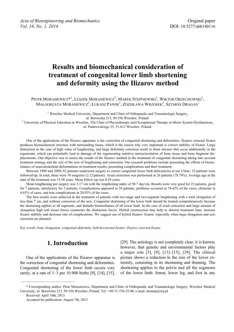
The basic fixator design for femur lengtheningconsisted of one proximal arch fixed with two Schanzscrews, a proximal ring fixed with two K-wires anda distal ring fixed with three K-wires. The basicfixator design for shank lengthening consisted of oneproximal ring with three K-wires, middle ring fixedwith two K-wires, and a distal ring fixed with twoK-wires. We had used a hybrid Ilizarov fixator,which has at middle or/and distal ring in femur con-figuration one or two Kirschner wires replaced byShantz screws (Fig. 2). In shank fixator constructionproximal ring had one Kirschner wire replaced byShantz screws.
Corticotomy was carried out at the distal femoralmetaphysis, at the proximal metaphysis of tibia, and at1/3 proximal of fibula in the lower leg, or at the apexof the deformity.
Fig. 2. Hybrid Ilizarov fixator for femur elongation
In the case of a significant shortening of the distalend of fibula, an additional elongation of the distalmetaphysis of this bone was performed in order torestore ankle stability, and to correct the proportionbetween tibia and fibula for improving the normal bio-mechanics of the ankle. Walking with partial weightbearing on the leg was initiated two days after surgery.Distraction and correction was initiated 6 days aftersurgery. Check-ups were carried out at intervals of2–6 weeks. The apparatus was removed when the pres-ence of at least 3 of 4 cortical layers was found.
The evaluation of the results of the treatment wasconducted with Paley’s [27] classification of compli-cations and a point scale for rating the lower limbdeveloped at our Clinic, which has been adopted bytreatment centers using Ilizarov method in our country[19], [20]. The following elements were taken intoconsideration in the point scale system: the size ofsegment shortening, locomotive abilities, axis defor-mation, lower limb muscle strength, range of motionin the joints of the lower limb, orthopedic aids, andthe subjective evaluation of the patient. In assessingthe results of treatment according to this scale, wecompared the differences between the number ofpoints obtained before and after treatment. In addition,an analysis of treatment outcomes including the inci-dence of complications was performed.
The comparison made by us was based on treat-ment strategy (single-stage, two-stage, multi-stage,single-segment, two-segment), the size of lengthening(<7 cm, >7 cm), and the correction or lack of correc-tion of the axis of lengthening of the limb.
P. MORASIEWICZ et al.136
No additional sources of financing were acceptedfor the purposes of this work.
For the statistical analysis of the results we used:Pearson’s correlation coefficient, Cramer’s V correla-tion coefficient, Spearman’s rho correlation coeffi-cient, and Kendall’s tau-b correlation coefficient. Allcoefficients were tested for their statistical correlationby tests available in the statistical package employed– SPSS Statistics 17.0. One-way analysis of varianceANOVA was used in the verification of the hypothe-sis of equality of mean values of the variable “resultof improvement” in the distribution of treatment strat-egy. The Shapiro–Wilk and Kolmogorov–Smirnovtests were used to confirm the hypothesis of normalitywhen the use of test data required normal distribution.For comparison of mean values of the variable “resultof improvement in groups” Student’s t test was used.Wilcoxon paired test was used to compare the effectof treatment on scoring. Statistical significance wasdefined at the level of α = 0.1, as p <= 0.05.
3. Results
The mean follow-up was 8.58 years (2.08–22.66).Average size of lengthening during one surgery was3.17 cm (0.5–7). The average size of the total lengthen-ing in the same patient was 6.73 cm (0.5–19). 11 patientshad total lengthening greater than 7 cm. The mean du-ration of one stage of treatment was 160 days. Theaverage lengthening index was 50.7 days/cm. Averagescore in the evaluation of lower extremity beforetreatment was 69 points and 90 points after treatment.The difference was statistically significant ( p < 0.001,Wilcoxon test).
Adverse events occurred 43 times in 24 patients(72.73%), on average 1.3 times per patient. Adverseevents included the occurrence of problems 32 times(74.42% of adverse events), difficulties 2 times(4.65% of adverse events), and complications 9 times(20.93% of adverse events).
The knee joint range of motion limitation occurred12 times (27.91% of adverse events). In 11 cases,contracture was abolished or significantly reducedfollowing intensive rehabilitation, while one patientrequired surgery to lengthen the tendon of rectusfemoris muscle with extensive plastic surgery anddissection of vastus intermedius muscle. Infectionsaround implants appeared 7 times (16.28% of adverseevents). In 6 cases the infection was controlled withlocal and systemic administration of antibiotics. Onepatient required surgical resection of the Kirschner
wire and debridement of the site of infection. Defor-mation within the regenerate occurred six times(13.95% of adverse events), in 4 cases requiring theadjustment of the apparatus, and in two cases deform-ity correction was achieved after re-operation usingIlizarov method. Delayed union occurred 5 times(11.63% of adverse events). In these patients, union ofthe regenerate was achieved after compression of re-generate in 1 case, in 2 cases after removal of the ap-paratus and immobilization in a cast, and in 2 casesafter the removal of the apparatus and limb immobili-zation in a brace. Fracture of regenerate occurred5 times (11.63% of adverse events). Bone union wasachieved after immobilization of the limb in a cast in4 cases and after a closed repositioning and stabiliza-tion using Ender rods in one case. Deep vein thrombo-sis was observed two times (4.65% of adverse events);in each case it resolved following drug therapy. Sub-luxation of the knee occurred two times (4.65% ofadverse events), requiring the repositioning and stabi-lization using the Ilizarov apparatus attached to thelower leg. Premature union of the regenerate occurred1 time (2.33% of adverse events); this patient wastreated with repeated surgical dissection of the regen-erate and additional distraction. Vascular injury oc-curred 1 time (2.33% of adverse events) and requiredsurgery to repair the vessel (femoral artery). Therewas no damage to the nerves. Breakage of Kirschnerwire occurred twice (4.65% of adverse events) and inboth cases required surgical re-stabilization of Ilizarovapparatus by exchanging Kirschner wire.
Depending on the treatment strategy statisticallysignificant difference in the number of complica-tions and outcome improved were not found in vari-ous groups ( p = 0.31 one-way analysis of varianceANOVA).
In the group with lengthening of >7 cm results af-ter the treatment had improved by an average of 26.73points, while in the case of patients with lengtheningof <7 cm by 18.82 points. As shown by Student’st test, the difference between the two groups is statis-tically significant ( p = 0.033). Lengthening > 7 cmhad an effect on the occurrence of adverse events.Amongst the variables “total lengthening” and “numberof complications” there is a negligible significant sto-chastic dependence at a level of 10% ( p = 0.078) andequal to 0.311 (Spearman’s rho coefficient).
Correction of the axis also influenced the occur-rence of adverse events. In the group with axis cor-rection the outcome was improved after treatment byan average of 21.54 points, while for patients withoutaxis correction by 21.14 points. Cramer’s V correla-tion coefficient for these variables is 0.348 and is sta-
Results and biomechanical consideration of treatment of congenital lower limb shortening and deformity... 137
tistically significant (p = 0.046). The mean differencebetween the group with correction and the groupwithout correction is not statistically significant p =0.929 (Student’s t-test).
4. Discussion
In previous studies, most authors separated con-genital femoral shortening from congenital shorteningof the fibula and congenital shortening of the tibia,presenting the results of the methods and results oftreatment of these diseases in separate publications[3], [9], [10], [12], [14], [16], [25], [26].
We believe that congenital shortening of the lowerlimb should be treated comprehensively because theshortening applies to all segments of the limb, anddisturbs biomechanics of all lower limb. Often, in pa-tients with a hypoplastic fibula or tibia, the shortenedfemoral segment also requires lengthening. A similarsituation occurs in the case of congenital shortening ofthe femur, where in many cases, the shortened lowerleg also requires lengthening.
As a result of the character and extent of defect,a large proportion of patients with congenital short-ening and deformity of the lower limb require two-segment or multi-stage treatment in order to restorethe limb axis and achieve equal limb length to createnormal, or similar to normal biomechanics conditions.Morasiewicz shows that Ilizarov patients with con-
genital aetiology had larger gait parametr asymmetrythan patients with posttraumatic or postseptic aetiol-ogy. She suggests that patients with congenital aetiol-ogy, from birthday put compensatory mechanism,which does not allow for gait symmetry, despite com-plete leg deformity correction and egalisation. In thegroup of patients with congenital shortening and de-formity, complete leg deformity correction and egali-sation, do not restore primary limb function, but givenew biomechanics conditions for obtaining correctlimb function [21].
The best results were achieved in patients withtwo-stage and two-segment elongation with a totalelongation of less than 7 cm and without correction ofthe axis.
Lengthening above 7 cm with axis correction andmulti-stage elongation had slightly worse results. Thegreatest improvement was achieved in patients withlengthening above 7 cm. A significantly lower inci-dence of complications was recorded in the groupwithout correction of the axis. After osteotomy, lowerlimb biomechanical configuration undergoes distur-bance. Bone fragment cannot transfer limb load, mus-cle induced bone fragment displacements.
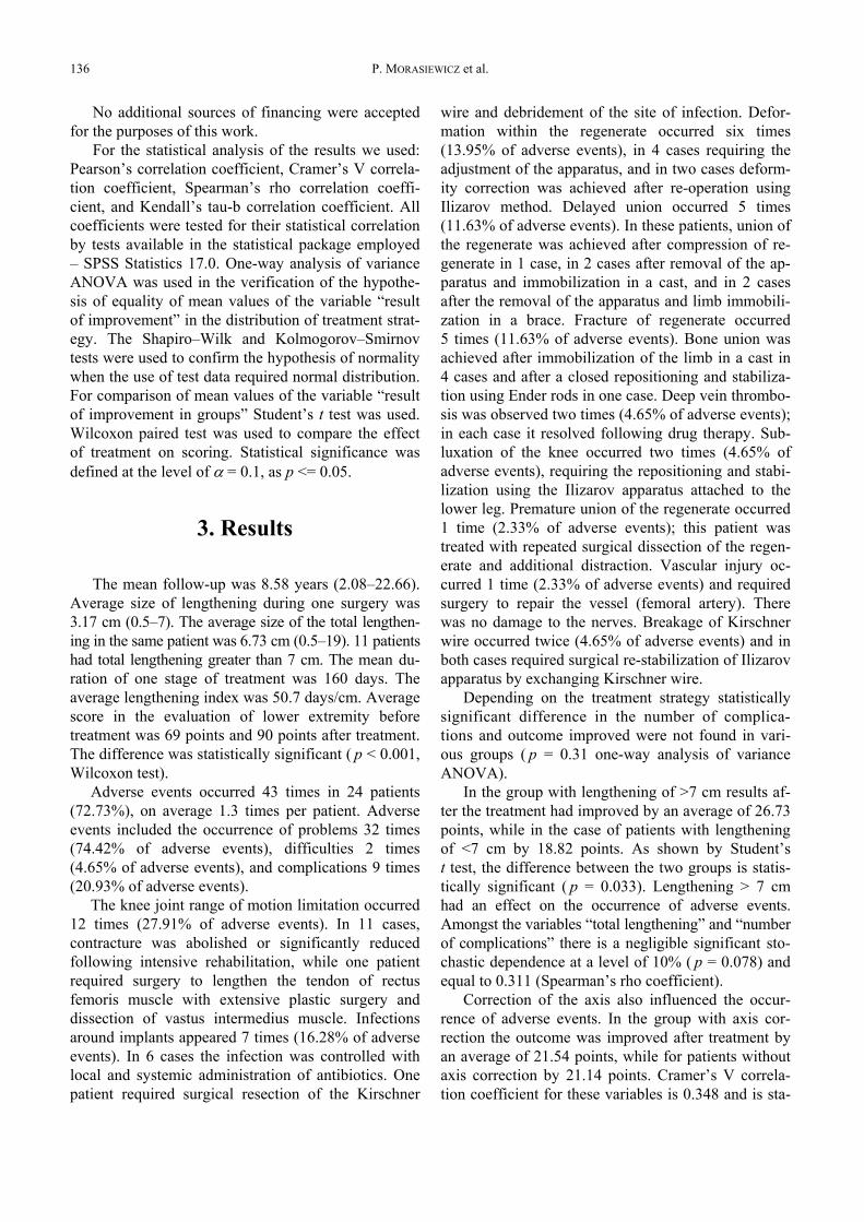
Distraction forces increased during rise of a valueof lengthening [2], (Fig. 3a–b). Worse results in thegroup with lengthening above 7 cm can be due togreater soft tissue forces and stress transfer in fixatorwhich have unfavourable influence on bone fragmentdisplacements and regenerate formation. During elon-gation and axis correction the influence of soft tissue
a) b)
Fig. 3a, b. Increasing of distraction forces during lengthening
P. MORASIEWICZ et al.138
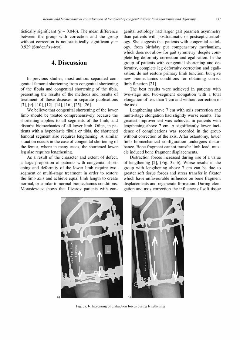
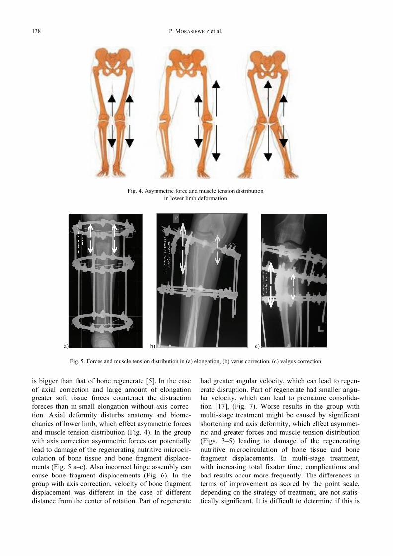
is bigger than that of bone regenerate [5]. In the caseof axial correction and large amount of elongationgreater soft tissue forces counteract the distractionforeces than in small elongation without axis correc-tion. Axial deformity disturbs anatomy and biome-chanics of lower limb, which effect asymmetric forcesand muscle tension distribution (Fig. 4). In the groupwith axis correction asymmetric forces can potentiallylead to damage of the regenerating nutritive microcir-culation of bone tissue and bone fragment displace-ments (Fig. 5 a–c). Also incorrect hinge assembly cancause bone fragment displacements (Fig. 6). In thegroup with axis correction, velocity of bone fragmentdisplacement was different in the case of differentdistance from the center of rotation. Part of regenerate
had greater angular velocity, which can lead to regen-erate disruption. Part of regenerate had smaller angu-lar velocity, which can lead to premature consolida-tion [17], (Fig. 7). Worse results in the group withmulti-stage treatment might be caused by significantshortening and axis deformity, which effect asymmet-ric and greater forces and muscle tension distribution(Figs. 3–5) leading to damage of the regeneratingnutritive microcirculation of bone tissue and bonefragment displacements. In multi-stage treatment,with increasing total fixator time, complications andbad results occur more frequently. The differences interms of improvement as scored by the point scale,depending on the strategy of treatment, are not statis-tically significant. It is difficult to determine if this is
Fig. 4. Asymmetric force and muscle tension distributionin lower limb deformation
a) b) c)
Fig. 5. Forces and muscle tension distribution in (a) elongation, (b) varus correction, (c) valgus correction
Results and biomechanical consideration of treatment of congenital lower limb shortening and deformity... 139
not a type 2 error, owing to the small size of the sub-groups. Too small numbers of individuals in the groupsdid not allow statistical analysis of the dependence ofthe development of adverse events based on treatmentstrategy. In the future, studies with more numerousgroups are indicated.
Fig. 6. Incorrect hinge assembly can cause bonefragment displacement
Fig. 7. Different velocity of bone fragment displacementduring axis correction
Lengthening index is high, compared to patientstreated with the Ilizarov method due to post-trauma orpost-inflammatory shortening and distortion. This isdue to inferior quality of bone tissue, which probablyhas less osteogenic potential caused by congenitaldeformities.
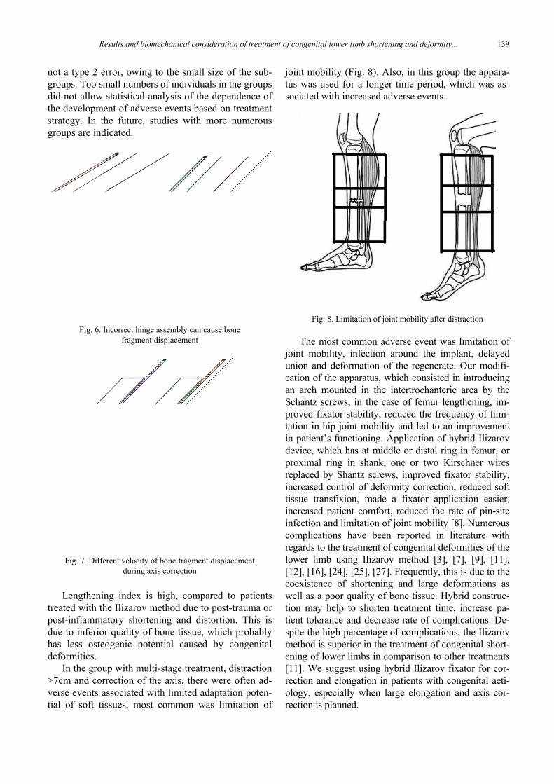
In the group with multi-stage treatment, distraction>7cm and correction of the axis, there were often ad-verse events associated with limited adaptation poten-tial of soft tissues, most common was limitation of
joint mobility (Fig. 8). Also, in this group the appara-tus was used for a longer time period, which was as-sociated with increased adverse events.
Fig. 8. Limitation of joint mobility after distraction
The most common adverse event was limitation ofjoint mobility, infection around the implant, delayedunion and deformation of the regenerate. Our modifi-cation of the apparatus, which consisted in introducingan arch mounted in the intertrochanteric area by theSchantz screws, in the case of femur lengthening, im-proved fixator stability, reduced the frequency of limi-tation in hip joint mobility and led to an improvementin patient’s functioning. Application of hybrid Ilizarovdevice, which has at middle or distal ring in femur, orproximal ring in shank, one or two Kirschner wiresreplaced by Shantz screws, improved fixator stability,increased control of deformity correction, reduced softtissue transfixion, made a fixator application easier,increased patient comfort, reduced the rate of pin-siteinfection and limitation of joint mobility [8]. Numerouscomplications have been reported in literature withregards to the treatment of congenital deformities of thelower limb using Ilizarov method [3], [7], [9], [11],[12], [16], [24], [25], [27]. Frequently, this is due to thecoexistence of shortening and large deformations aswell as a poor quality of bone tissue. Hybrid construc-tion may help to shorten treatment time, increase pa-tient tolerance and decrease rate of complications. De-spite the high percentage of complications, the Ilizarovmethod is superior in the treatment of congenital short-ening of lower limbs in comparison to other treatments[11]. We suggest using hybrid Ilizarov fixator for cor-rection and elongation in patients with congenital aeti-ology, especially when large elongation and axis cor-rection is planned.
P. MORASIEWICZ et al.140
According to our experience and results, the Ilizarovmethod is effective and has an acceptable rate of com-plications. This method allows not only to lengthen,but also to correct the axis of the limb in many planesand restore biomechanics conditions [11], [24], [25].Modular system allows the Ilizarov apparatus, withoutany additional surgery, to change the design of thestabilizer during the treatment in order to perform axiscorrections, whether primary or developed in thecourse of the treatment.
Acknowledgements
No additional sources of financing were accepted for the pur-poses of this work.
References
[1] ACHTERMAN C., KALAMCHI A., Congenital deficiency of thefibula, J. Bone Joint Surg., 1979, 61-B, 133–137.
[2] ARONSON J., Basic Science and Biological Principles of Dis-traction Osteogenesis, [in:] S. Rozbruch, S. Ilizarov (eds.),Limb Lengthening and Reconstruction Surgery, New York,Informa Healthcare, 2007, 19–42.
[3] ASTON W.J.S., CALDER P.R., BAKER D. et al., Lengthening ofthe congenital short femur using Ilizarov technique, J. BoneJoint Surg., 2009, 91-B, 962–967.
[4] BEVAN W., MILLAR E.A., A Review of Proximal FocalFemoral deficiencies, J. Bone Joint Surg., 1967, 49, 1376–1388.
[5] BĘDZIŃSKI R., FILIPIAK J., Experimental analysis of externalfixators for femoral bone elongation, Acta Bioeng. Biomech.,1999, 1(2), 93–105.
[6] BODEN S., FALLON M., DAVIDSON R., MENNUTI M. et al.,Proximal femoral focal deficiency. Evidence for a defect inproliferation and maturation of chondrocytes, J. Bone JointSurg., 1989, 71, 1119–1129.
[7] CHOI I., KUMAR S., BOWEN J., Amputation or limb-lengthening for partial or total absence of the fibula, J. BoneJoint Surg., 1990, 72, 1391–1399.
[8] FRAGOMEN A., BLYAKHER A., ILIZAROV S., Mechanical Prin-ciples of the Ilizarov Method, [in:] S. Rozbruch, S. Ilizarov(eds.), Limb Lengthening and Reconstruction Surgery, NewYork, Informa Healthcare, 2007, 43–52.
[9] GANGER R., GRILL F., LEHNER A. et al., Kongenitaler Fe-murdefekt, Orthopade, 1999, 12, 1045–1057.
[10] GILLESPIE R., TORODE I.P., Classification and management ofCongenital Abnormalities of the Femur, J. Bone Joint Surg.,1983, 65-B, 557–584.
[11] GRILL F., Correction of Complicated Extremity Deformitiesby External Fixation, Clin. Orthop. Rel. Res., 1989, 241,166–176.
[12] GRILL F., DUNGL P., Lengthening for Congenital Short Fe-mur, J. Bone Joint Surg., 1991, 73-B, 439–447.
[13] HAMANISHI C., Congenital Short Femur, J. Bone Joint Surg.,1980, 62-B, 307–320.
[14] HEFTI F., Defektmissbildungen an den unteren Extremitaten,Orthopade, 2008, 4, 381–401.
[15] KALAMCHI A., Congenital Lower limb Deficiencies,Springer, New York, 1989.
[16] KARGER C., GUILLE J.T., BOWEN J.R., Lengthening of Con-genital Lower Limb Deficiencies, Clin. Orthop. Rel. Res.,1993, 291, 236–245.
[17] KONERA W., SNELA S., GREGOSIEWICZ A., Limb elongationwith simultaneous axis correction with Ilizarov method, Chir.Narządów Ruchu Ortop. Pol., 1994, Supl. 1, 201–204.
[18] LEHNER A., GRILL F., The childhood ankle joint. Deformities,abnormalities and clinical variations, Radiologe, 1999, 1, 69–73.
[19] MORASIEWICZ L., ORZECHOWSKI W., A point system forevaluating patients with limb shortening and deformity,Ortop. Traumatol. Rehabil., 2002, 4, 305–309.
[20] MORASIEWICZ L., Establishment criteria of evaluation ofIlizarov method treatment, VI ASAMI Conference, Zakopane20–22.11.1992.
[21] MORASIEWICZ M., KOPROWSKI P., WRZOSEK Z. et al., Gaitanalysis in patients after lengthening and correction of tibiawith Ilizarov technique, Fizoter., 2010, 18, 9–18.
[22] MORASIEWICZ P., FILIPIAK J., KONIETZKO M. et al., The impactof the type of derotation mechanism on the stiffness of theIlizarov fixator, Acta Bioeng. Biomech., 2012, 14, 67–73.
[23] MORASIEWICZ P., FILIPIAK J., KRYSZTOFORSKI K. et al.,Biomechanical aspects of lower limb torsional deformationcorrection with the Ilizarov external fixator, Ann. Biomed.Eng., 2014, 42(3), 613-8, DOI: 10.1007/s10439-013-0911-6.
[24] NAUDIE D., HAMDY R., FASSIER F. et al., Complications ofLimb-Lengthening in Children Who Have an UnderlyingBone Disorder, J. Bone Joint Surg., 1998, 80-A, 18–24.
[25] NAUDIE D., HAMDY R., FASSIER F. et al., Management offibular hemimelia, J. Bone Joint Surg, 1997, 79-B, 58–65.
[26] NOONAN K.J., LEYES M., FORRIOL F. et al., Distraction Osteo-genesis of the Lower Extremity with Use of Monolateral Exter-nal Fixation, J. Bone Joint Surg., 1998, 80-A, 793–805.
[27] PALEY D., Problems, Obstacles, and Complications of LimbLengthening by the Ilizarov Technique, Clin. Orthop. Rel.Res., 1990, 250, 81–104.
[28] PAPPAS A.M., MILLER J.T., WORCESTER E.D., Congenital Ball-and-Socket Ankle Joints and Related Lower-Extremity Malfor-mations, J. Bone Joint Surg., 1982, 64-A, 672–679.
[29] ROGALA E.J., WYNNE-DAVIES R., LITTLEJOHN A. et al., Con-genital limb anomalies: frequency and aetiological factors,J. Med. Genet., 1974, 11, 221–233.