Protozoologica - Wydawnictwo Uniwersytetu Jagiellońskiego · Penza, Russia; E-mail:...
14
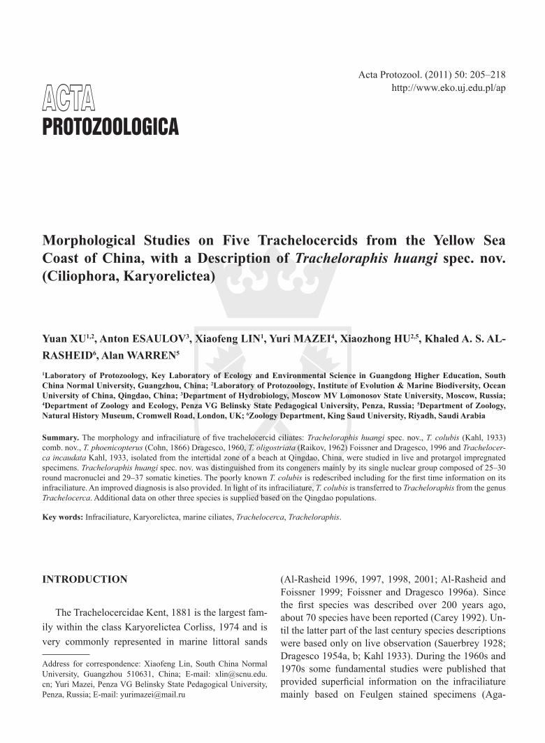
Acta Protozool. (2011) 50: 205–218 http://www.eko.uj.edu.pl/ap Address for correspondence: Xiaofeng Lin, South China Normal University, Guangzhou 510631, China; E-mail: [email protected]. cn; Yuri Mazei, Penza VG Belinsky State Pedagogical University, Penza, Russia; E-mail: [email protected] ACTA PROTOZOOLOGICA Morphological Studies on Five Trachelocercids from the Yellow Sea Coast of China, with a Description of Tracheloraphis huangi spec. nov. (Ciliophora, Karyorelictea) Yuan XU 1,2 , Anton ESAULOV 3 , Xiaofeng LIN 1 , Yuri MAZEI 4 , Xiaozhong HU 2,5 , Khaled A. S. AL- RASHEID 6 , Alan WARREN 5 1 Laboratory of Protozoology, Key Laboratory of Ecology and Environmental Science in Guangdong Higher Education, South China Normal University, Guangzhou, China; 2 Laboratory of Protozoology, Institute of Evolution & Marine Biodiversity, Ocean University of China, Qingdao, China; 3 Department of Hydrobiology, Moscow MV Lomonosov State University, Moscow, Russia; 4 Department of Zoology and Ecology, Penza VG Belinsky State Pedagogical University, Penza, Russia; 5 Department of Zoology, Natural History Museum, Cromwell Road, London, UK; 6 Zoology Department, King Saud University, Riyadh, Saudi Arabia Summary. The morphology and infraciliature of five trachelocercid ciliates: Tracheloraphis huangi spec. nov., T. colubis (Kahl, 1933) comb. nov., T. phoenicopterus (Cohn, 1866) Dragesco, 1960, T. oligostriata (Raikov, 1962) Foissner and Dragesco, 1996 and Trachelocer- ca incaudata Kahl, 1933, isolated from the intertidal zone of a beach at Qingdao, China, were studied in live and protargol impregnated specimens. Tracheloraphis huangi spec. nov. was distinguished from its congeners mainly by its single nuclear group composed of 25–30 round macronuclei and 29–37 somatic kineties. The poorly known T. colubis is redescribed including for the first time information on its infraciliature. An improved diagnosis is also provided. In light of its infraciliature, T. colubis is transferred to Tracheloraphis from the genus Trachelocerca. Additional data on other three species is supplied based on the Qingdao populations. Key words: Infraciliature, Karyorelictea, marine ciliates, Trachelocerca, Tracheloraphis. INTRODUCTION The Trachelocercidae Kent, 1881 is the largest fam- ily within the class Karyorelictea Corliss, 1974 and is very commonly represented in marine littoral sands (Al-Rasheid 1996, 1997, 1998, 2001; Al-Rasheid and Foissner 1999; Foissner and Dragesco 1996a). Since the first species was described over 200 years ago, about 70 species have been reported (Carey 1992). Un- til the latter part of the last century species descriptions were based only on live observation (Sauerbrey 1928; Dragesco 1954a, b; Kahl 1933). During the 1960s and 1970s some fundamental studies were published that provided superficial information on the infraciliature mainly based on Feulgen stained specimens (Aga-
Transcript of Protozoologica - Wydawnictwo Uniwersytetu Jagiellońskiego · Penza, Russia; E-mail:...
Acta Protozool. (2011) 50: 205–218 http://www.eko.uj.edu.pl/ap
Address for correspondence: Xiaofeng Lin, South China Normal University, Guangzhou 510631, China; E-mail: [email protected]; Yuri Mazei, Penza VG Belinsky State Pedagogical University, Penza, Russia; E-mail: [email protected]
ActAProtozoologica
Morphological Studies on Five Trachelocercids from the Yellow Sea Coast of China, with a Description of Tracheloraphis huangi spec. nov. (Ciliophora, Karyorelictea)
Yuan XU1,2, Anton ESAULOV3, Xiaofeng LIN1, Yuri MAZEI4, Xiaozhong HU2,5, Khaled A. S. AL-RASHEID6, Alan WARREN5
1Laboratory of Protozoology, Key Laboratory of Ecology and Environmental Science in Guangdong Higher Education, South China Normal University, Guangzhou, China; 2Laboratory of Protozoology, Institute of Evolution & Marine Biodiversity, Ocean University of China, Qingdao, China; 3Department of Hydrobiology, Moscow MV Lomonosov State University, Moscow, Russia; 4Department of Zoology and Ecology, Penza VG Belinsky State Pedagogical University, Penza, Russia; 5Department of Zoology, Natural History Museum, Cromwell Road, London, UK; 6Zoology Department, King Saud University, Riyadh, Saudi Arabia
Summary. The morphology and infraciliature of five trachelocercid ciliates: Tracheloraphis huangi spec. nov., T. colubis (Kahl, 1933) comb. nov., T. phoenicopterus (Cohn, 1866) Dragesco, 1960, T. oligostriata (Raikov, 1962) Foissner and Dragesco, 1996 and Trachelocer-ca incaudata Kahl, 1933, isolated from the intertidal zone of a beach at Qingdao, China, were studied in live and protargol impregnated specimens. Tracheloraphis huangi spec. nov. was distinguished from its congeners mainly by its single nuclear group composed of 25–30 round macronuclei and 29–37 somatic kineties. The poorly known T. colubis is redescribed including for the first time information on its infraciliature. An improved diagnosis is also provided. In light of its infraciliature, T. colubis is transferred to Tracheloraphis from the genus Trachelocerca. Additional data on other three species is supplied based on the Qingdao populations.
Key words: Infraciliature, Karyorelictea, marine ciliates, Trachelocerca, Tracheloraphis.
INTRODUCTION
The Trachelocercidae Kent, 1881 is the largest fam-ily within the class Karyorelictea Corliss, 1974 and is very commonly represented in marine littoral sands
(Al-Rasheid 1996, 1997, 1998, 2001; Al-Rasheid and Foissner 1999; Foissner and Dragesco 1996a). Since the first species was described over 200 years ago, about 70 species have been reported (Carey 1992). Un-til the latter part of the last century species descriptions were based only on live observation (Sauerbrey 1928; Dragesco 1954a, b; Kahl 1933). During the 1960s and 1970s some fundamental studies were published that provided superficial information on the infraciliature mainly based on Feulgen stained specimens (Aga-
Y. Xu et al.206
maliev 1966; Borror 1963; Dragesco 1960, 1963, 1965; Dragesco and Raikov 1966; Kovaleva 1966; Kovale-va and Golemansky 1979; Raikov 1957, 1962, 1963; Raikov and Kovaleva 1968). Subsequently, the generic classification of trachelocercids became available. But this classification was still limited as it was based main-ly on characters observed in vivo such as the absence or presence of the glabrous stripe (Dragesco 1960). Dragesco and Dragesco-Kernéis (1986) and Wilbert (1986) revealed for the first time using silver impreg-nation techniques the infraciliature of trachelocercids, especially the oral ciliary pattern. Subsequent studies addressed many of the confusing issues associated with trachelocercid systematics with some species being me-ticulously redescribed using modern techniques. Fur-thermore, new standards for genus- and species-level diagnoses and classification were established based mainly on the infraciliature, and in particular the oral ciliature (Foissner 1996, 1997a, 1997b, 1998; Foiss-ner and Al-Rasheid 1999a, b; Foissner and Dragesco 1996a, b). However, to date the infraciliature has been described for only 20 out of the ~ 70 trachelocercid spe-cies. Thus, the remaining ~ 50 species are all question-able in terms of their validity and generic placement (Dragesco and Dragesco-Kernéis 1986; Foissner 1996, 1997b; Foissner and Al-Rasheid 1999a; Foissner and Dragesco 1996a, b; Xu et al. 2011).
In recent years several reports on the morphology and phylogeny of karyorelicteans have been published (Alekperov et al. 2007, Andreoli et al. 2009, Gao et al. 2010, Mazei et al. 2009, Xu et al. 2011). In the present study, five trachelocercids isolated from the intertidal zone of a beach at Qingdao, China, were investigated both in vivo and following protargol impregnation.
MATERIALS AND METHODS
Ciliates were sampled from the intertidal zone of the No. 1 sandy beach at Qingdao (36°06′N; 120°34′E), China.
Tracheloraphis huangi spec. nov. was collected in November 2005, when the water temperature was 18°C and salinity was 29‰. Tracheloraphis oligostriata was collected in April 2010, with wa-ter temperature 11°C, salinity 20‰. Tracheloraphis colubis (Kahl, 1933) comb. nov., T. phoenicopterus (Cohn, 1866) Dragesco, 1960 and Trachelocerca incaudata (Kahl, 1933) Dragesco, 1960 were collected in May 2010, when the water temperature was about 20°C, and salinity was 30‰. Sampling methods were mainly ac-cording to Fan et al. (2010). Living cells were studied by bright field and differential interference microscopy (100 × to 1000 × magnifi-cations). The infraciliature was revealed by the protargol impreg-
nation method (Wilbert 1975) using the following fixative: 10 ml saturated, aqueous mercuric chloride and 3 ml Bouin’s solution, mixed just before use (Xu et al. 2011). Counts and measurements of stained specimens were performed at a magnification of 1000 ×. Drawings were made with the help of a camera lucida. Terminology is mainly according to Foissner (1996).
RESULTS AND DISCUSSION
Tracheloraphis huangi spec. nov. (Figs 1, 2; Table 1)
Diagnosis: Extended cells in vivo about 500–1200 × 60–100 μm in size. 29–37 somatic kineties. Glabrous stripe up to one third of cell width. Single nuclear group composed of ca. 25–30 macronuclei. Cortical granules minute and colorless.
Type locality: The intertidal zone of the No. 1 sandy beach at Qingdao (36°03′N; 120°20′E), China. Yellow Sea coast.
Type specimens: A protargol slide containing the holotype specimen marked with ink circle is deposited in the Natural History Museum, London, UK, with reg-istration number NHMUK 2011.4.27.1. One paratype slide with protargol-impregnated specimens is deposit-ed in the Laboratory of Protozoology, OUC, China (No. WYG2005111701).
Dedication: The species is named in honor of Prof. Zongguo Huang, The Third Institute of Oceanography State Oceanic Administration, China, in recognition of his contributions in the field of marine biodiversity.
Description: Fully extended cells about 500–1200 × 60–100 μm in vivo, filiform in shape; flexible and contractile (Figs 1A–D, 2A–D); cell distinctly tripar-tite, with neck, tail and trunk regions (Figs 1A, 2A, B); trunk flattened ca. 2–3 : 1 (Fig. 2C); head conspicu-ous, claviform (Figs 1A, 2G). Endoplasm grayish and opaque due to multiple refractile (crystalline?) inclu-sions about 2 × 4 μm in size (Figs 1F, 2A–E). Nuclear apparatus (capsule) in centre of trunk, containing about 25–30 globular macronuclei which form tight cluster ca. 30–40 μm in diameter (Figs 1G, 2H, I, O). Micronu-clei difficult to detect. Contractile vacuole not observed. Cortical granules globular, minute (about 0.5 μm in di-ameter), and colorless, forming narrow stripes between ciliary rows and densely distributed in glabrous stripe (Figs 1E, H, 2F). Locomotion by gliding, winding be-tween sand grains and organic debris.
Cell surface densely ciliated but with an unciliated zone, the glabrous stripe, which extends the whole
Taxonomy of Five Trachelocercids 207
Figs 1A–K. Tracheloraphis huangi spec. nov. from life (A–H) and after protargol impregnation (I–K). A – typical individual, noting the glabrous stripe occupying about one quarter of the body width; arrow shows the single nuclear group; B, C – shape variants; D – contracted cell; E, H – distribution of cortical granules between ciliary rows (arrowheads), around buccal edge (arrowheads) and in the glabrous stripe (double arrowheads) in the mid-body (E) and at the anterior end (H); F – ellipsoidal (crystalline?) inclusions; G – to show about thirty mac-ronuclei inside the nuclear group, noting the (protein) crystals and the nucleoli; I, J – anterior end, indicating the circumoral kinety, brosse and glabrous stripe bordered by the bristle kinety; arrowheads in (I) show anterior secant system; K – mid-body region, marking the gla-brous stripe, bristle kinety and anterior secant system (arrowheads). B – brosse, BK – bristle kinety, C – (protein) crystal, CK – circumoral kinety, GS – glabrous stripe, Ma – macronuclei, NG – nuclear group, NU – nucleoli. Scale bars: 400 μm (A–C), 200 μm (D), 30 μm (I, J).
body length in the midline of the left side; maximum width in trunk region approximately one third of body width (Fig. 1A). Entire infraciliature consisting of diki-netids. Somatic cilia about 10 μm long in vivo and ar-ranged in longitudinal rows. Anterior ends of ciliary rows curved to right and composed of densely spaced dikinetids (Figs 1I, J, 2K, M). Anterior and posterior secant system formed on left side of glabrous stripe where some kineties abut to the bristle kinety (Figs 1I,
K, 2P). Ciliary rows neighboring the right branch of the bristle kinety unshortened anteriorly and thus extend along the glabrous stripe. Glabrous stripe bordered by the bristle kinety, and the kinetids of which are more widely spaced and irregularly arranged than those of the somatic ciliary rows (Figs 1I, K, 2K, L, P). Oral infraciliature consisting of a circumoral kinety, which is interrupted by the inserted brosse kineties (Figs 1I, J, 2K, M).

Y. Xu et al.208
Comparison: As most species of the family Trach-elocercidae have only been studied based on live obser-vation, their generic classifications remain questionable based on the new generic definitions (Foissner 1996, 1997b; Foissner and Al-Rasheid 1999a; Foissner and
Dragesco 1996a, b). Therefore, the comparison between Tracheloraphis huangi spec. nov. and its most closely related species should not be limited to those within the genus Tracheloraphis. Of the 70 trachelocercid species, 34 have their macronuclei arranged in a single group
Figs 2A–P. Photomicrographs of Tracheloraphis huangi spec. nov. from life (A–I) and after protargol impregnation (J–P). A, B – extended individuals gliding; C, D – contracted individuals; E – a dividing cell; F – to show the distribution of cortical granules between the ciliary rows (arrowheads); G – anterior end of the body; H, I – nuclear group comprising about thirty macronuclei (arrowheads); J – infraciliature and nuclear group (arrow); K, M – anterior infraciliature of left and right sides, circumoral kinety, brosse, glabrous stripe and bristle kinety; L, N – mid-body of right and left sides respectively, showing the glabrous stripe bordered by the bristle kinety (arrowheads in L) and somatic kineties (N); O – nuclear group, noting the nuclei forming a tight cluster; P – left side of the glabrous stripe, marking the posterior secant system (arrowheads). B – brosse, BK – bristle kinety, CK – circumoral kinety, GS – glabrous stripe, SK – somatic kineties. Scale bars: 400 μm (A–C), 150 μm (D, J), 30 μm (M).
Taxonomy of Five Trachelocercids 209
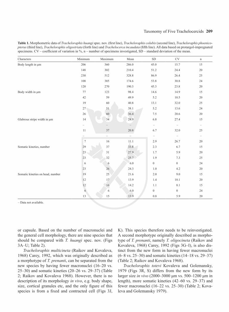
Table 1. Morphometric data of Tracheloraphis huangi spec. nov. (first line), Tracheloraphis colubis (second line), Tracheloraphis phoenico-pterus (third line), Tracheloraphis oligostriata (forth line) and Trachelocerca incaudata (fifth line). All data based on protargol-impregnated specimens. CV – coefficient of variation in %, n – number of specimens investigated, SD – standard deviation of the mean.
Characters Minimum Maximum Mean SD CV n
Body length in μm 206 360 286.0 45.0 15.7 15
148 302 210.4 51.2 24.4 20
230 512 328.8 86.9 26.4 25
108 385 174.6 53.8 30.8 24
120 270 190.3 45.3 23.8 20
Body width in μm 77 123 98.4 14.6 14.9 15
42 59 49.9 5.2 10.5 20
19 60 40.8 13.1 32.0 25
27 51 38.1 5.2 13.6 24
26 60 36.4 7.5 20.6 20
Glabrous stripe width in μm 14 34 24.9 6.8 27.4 15
– – – – – –
11 37 20.8 6.7 32.0 25
– – – – – –
7 16 11.1 2.9 26.7 20
Somatic kineties, number 29 37 33.8 2.3 6.7 15
23 31 27.9 1.7 5.9 20
23 32 25.7 1.9 7.3 25
6 6 6.0 0 0 24
23 26 24.3 1.0 4.2 20
Somatic kineties on head, number 19 25 21.6 2.0 9.0 15
12 17 13.9 1.4 10.1 20
12 16 14.2 1.1 8.1 15
6 6 6.0 0 0 24
13 15 13.9 0.8 5.9 20
– Data not available.
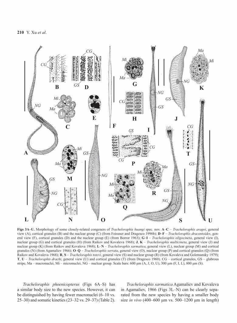
or capsule. Based on the number of macronuclei and the general cell morphology, there are nine species that should be compared with T. huangi spec. nov. (Figs 3A–U; Table 2).
Tracheloraphis multicineta (Raikov and Kovaleva, 1968) Carey, 1992, which was originally described as a morphotype of T. prenanti, can be separated from the new species by having fewer macronuclei (16–20 vs. 25–30) and somatic kineties (20–26 vs. 29–37) (Table 2; Raikov and Kovaleva 1968). However, there is no description of its morphology in vivo, e.g. body shape, size, cortical granules etc, and the only figure of this species is from a fixed and contracted cell (Figs 3J,
K). This species therefore needs to be reinvestigated. A second morphotype originally described as morpho-type of T. prenanti, namely T. oligocineta (Raikov and Kovaleva, 1968) Carey, 1992 (Figs 3G–I), is also dis-tinct from the new form in having fewer macronuclei (6–8 vs. 25–30) and somatic kineties (14–18 vs. 29–37) (Table 2; Raikov and Kovaleva 1968).
Tracheloraphis totevi Kovaleva and Golemansky, 1979 (Figs 3R, S) differs from the new form by its larger size in vivo (2000–3000 μm vs. 500–1200 μm in length), more somatic kineties (42–60 vs. 29–37) and fewer macronuclei (16–22 vs. 25–30) (Table 2; Kova-leva and Golemansky 1979).
Y. Xu et al.210
Figs 3A–U. Morphology of some closely-related congeners of Tracheloraphis huangi spec. nov. A–C – Tracheloraphis aragoi, general view (A), cortical granules (B) and the nuclear group (C) (from Foissner and Dragesco 1996b); D–F – Tracheloraphis dracontoides, gen-eral view (F), cortical granules (D) and the nuclear group (E) (from Borror 1963); G–I – Tracheloraphis oligocineta, general view (I), nuclear group (G) and cortical granules (H) (from Raikov and Kovaleva 1968); J, K – Tracheloraphis multicineta, general view (J) and nuclear group (K) (from Raikov and Kovaleva 1968); L–N – Tracheloraphis sarmatica, general view (L), nuclear group (M) and cortical granules (N) (from Agamaliev 1966); O–Q – Tracheloraphis serrata, general view (O), nuclear group (P) and cortical granules (Q) (from Raikov and Kovaleva 1968); R, S – Tracheloraphis totevi, general view (S) and nuclear group (R) (from Kovaleva and Golemansky 1979); T, U – Tracheloraphis drachi, general view (U) and cortical granules (T) (from Dragesco 1960). CG – cortical granules, GS – glabrous stripe, Ma – macronuclei, Mi – micronuclei, NG – nuclear group. Scale bars: 600 μm (A, J, O, U), 300 μm (F, I, L), 800 μm (S).
Tracheloraphis phoenicopterus (Figs 6A–S) has a similar body size to the new species. However, it can be distinguished by having fewer macronuclei (6–10 vs. 25–30) and somatic kineties (23–32 vs. 29–37) (Table 2).
Tracheloraphis sarmatica Agamaliev and Kovaleva in Agamaliev, 1966 (Figs 3L–N) can be clearly sepa-rated from the new species by having a smaller body size in vivo (400–600 μm vs. 500–1200 μm in length)
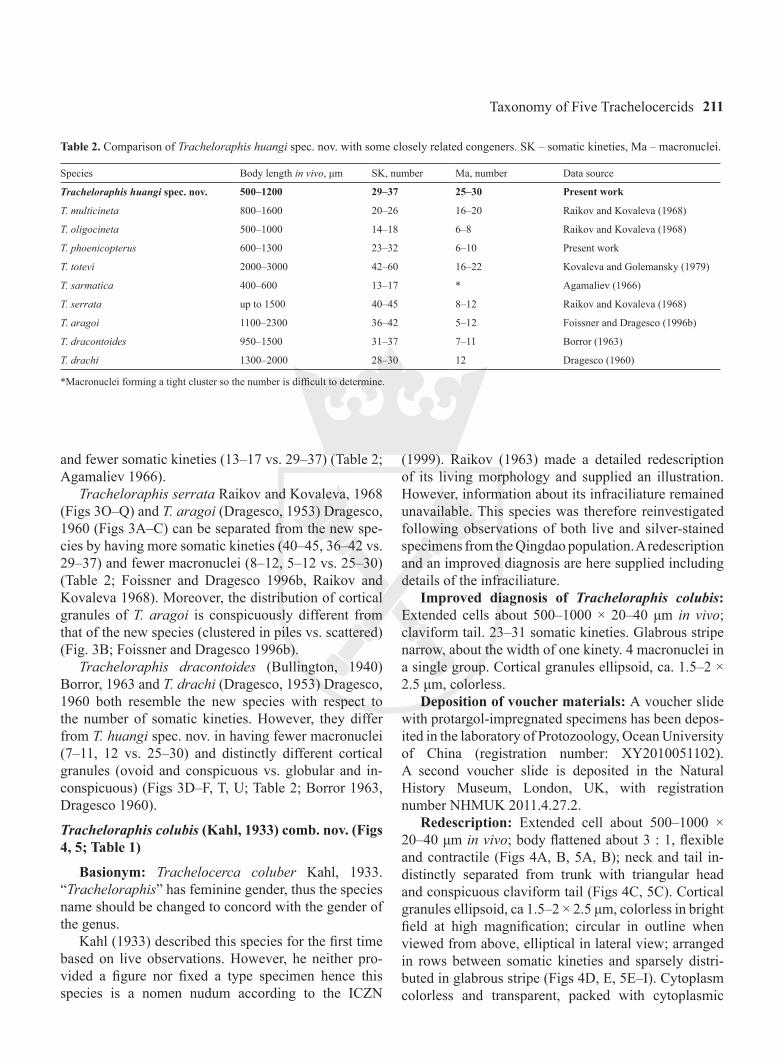
Taxonomy of Five Trachelocercids 211
Table 2. Comparison of Tracheloraphis huangi spec. nov. with some closely related congeners. SK – somatic kineties, Ma – macronuclei.
Species Body length in vivo, μm SK, number Ma, number Data source
Tracheloraphis huangi spec. nov. 500–1200 29–37 25–30 Present work
T. multicineta 800–1600 20–26 16–20 Raikov and Kovaleva (1968)
T. oligocineta 500–1000 14–18 6–8 Raikov and Kovaleva (1968)
T. phoenicopterus 600–1300 23–32 6–10 Present work
T. totevi 2000–3000 42–60 16–22 Kovaleva and Golemansky (1979)
T. sarmatica 400–600 13–17 * Agamaliev (1966)
T. serrata up to 1500 40–45 8–12 Raikov and Kovaleva (1968)
T. aragoi 1100–2300 36–42 5–12 Foissner and Dragesco (1996b)
T. dracontoides 950–1500 31–37 7–11 Borror (1963)
T. drachi 1300–2000 28–30 12 Dragesco (1960)
*Macronuclei forming a tight cluster so the number is difficult to determine.
and fewer somatic kineties (13–17 vs. 29–37) (Table 2; Agamaliev 1966).
Tracheloraphis serrata Raikov and Kovaleva, 1968 (Figs 3O–Q) and T. aragoi (Dragesco, 1953) Dragesco, 1960 (Figs 3A–C) can be separated from the new spe-cies by having more somatic kineties (40–45, 36–42 vs. 29–37) and fewer macronuclei (8–12, 5–12 vs. 25–30) (Table 2; Foissner and Dragesco 1996b, Raikov and Kovaleva 1968). Moreover, the distribution of cortical granules of T. aragoi is conspicuously different from that of the new species (clustered in piles vs. scattered) (Fig. 3B; Foissner and Dragesco 1996b).
Tracheloraphis dracontoides (Bullington, 1940) Borror, 1963 and T. drachi (Dragesco, 1953) Dragesco, 1960 both resemble the new species with respect to the number of somatic kineties. However, they differ from T. huangi spec. nov. in having fewer macronuclei (7–11, 12 vs. 25–30) and distinctly different cortical granules (ovoid and conspicuous vs. globular and in-conspicuous) (Figs 3D–F, T, U; Table 2; Borror 1963, Dragesco 1960).
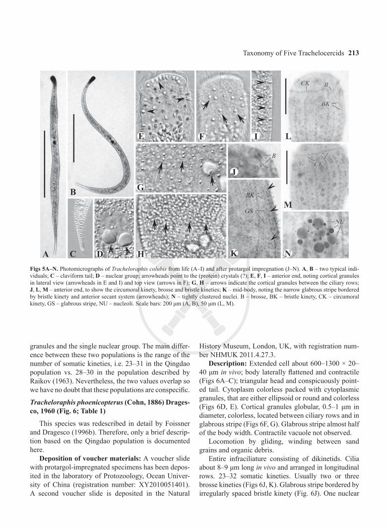
Tracheloraphis colubis (Kahl, 1933) comb. nov. (Figs 4, 5; Table 1)
Basionym: Trachelocerca coluber Kahl, 1933. “Tracheloraphis” has feminine gender, thus the species name should be changed to concord with the gender of the genus.
Kahl (1933) described this species for the first time based on live observations. However, he neither pro-vided a figure nor fixed a type specimen hence this species is a nomen nudum according to the ICZN
(1999). Raikov (1963) made a detailed redescription of its living morphology and supplied an illustration. However, information about its infraciliature remained unavailable. This species was therefore reinvestigated following observations of both live and silver-stained specimens from the Qingdao population. A redescription and an improved diagnosis are here supplied including details of the infraciliature.
Improved diagnosis of Tracheloraphis colubis: Extended cells about 500–1000 × 20–40 μm in vivo; claviform tail. 23–31 somatic kineties. Glabrous stripe narrow, about the width of one kinety. 4 macronuclei in a single group. Cortical granules ellipsoid, ca. 1.5–2 × 2.5 μm, colorless.
Deposition of voucher materials: A voucher slide with protargol-impregnated specimens has been depos-ited in the laboratory of Protozoology, Ocean University of China (registration number: XY2010051102). A second voucher slide is deposited in the Natural History Museum, London, UK, with registration number NHMUK 2011.4.27.2.
Redescription: Extended cell about 500–1000 × 20–40 μm in vivo; body flattened about 3 : 1, flexible and contractile (Figs 4A, B, 5A, B); neck and tail in-distinctly separated from trunk with triangular head and conspicuous claviform tail (Figs 4C, 5C). Cortical granules ellipsoid, ca 1.5–2 × 2.5 μm, colorless in bright field at high magnification; circular in outline when viewed from above, elliptical in lateral view; arranged in rows between somatic kineties and sparsely distri-buted in glabrous stripe (Figs 4D, E, 5E–I). Cytoplasm colorless and transparent, packed with cytoplasmic

Y. Xu et al.212
Figs 4A–M. Tracheloraphis colubis from life (A–E, J–M), after protargol impregnation (F–I). A – typical individual, noting the nuclear group; B – showing the flexibility of the cell; C – posterior end, showing the rounded, claviform tail; D – anterior part, marking the arrange-ment of the cortical granules, the outlines of which are round in top view (arrows) and elliptical in lateral view (arrowheads); E – lateral view, showing the elliptical cortical granules at the cell margin; F, G – infraciliature of anterior end to mark circumoral kinety, brosse, gla-brous stripe and bristle kineties; H – infraciliature of mid-body, showing the glabrous stripe bordered by bristle kinety and anterior secant system (arrowheads); I – nuclear group composed of macronuclei and micronuclei (from Raikov 1963); J, K – to show the distribution of cortical granules (from Raikov 1963); L, M – general view (from Raikov 1963). B – brosse, BK – bristle kinety, CK – circumoral kinety, GS – glabrous stripe, Ma – macronuclei, Mi – micronuclei, NG – nuclear group, NU – nucleoli. Scale bars: 200 μm (A, B, L, M), 50 μm (C, D, F, G), 10 μm (I).
granules that are ellipsoid, about 1–3 μm long and colorless. Contractile vacuole not observed.
Locomotion by gliding, winding between sand grains and organic debris.
Entire infraciliature consisting of dikinetids. Cilia about 10 μm long in vivo and arranged in longitudinal rows. Usually one or two brosse kineties (Figs 4F, 5J, L). Glabrous stripe very narrow, bordered by irregularly spaced bristle kinety (Figs 4H, 5K). Anterior and posterior secant system formed on left side of glabrous stripe where some kineties abut to the bristle kinety (Figs 4H, 5K). Four macronuclei in a single group (Figs 5D, N); micronuclei difficult to detect.
Discussion: Kahl (1933) assigned this species to the genus Trachelocerca based on its curved posterior end. Later, Raikov (1963) redescribed it and retained it in the genus Trachelocerca because of its narrow glabrous stripe. However, these characters are of very limited value for the generic classification of trachelocercids (Foissner and Dragesco 1996b). According to present study, this species has the circumoral kinety interrupted by a brosse and thus should be transferred to the genus Tracheloraphis (Foissner and Dragesco 1996b).
The Qingdao population matches the population described by Raikov (1963) in most characters, par-ticularly the claviform tail, the large ellipsoid cortical
Taxonomy of Five Trachelocercids 213
Figs 5A–N. Photomicrographs of Tracheloraphis colubis from life (A–I) and after protargol impregnation (J–N). A, B – two typical indi-viduals; C – claviform tail; D – nuclear group; arrowheads point to the (protein) crystals (?); E, F, I – anterior end, noting cortical granules in lateral view (arrowheads in E and I) and top view (arrows in F); G, H – arrows indicate the cortical granules between the ciliary rows; J, L, M – anterior end, to show the circumoral kinety, brosse and bristle kineties; K – mid-body, noting the narrow glabrous stripe bordered by bristle kinety and anterior secant system (arrowheads); N – tightly clustered nuclei. B – brosse, BK – bristle kinety, CK – circumoral kinety, GS – glabrous stripe, NU – nucleoli. Scale bars: 200 μm (A, B), 50 μm (L, M).
granules and the single nuclear group. The main differ-ence between these two populations is the range of the number of somatic kineties, i.e. 23–31 in the Qingdao population vs. 28–30 in the population described by Raikov (1963). Nevertheless, the two values overlap so we have no doubt that these populations are conspecific.
Tracheloraphis phoenicopterus (Cohn, 1886) Drages-co, 1960 (Fig. 6; Table 1)
This species was redescribed in detail by Foissner and Dragesco (1996b). Therefore, only a brief descrip-tion based on the Qingdao population is documented here.
Deposition of voucher materials: A voucher slide with protargol-impregnated specimens has been depos-ited in the laboratory of Protozoology, Ocean Univer-sity of China (registration number: XY2010051401). A second voucher slide is deposited in the Natural
History Museum, London, UK, with registration num-ber NHMUK 2011.4.27.3.
Description: Extended cell about 600–1300 × 20–40 μm in vivo; body laterally flattened and contractile (Figs 6A–C); triangular head and conspicuously point-ed tail. Cytoplasm colorless packed with cytoplasmic granules, that are either ellipsoid or round and colorless (Figs 6D, E). Cortical granules globular, 0.5–1 μm in diameter, colorless, located between ciliary rows and in glabrous stripe (Figs 6F, G). Glabrous stripe almost half of the body width. Contractile vacuole not observed.
Locomotion by gliding, winding between sand grains and organic debris.
Entire infraciliature consisting of dikinetids. Cilia about 8–9 μm long in vivo and arranged in longitudinal rows. 23–32 somatic kineties. Usually two or three brosse kineties (Figs 6J, K). Glabrous stripe bordered by irregularly spaced bristle kinety (Fig. 6J). One nuclear
Y. Xu et al.214
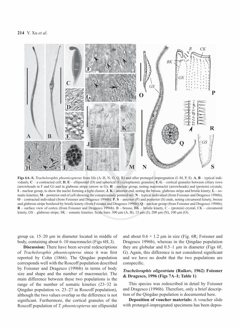
Figs 6A–S. Tracheloraphis phoenicopterus from life (A–H, N, O, Q, R) and after protargol impregnation (I–M, P, S). A, B – typical indi-viduals; C – a contracted cell; D, E – ellipsoidal (D) and spherical (E) cytoplasmic granules; F, G – cortical granules between ciliary rows (arrowheads in F and G) and in glabrous stripe (arrow in G); H – nuclear group, noting macronuclei (arrowheads) and (protein) crystals; I – nuclear group, to show the nuclei forming a tight cluster; J, K – anterior end, noting the brosse, glabrous stripe and bristle kinety; L – so-matic kineties; M – posterior end of cell showing the conspicuously pointed tail; N – typical individual (from Foissner and Dragesco 1996b); O – contracted individual (from Foissner and Dragesco 1996b); P, S – anterior (P) and posterior (S) ends, noting circumoral kinety, brosse and glabrous stripe bordered by bristle kinety (from Foissner and Dragesco 1996b); Q – nuclear group (from Foissner and Dragesco 1996b); R – surface view of cortex (from Foissner and Dragesco 1996b). B – brosse, BK – bristle kinety, C – (protein) crystal, CK – circumoral kinety, GS – glabrous stripe, SK – somatic kineties. Scale bars: 300 μm (A, B), 15 μm (J), 200 μm (N), 100 μm (O).
group ca. 15–20 μm in diameter located in middle of body, containing about 6–10 macronuclei (Figs 6H, I).
Discussion: There have been several redescriptions of Tracheloraphis phoenicopterus since it was first reported by Cohn (1866). The Qingdao population corresponds well with the Roscoff population described by Foissner and Dragesco (1996b) in terms of body size and shape and the number of macronuclei. The main difference between these two populations is the range of the number of somatic kineties (23–32 in Qingdao population vs. 23–27 in Roscoff population), although the two values overlap so the difference is not significant. Furthermore, the cortical granules of the Roscoff population of T. phoenicopterus are ellipsoidal
and about 0.6 × 1.2 μm in size (Fig. 6R; Foissner and Dragesco 1996b), whereas in the Qingdao population they are globular and 0.5–1 μm in diameter (Figs 6F, G). Again, this difference is not considered significant and we have no doubt that the two populations are conspecific.
Tracheloraphis oligostriata (Raikov, 1962) Foissner & Dragesco, 1996 (Figs 7A–I; Table 1)
This species was redescribed in detail by Foissner and Dragesco (1996b). Therefore, only a brief descrip-tion of the Qingdao population is documented here.
Deposition of voucher materials: A voucher slide with protargol-impregnated specimens has been depos-
Taxonomy of Five Trachelocercids 215
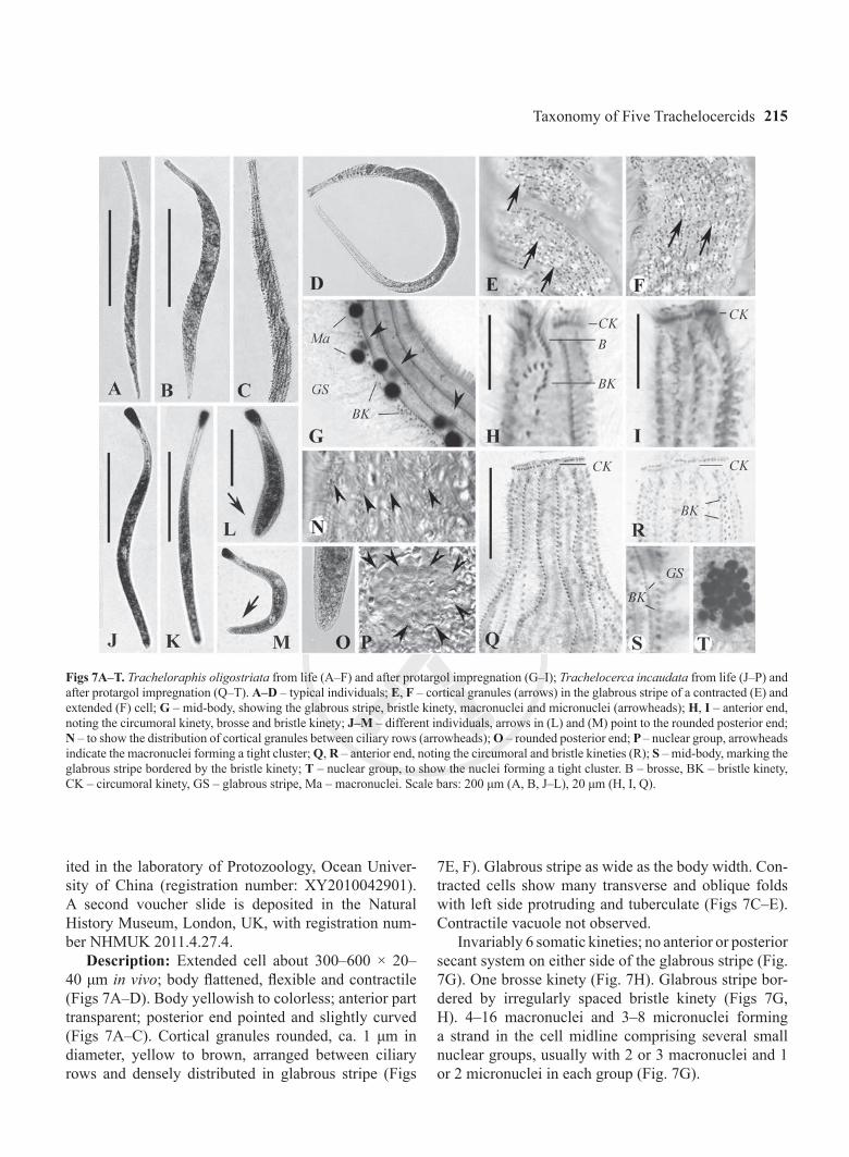
Figs 7A–T. Tracheloraphis oligostriata from life (A–F) and after protargol impregnation (G–I); Trachelocerca incaudata from life (J–P) and after protargol impregnation (Q–T). A–D – typical individuals; E, F – cortical granules (arrows) in the glabrous stripe of a contracted (E) and extended (F) cell; G – mid-body, showing the glabrous stripe, bristle kinety, macronuclei and micronuclei (arrowheads); H, I – anterior end, noting the circumoral kinety, brosse and bristle kinety; J–M – different individuals, arrows in (L) and (M) point to the rounded posterior end; N – to show the distribution of cortical granules between ciliary rows (arrowheads); O – rounded posterior end; P – nuclear group, arrowheads indicate the macronuclei forming a tight cluster; Q, R – anterior end, noting the circumoral and bristle kineties (R); S – mid-body, marking the glabrous stripe bordered by the bristle kinety; T – nuclear group, to show the nuclei forming a tight cluster. B – brosse, BK – bristle kinety, CK – circumoral kinety, GS – glabrous stripe, Ma – macronuclei. Scale bars: 200 μm (A, B, J–L), 20 μm (H, I, Q).
ited in the laboratory of Protozoology, Ocean Univer-sity of China (registration number: XY2010042901). A second voucher slide is deposited in the Natural History Museum, London, UK, with registration num-ber NHMUK 2011.4.27.4.
Description: Extended cell about 300–600 × 20–40 μm in vivo; body flattened, flexible and contractile (Figs 7A–D). Body yellowish to colorless; anterior part transparent; posterior end pointed and slightly curved (Figs 7A–C). Cortical granules rounded, ca. 1 μm in diameter, yellow to brown, arranged between ciliary rows and densely distributed in glabrous stripe (Figs
7E, F). Glabrous stripe as wide as the body width. Con-tracted cells show many transverse and oblique folds with left side protruding and tuberculate (Figs 7C–E). Contractile vacuole not observed.
Invariably 6 somatic kineties; no anterior or posterior secant system on either side of the glabrous stripe (Fig. 7G). One brosse kinety (Fig. 7H). Glabrous stripe bor-dered by irregularly spaced bristle kinety (Figs 7G, H). 4–16 macronuclei and 3–8 micronuclei forming a strand in the cell midline comprising several small nuclear groups, usually with 2 or 3 macronuclei and 1 or 2 micronuclei in each group (Fig. 7G).
Y. Xu et al.216
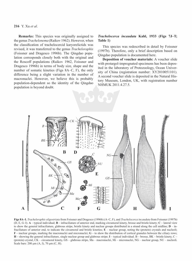
Remarks: This species was originally assigned to the genus Trachelonema (Raikov 1962). However, when the classification of trachelocercid karyorelictids was revised, it was transferred to the genus Tracheloraphis (Foissner and Dragesco 1996b). The Qingdao popu-lation corresponds closely both with the original and the Roscoff populations (Raikov 1962, Foissner and Dragesco 1996b) in terms of body size, shape and the number of somatic kineties (Figs 8A–C, F), the only difference being a slight variation in the number of macronuclei. However, we believe this is probably population-dependent so the identity of the Qingdao population is beyond doubt.
Trachelocerca incaudata Kahl, 1933 (Figs 7J–T; Table 1)
This species was redescribed in detail by Foissner (1997b). Therefore, only a brief description based on Qingdao population is documented here.
Deposition of voucher materials: A voucher slide with protargol-impregnated specimens has been depos-ited in the laboratory of Protozoology, Ocean Univer-sity of China (registration number: XY2010051101). A second voucher slide is deposited in the Natural His-tory Museum, London, UK, with registration number NHMUK 2011.4.27.5.
Figs 8A–I. Tracheloraphis oligostriata from Foissner and Dragesco (1996b) (A–C, F), and Trachelocerca incaudata from Foissner (1997b) (D, E, G–I). A – typical individual; B – infraciliature of anterior end, marking circumoral kinety, brosse and bristle kinety; C – lateral view to show the general infraciliature, glabrous stripe, bristle kinety and nuclear groups distributed in a strand along the cell midline; D – in-fraciliature of anterior end, to indicate the circumoral and bristle kineties; E – nuclear group, noting the (protein) crystals and nucleoli; F – nuclear groups, marking the macronuclei and micronuclei; G – to show the distribution of cortical granules between the ciliary rows; H – showing the general infraciliature, single nuclear group and glabrous stripe; I – typical individual. B – brosse, BK – bristle kinety, C – (protein) crystal, CK – circumoral kinety, GS – glabrous stripe, Ma – macronuclei, Mi – micronuclei, NG – nuclear group, NU – nucleoli. Scale bars: 200 μm (A, I), 70 μm (C, H).

Taxonomy of Five Trachelocercids 217
Description: Extended cell about 300–600 × 20–40 μm in vivo; body claviform and contractile; head and neck areas distinct, posterior end rounded (Figs 7J–M, O). Body grey to blackish in color due to numerous inclusions (Figs 7J–L); cortical granules rounded, ca. 0.5 μm in diameter, colorless, located between cili-ary rows and in glabrous stripe (Fig. 7N). Glabrous stripe narrow, about the width of 2 or 3 ciliary rows. Contractile vacuole not observed.
Twenty-three to 26 somatic kineties with cilia about 10 μm long in vivo; with anterior and posterior secant system on the left side of the glabrous stripe (Fig. 7Q). Glabrous stripe bordered by irregularly spaced bristle kinety (Figs 7R, S). Single nuclear group, ca. 15 μm in diameter, composed of 6–8 macronuclei (Fig. 7T).
Remarks: The original description of Trachelocer-ca incaudata by Kahl (1933) was rather brief and de-tailed data were not available until it was redescribed by Foissner (1997b). The Qingdao population corresponds well with Foissner’s (1997b) Roscoff population (Figs 8D, E, G–I) in terms of body size and shape, the width of the glabrous stripe and the number of macronuclei. The only minor difference is the range of the number of somatic kineties, i.e. 23–26 vs. 25–40 in the Roscoff population (Foissner 1997b). Nevertheless, since these values overlap we do not consider this to be a signifi-cant difference. Therefore, we have no doubt that the two populations are conspecific.
Acknowledgements. This work was supported by the Russian Foundation for Basic Research (Project number: 10-04-00496-a), the National Natural Science Foundation of China (project number: 30870280), and the Center of Biodiversity Research, King Saud University, Saudi Arabia.
REFERENCES
Agamaliev F. G. (1966) New species of psammobiotic ciliates of the western coast of the Caspian Sea. Acta Protozool. 4: 169–183
Alekperov I., Buskey E., Snegovaya N. (2007) The free-living cilia-tes of the Mexican Gulf coast near Port Aransas city and its suburbs (South Texas, USA). Protistology 5: 101–130
Al-Rasheid K. A. S. (1996) Records of free-living ciliates in Saudi Arabia. I. Marine interstitial ciliates of the Arabian gulf islands of Al-Bātinah and Abū Ali. Arab Gulf J. Scient. Res. 14: 747–765
Al-Rasheid K. A. S. (1997) Records of free-living ciliates in Saudi Arabia. III. Marine interstitial ciliates of the Arabian gulf island of Tarut. Arab Gulf J. Scient. Res. 15: 733–766
Al-Rasheid K. A. S. (1998) Records of marine interstitial karyore-lictid ciliates from Jubail Marine Wildlife Sanctuary in the Gulf-Shore of Saudi Arabia. Arab Gulf J. Scient. Res. 16: 595–610
Al-Rasheid K. A. S. (2001) New records of interstitial ciliates (Pro-tozoa Ciliophora) from the Saudi coasts of the Red Sea. Trop. Zool. 14: 133–156
Al-Rasheid K. A. S., Foissner W. (1999) Apical feeding in the ka-ryorelictids (Protozoa, Ciliophora) Sultanophrys arabica and Tracheloraphis sp. J. Eukaryot. Microbiol. 46: 458–463
Andreoli l., Mangini L., Ferrantini F., Santangelo G., Verni F., Pe-troni G. (2009) Molecular phylogeny of unculturable Karyore-lictea (Alveolata, Ciliophora). Zool. Scr. 38: 651–662
Borror A. C. (1963) Morphology and ecology of the benthic ciliated protozoa of Alligator Harbor, Florida. Arch. Protistenkd. 106: 465–534
Carey P. G. (1992) Marine interstitial ciliates. Chapman & Hall, London, New York, Tokyo, Melbourne, Madras
Cohn F. (1866) Neue infusorien im Seeaquarium. Z. wiss. Zool. 16: 253–302
Dragesco J. (1954a) Diagnoses préliminaires de quelques ciliés psammophiles nouveaux. Bull. Soc. Zool. Fr. 79: 57–62
Dragesco J. (1954b) Diagnoses préliminaires de quelques ciliés nouveaux des sables de Banyuls-Sur-Mer (I). Vie et Milieu 4: 633–637
Dragesco J. (1960) Ciliés mésopsammiques littoraux. Systématique, morphologie, écologie. Trav. Stn Biol. Roscoff (N. S.) 12: 1–356
Dragesco J. (1963) Compléments à la connaissance des ciliés mé-sopsammiques de Roscoff. I. Holotriches. Cah. Biol. Mar. 4: 91–119
Dragesco J. (1965) Ciliés mésopsammiques d’afrique noire. Cah. Biol. Mar. 6: 357–399
Dragesco J., Dragesco-Kernéis A. (1986) Ciliés libres de l’Afrique intertropicale. Faune Tropicale 26: 1–559
Dragesco J., Raikov I. B. (1966) L’appareil nucléaire, la division et quelques stades de la conjugaison de Tracheloraphis margari-tatus (Kahl) et T. caudatus sp. nov. (Ciliata, Holotricha). Arch. Protistenkd. 109: 99–113
Fan X., Chen X., Song W., Al-Rasheid K. A. S., Warren A. (2010) Two new marine scuticociliates, Sathrophilus planus n. sp. and Pseudoplatynematum dengi n. sp., with improved definition of Pseudoplatynematum (Ciliophora, Oligohymenophora). Eur. J. Protistol. 46: 212–220
Foissner W. (1996) Updating the trachelocercids (Ciliophora, Kary-orelictea). II. Prototrachelocerca nov. gen. (Prototrachelocerci-dae nov. fam.), with a redescription of P. fasciolata (Sauerbrey, 1928) nov. comb. and P. caudata (Dragesco & Raikov, 1966) nov. comb. Eur. J. Protistol. 32: 336–355
Foissner W. (1997a) Updating the trachelocercids (Ciliophora, Karyorelictea). IV. Transfer of Trachelocerca entzi Kahl, 1927 to the Gymnostomatea as a new genus, Trachelotractus gen. n. (Helicoprorodontidae). Acta Protozool. 36: 63–74
Foissner W. (1997b) Updating the trachelocercids (Ciliophora, Karyorelictea). V. Redescription of Kovalevaia sulcata (Kova-leva, 1966) gen. n., comb. n. and Trachelocerca incaudata Kahl, 1933. Acta Protozool. 36: 197–219
Foissner W. (1998) The karyorelictids (Protozoa: Ciliophora), a unique and enigmatic assemblage of marine, interstitial ciliates: a review emphasizing ciliary patterns and evolution. In: Evolutionary relationships among protozoa, (Eds. G. H. Coombs, K. Vickerman, M.A. Sleigh, A. Warren). Chapman & Hall, London, 305–325
Foissner W., Al-Rasheid, K. A. S. (1999a) Updating the trachelocer-cids (Ciliophora, Karyorelictea). VI. A detailed description of Sultanophrys arabica nov. gen., nov. spec. (Sultanophryideae nov. fam.). Eur. J. Protistol. 35: 146–160
Foissner W., Al-Rasheid K. A. S. (1999b) Ontogenesis in a trache-locercid ciliate (Ciliophora, Karyorelictea), Sultanophrys ara-
Y. Xu et al.218
bica, with an account of evolution at the base of the ciliate tree. Acta Protozool. 38: 273–290
Foissner W., Dragesco J. (1996a) Updating the trachelocercids (Ciliophora, Karyorelictea). I. A detailed description of the in-fraciliature of Trachelolophos gigas n. g., n. sp. and T. filum (Dragesco and Dragesco-Kernéis, 1986) n. comb. J. Eukaryot. Microbiol. 43: 12–25
Foissner W., Dragesco J. (1996b) Updating the trachelocercids (Ci-liophora, Karyorelictea). III. Redefinition of the genera Trache-locerca Ehrenberg and Tracheloraphis Dragesco, and evolution in trachelocercid ciliates. Arch. Protistenkd. 147: 43–91
Gao S., Strüder-Kypke M. C., Al-Rasheid K. A. S., Lin X., Song W. (2010) Molecular phylogeny of three ambiguous ciliate genera: Kentrophoros, Trachelolophos and Trachelotractus (Alveolata, Ciliophora). Zool. Scr. 39: 305–313
ICZN (International Commission on Zoological Nomenclature) (1999) International code of zoological nomenclature. Interna-tional Trust for Zoological Nomenclature, London
Kahl A. (1933) Ciliata libera et ectocommensalia. Tierwelt Nordund Ostsee 23 (Teil II, C3), 29–146
Kovaleva V. G. (1966) Infusoria of the mesopsammon in sand bays of the Black Sea. Zool. Zh. 45: 1600–1611
Kovaleva V. G., Golemansky V. G. (1979) Psammobiotic ciliates of the Bulgarian coast of the Black Sea. Acta Protozool. 18: 265–284
Mazei Y., Gao S., Warren A., Li L., Li J., Song W., Esaulov A. (2009) A reinvestigation of the marine ciliate Trachelocerca ditis (Wright, 1982) Foissner and Dragesco, 1996 (Ciliophora, Karyorelictea) from the Yellow Sea and an assessment of its
phylogenetic position inferred from the small subunit rRNA gene sequence. Acta Protozool. 48: 213–221
Raikov I. B. (1957) Nuclear apparatus and its reorganization dur-ing the fission cycle in the infusoria Trachelocerca margaritata (Kahl) and T. dogieli sp. n. (Holotricha). Zool. Zh. 36: 344–359
Raikov I. B. (1962) Les cilié mésopsammiques du littoral de la Mer Blanche (U.R.S.S.) avec une description de quelques espèces nouvelles ou peu connues. Cah. Biol. mar. 3: 325–361
Raikov I. B. (1963) Ciliates of the mesopsammon of the Ussuri Gulf (Japan Sea). Zool. Zh. 42: 1753–1767
Raikov I. B., Kovaleva V. G. (1968) Complements to the fauna of psammobiotic ciliates of the Japan Sea (Posjet gulf). Acta Pro-tozool. 6: 309–333
Sauerbrey E. (1928) Beobachtungen über einige neue oder wenig bekannte marine Ciliaten. Arch. Protistenkd. 62: 355–407
Wilbert N. (1975) Eine verbesserte Technik der Protargolimprägna-tion für Ciliaten. Mikrokosmos 64: 171–179
Wilbert N. (1986) Die orale infraciliature von Tracheloraphis do-gieli Raikov, 1957 (Ciliophora, Gymnostomata, Karyorelicti-da). Arch. Protistenkd. 132: 191–195
Xu Y., Huang J., Warren A., Al-Rasheid K. A. S., Al-Farraj S. A., Song W. (2011) Morphological and molecular information of a new species of Geleia (Ciliophora, Karyorelictea), with re-descriptions of two Kentrophoros species from China. Eur. J. Protistol. 47: 172–185
Received on 6th June, 2011; revised on 21st June, 2011; accepted on 14th July, 2011