Oral squamous cell carcinoma after dental implant treatment
11
r e v e s p c i r o r a l m a x i l o f a c . 2 0 1 8; 4 0(4) :176–186 www.elsevier.es/recom Revista Española de Cirugía Oral y Maxilofacial Review Oral squamous cell carcinoma after dental implant treatment Philip J. Brabyn * , Luis Naval, Ian Zylberberg, Mario Fernando Mu ˜ noz-Guerra Oral and Maxillofacial Surgery Department, Hospital Universitario La Princesa, Madrid, Spain a r t i c l e i n f o Keywords: Oral squamous cell carcinoma Peri-implantitis Dental implants a b s t r a c t Peri-implantitis is an inflammatory response of the soft tissue surrounding osteointe- grated implants. Squamous cell carcinoma can be sometimes confused clinically with peri-implantitis, and there have been various cases published of squamous cell carcinoma development in areas surrounding dental implants. Between 2008 and 2017, 6 cases of SCC surrounding implants were reported. 66.6% had a previous history of OSCC and association with risk factors (tobacco or alcohol consumption) was present in three patients. A literature search retrieved 54 cases (25 articles) published between 1996 and 2017. 42.6% of the patients had a previous history of OSCC, 42.6% of them also had risk factors, and 51.9% of the patients had some type of pre-malignant lesion. Of the 18 patients that had no past oncological history or pre-malignant lesion (33.3%), 8 of them did not have any risk factors either. The incidence rate of oral squamous cell carcinoma surrounding implants seems to be higher in patients with previous oral tumors. Therefore, a close follow-up of these at-risk patients (tobacco or alcohol consumption, or previous history of cancer) should be carried out, especially those that present peri-implantitis. © 2018 SECOM. Published by Elsevier Espa ˜ na, S.L.U. This is an open access article under the CC BY-NC-ND license (http://creativecommons.org/licenses/by-nc-nd/4.0/). Carcinoma epidermoide intraoral después del tratamiento implantológico dental Palabras clave: Carcinoma epidermoide intraoral Periimplantitis Implantes dentales r e s u m e n La periimplantitis es una respuesta inflamatoria del tejido blando de alrededor de los implantes osteointegrados. El carcinoma epidermoide en ocasiones se puede confundir clínicamente con la periimplantitis, y se han reportado numerosos casos del desarrollo de carcinoma epidermoide en una región de la cavidad oral asiento de un tratamiento implantológico. ∗ Corresponding author. E-mail address: [email protected] (P.J. Brabyn). https://doi.org/10.1016/j.maxilo.2018.02.003 1130-0558/© 2018 SECOM. Published by Elsevier Espa ˜ na, S.L.U. This is an open access article under the CC BY-NC-ND license (http:// creativecommons.org/licenses/by-nc-nd/4.0/). Document downloaded from http://www.elsevier.es, day 01/10/2018. This copy is for personal use. Any transmission of this document by any media or format is strictly prohibited. Document downloaded from http://www.elsevier.es, day 01/10/2018. This copy is for personal use. Any transmission of this document by any media or format is strictly prohibited.
Transcript of Oral squamous cell carcinoma after dental implant treatment
r e v e s p c i r o r a l m a x i l o f a c . 2 0 1 8;4 0(4):176–186
www.elsev ier .es / recom
Revista Española de
Cirugía Oral yMaxilofacial
Review
Oral squamous cell carcinoma after dental implant
treatment
Philip J. Brabyn ∗, Luis Naval, Ian Zylberberg, Mario Fernando Munoz-Guerra
Oral and Maxillofacial Surgery Department, Hospital Universitario La Princesa, Madrid, Spain
a r t i c l e i n f o
Keywords:
Oral squamous cell carcinoma
Peri-implantitis
Dental implants
a b s t r a c t
Peri-implantitis is an inflammatory response of the soft tissue surrounding osteointe-
grated implants. Squamous cell carcinoma can be sometimes confused clinically with
peri-implantitis, and there have been various cases published of squamous cell carcinoma
development in areas surrounding dental implants.
Between 2008 and 2017, 6 cases of SCC surrounding implants were reported. 66.6% had a
previous history of OSCC and association with risk factors (tobacco or alcohol consumption)
was present in three patients.
A literature search retrieved 54 cases (25 articles) published between 1996 and 2017. 42.6%
of the patients had a previous history of OSCC, 42.6% of them also had risk factors, and
51.9% of the patients had some type of pre-malignant lesion. Of the 18 patients that had
no past oncological history or pre-malignant lesion (33.3%), 8 of them did not have any risk
factors either.
The incidence rate of oral squamous cell carcinoma surrounding implants seems to be
higher in patients with previous oral tumors. Therefore, a close follow-up of these at-risk
patients (tobacco or alcohol consumption, or previous history of cancer) should be carried
out, especially those that present peri-implantitis.
© 2018 SECOM. Published by Elsevier Espana, S.L.U. This is an open access article under
the CC BY-NC-ND license (http://creativecommons.org/licenses/by-nc-nd/4.0/).
Carcinoma epidermoide intraoral después del tratamiento implantológicodental
Palabras clave:
Carcinoma epidermoide intraoral
Periimplantitis
Implantes dentales
r e s u m e n
La periimplantitis es una respuesta inflamatoria del tejido blando de alrededor de los
implantes osteointegrados. El carcinoma epidermoide en ocasiones se puede confundir
clínicamente con la periimplantitis, y se han reportado numerosos casos del desarrollo
de carcinoma epidermoide en una región de la cavidad oral asiento de un tratamiento
implantológico.
∗ Corresponding author.E-mail address: [email protected] (P.J. Brabyn).
https://doi.org/10.1016/j.maxilo.2018.02.0031130-0558/© 2018 SECOM. Published by Elsevier Espana, S.L.U. This is an open access article under the CC BY-NC-ND license (http://creativecommons.org/licenses/by-nc-nd/4.0/).
Document downloaded from http://www.elsevier.es, day 01/10/2018. This copy is for personal use. Any transmission of this document by any media or format is strictly prohibited.Document downloaded from http://www.elsevier.es, day 01/10/2018. This copy is for personal use. Any transmission of this document by any media or format is strictly prohibited.
r e v e s p c i r o r a l m a x i l o f a c . 2 0 1 8;4 0(4):176–186 177
Entre los anos 2008 y 2017 se diagnosticaron 6 casos de carcinoma epidermoide alrededor de
implantes. El 66,6% de los casos presentaban una historia previa de carcinoma epidermoide
intraoral, y la asociación con factores de riesgo (tabaco o alcohol) estaba presente en 3
pacientes.
De la revisión de la literatura se encontraron 54 casos (en 25 artículos) publicados entre
1996 y 2017. Un 42,6% de los pacientes tenían historia previa de carcinoma epidermoide
intraoral, el 42,6% de ellos también presentaban factores de riesgo y un 51,9% tenían alguna
lesión premaligna. De los 18 pacientes sin historia oncológica previa ni presencia de lesión
premaligna (33,3%), 8 tampoco tenían ningún factor de riesgo.
La incidencia de carcinoma epidermoide intraoral alrededor de implantes dentales
parece ser mayor en pacientes con tumores orales previos. Por ello se debe recomendar
un seguimiento cercano de estos pacientes de riesgo (fumadores y consumo de alcohol, o
historia previa de cáncer), especialmente estos pacientes que presentan periimplantitis.
© 2018 SECOM. Publicado por Elsevier Espana, S.L.U. Este es un artıculo Open Access
bajo la licencia CC BY-NC-ND (http://creativecommons.org/licenses/by-nc-nd/4.0/).
Introduction
Oral cancer is one of the most frequent cancers of the head and
neck region, and represents between 2 and 4% of all malignant
tumors. Approximately 90% of oral malignancies are squa-
mous cell carcinoma (SCC). Oral SCC (OSCC) is more frequent
in males and in patients older than 60 years, and its etiol-
ogy is multifactorial. It is frequently associated with habits
of tobacco or alcohol consumption or bad oral hygiene, but
other factors, such as infections (viral o bacterial) or immuno-
suppression can also be implicated.1,2 This type of cancer
originates from the stratified squamous epithelium of the oral
cavity, and irritative or traumatic factors seem to play a role
in its development.2
OSCC has a local recurrence rate of about 20%.3 Second
primary tumors of the mouth are also not infrequent, and
there is currently an increase in the female population and in
patients younger that 40, even in non-smokers. When a sus-
picious lesion appears in a patient with a past history of oral
tumors, a differential diagnosis between local recurrence and
second primary should take place. To exclude the possibility
of local recurrence the following must be considered: a second
primary tumor has to be at least 2 cm away from the primary
tumor, and 3 years must have passed from the diagnosis of
the primary tumor.4
Osteointegrated implants are a safe and efficient technique
for dental rehabilitation, and also for oral rehabilitation after
surgical resection of oral tumors.4,5 Dental implants (DI) are
used more and more by implantologists and maxillofacial sur-
geons due to their success in these past decades, but they are
not free of complications. One of the most common complica-
tions of DI is peri-implantitis (PI), which is in an inflammatory
process that affects the soft tissues and the bone surrounding
the implants. Its cause is multifactorial, and usually presents
itself as a swelling of the gum (erythema, hyperplasia or ulcer),
with formation of peri-implant pockets due to surrounding
bone loss.1,4–8 Clinically, OSCC could be confused with PI (gin-
gival inflammation, tendency to bleed and bone loss), so a
correct differential diagnosis is necessary and one must recur
to a histologic diagnosis in many cases.7,8 A biopsy is recom-
mended mainly in highly-suspicious cases, for example in a
long-lasting swelling of an area surrounding a dental implant
that has not healed after conventional treatment,9 or if its
appearance is sudden and severe.4
The objective of this article is to revise a cohort of patients
with a past history of cancer who were rehabilitated with
dental implants (DI). Additionally, we detected 2 cases of carci-
noma that appeared in patients that did not have a past history
of cancer. A systematic review of published articles and case
reports was also done to find an association between dental
implants and OSCC.
Materials and methods
A retrospective study was done of cases of implant-related
malignancies diagnosed in our center between 2008 and
2017. Data of age, gender, risk factors, clinical presentation,
tumor location, previous treatments, follow-up and time-
lapse between implant placement and tumor diagnosis was
also retrieved. The patients were free of tumor, or had no
apparent lesion at the time of the implant placement.
A systematic review of articles and case reports published
in medical literature was conducted (up to May 2017) using
Medline (PubMed), Cochrane Database and Google Scholar,
using the search terms “cancer”, “squamous cell carcinoma”,
“oral cancer” and “dental implants”, “dental rehabilitation”
“dental implant complications”. The Boolean operator “AND”
was used to find an association between dental implants
and OSCC. Searches were also carried out of the articles
listed in the Bibliography of the articles reviewed to identify
relevant studies that might have been omitted. The search
was restricted to articles published in English or Spanish, or
abstracts in English.
Results
Between 2008 and 2017, a total of 6 cases of implant-related
SCC reported. There were only four cases (66.6%) in which the
patient had a previous history of OSCC, all of them treated and
in follow-up in our center. Two patients developed malignan-
cies surrounding implants with no past history of cancer or
Document downloaded from http://www.elsevier.es, day 01/10/2018. This copy is for personal use. Any transmission of this document by any media or format is strictly prohibited.Document downloaded from http://www.elsevier.es, day 01/10/2018. This copy is for personal use. Any transmission of this document by any media or format is strictly prohibited.
178
r
e
v
e
s
p
c
i
r
o
r
a
l
m
a
x
i
l
o
f
a
c
.
2
0
1
8;4
0(4
):17
6–1
86
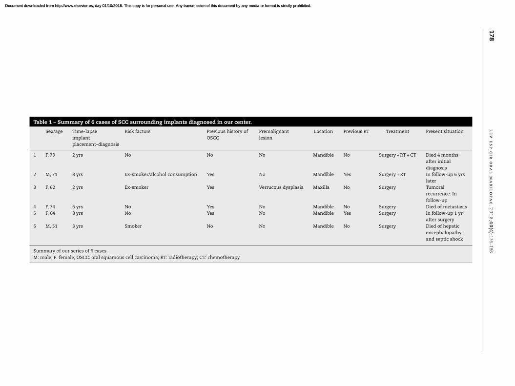
Table 1 – Summary of 6 cases of SCC surrounding implants diagnosed in our center.
Sex/age Time-lapse
implant
placement–diagnosis
Risk factors Previous history of
OSCC
Premalignant
lesion
Location Previous RT Treatment Present situation
1 F, 79 2 yrs No No No Mandible No Surgery + RT + CT Died 4 months
after initial
diagnosis
2 M, 71 8 yrs Ex-smoker/alcohol consumption Yes No Mandible Yes Surgery + RT In follow-up 6 yrs
later
3 F, 62 2 yrs Ex-smoker Yes Verrucous dysplasia Maxilla No Surgery Tumoral
recurrence. In
follow-up
4 F, 74 6 yrs No Yes No Mandible No Surgery Died of metastasis
5 F, 64 8 yrs No Yes No Mandible Yes Surgery In follow-up 1 yr
after surgery
6 M, 51 3 yrs Smoker No No Mandible No Surgery Died of hepatic
encephalopathy
and septic shock
Summary of our series of 6 cases.
M: male; F: female; OSCC: oral squamous cell carcinoma; RT: radiotherapy; CT: chemotherapy.
Document downloaded from http://www.elsevier.es, day 01/10/2018. This copy is for personal use. Any transmission of this document by any media or format is strictly prohibited.Document downloaded from http://www.elsevier.es, day 01/10/2018. This copy is for personal use. Any transmission of this document by any media or format is strictly prohibited.
r e v e s p c i r o r a l m a x i l o f a c . 2 0 1 8;4 0(4):176–186 179
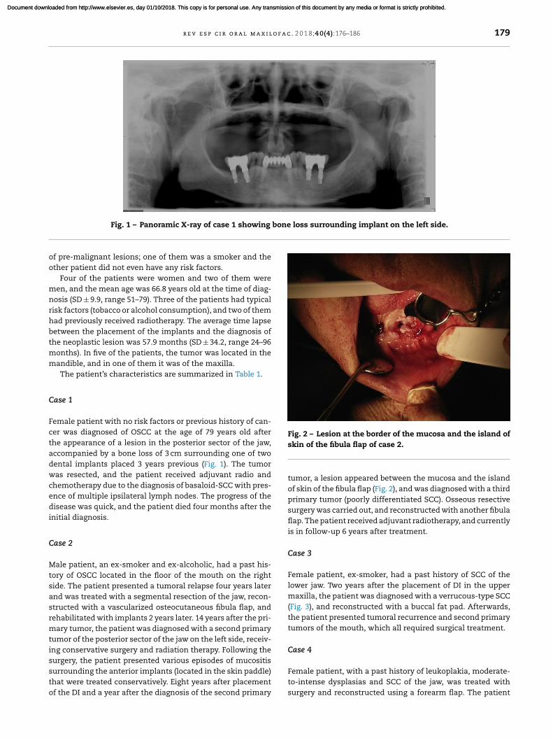
Fig. 1 – Panoramic X-ray of case 1 showing bone loss surrounding implant on the left side.
of pre-malignant lesions; one of them was a smoker and the
other patient did not even have any risk factors.
Four of the patients were women and two of them were
men, and the mean age was 66.8 years old at the time of diag-
nosis (SD ± 9.9, range 51–79). Three of the patients had typical
risk factors (tobacco or alcohol consumption), and two of them
had previously received radiotherapy. The average time lapse
between the placement of the implants and the diagnosis of
the neoplastic lesion was 57.9 months (SD ± 34.2, range 24–96
months). In five of the patients, the tumor was located in the
mandible, and in one of them it was of the maxilla.
The patient’s characteristics are summarized in Table 1.
Case 1
Female patient with no risk factors or previous history of can-
cer was diagnosed of OSCC at the age of 79 years old after
the appearance of a lesion in the posterior sector of the jaw,
accompanied by a bone loss of 3 cm surrounding one of two
dental implants placed 3 years previous (Fig. 1). The tumor
was resected, and the patient received adjuvant radio and
chemotherapy due to the diagnosis of basaloid-SCC with pres-
ence of multiple ipsilateral lymph nodes. The progress of the
disease was quick, and the patient died four months after the
initial diagnosis.
Case 2
Male patient, an ex-smoker and ex-alcoholic, had a past his-
tory of OSCC located in the floor of the mouth on the right
side. The patient presented a tumoral relapse four years later
and was treated with a segmental resection of the jaw, recon-
structed with a vascularized osteocutaneous fibula flap, and
rehabilitated with implants 2 years later. 14 years after the pri-
mary tumor, the patient was diagnosed with a second primary
tumor of the posterior sector of the jaw on the left side, receiv-
ing conservative surgery and radiation therapy. Following the
surgery, the patient presented various episodes of mucositis
surrounding the anterior implants (located in the skin paddle)
that were treated conservatively. Eight years after placement
of the DI and a year after the diagnosis of the second primary
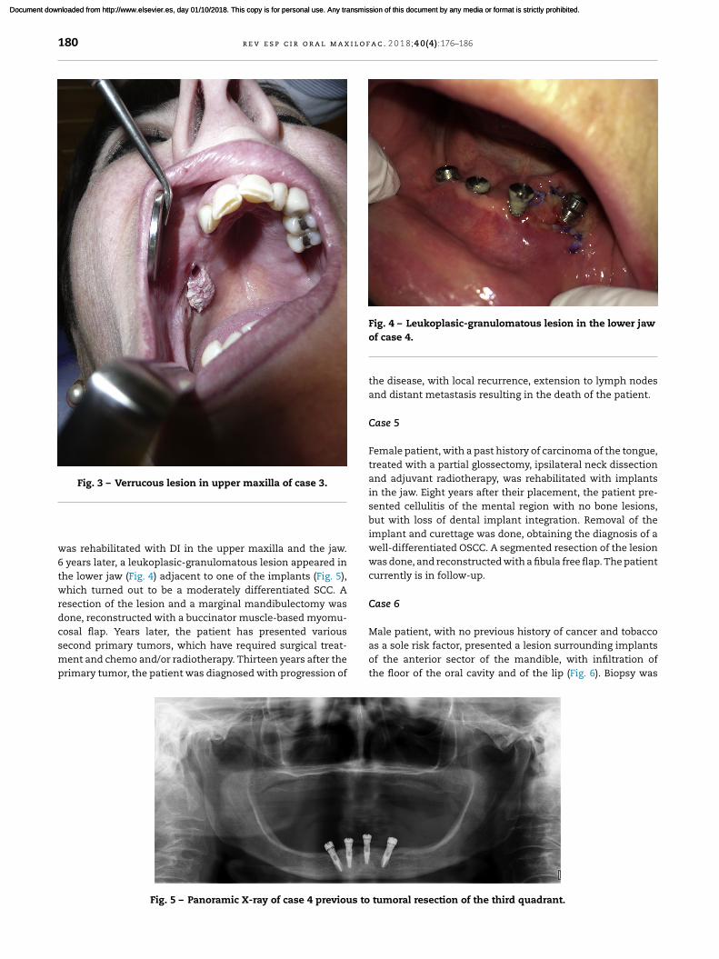
Fig. 2 – Lesion at the border of the mucosa and the island of
skin of the fibula flap of case 2.
tumor, a lesion appeared between the mucosa and the island
of skin of the fibula flap (Fig. 2), and was diagnosed with a third
primary tumor (poorly differentiated SCC). Osseous resective
surgery was carried out, and reconstructed with another fibula
flap. The patient received adjuvant radiotherapy, and currently
is in follow-up 6 years after treatment.
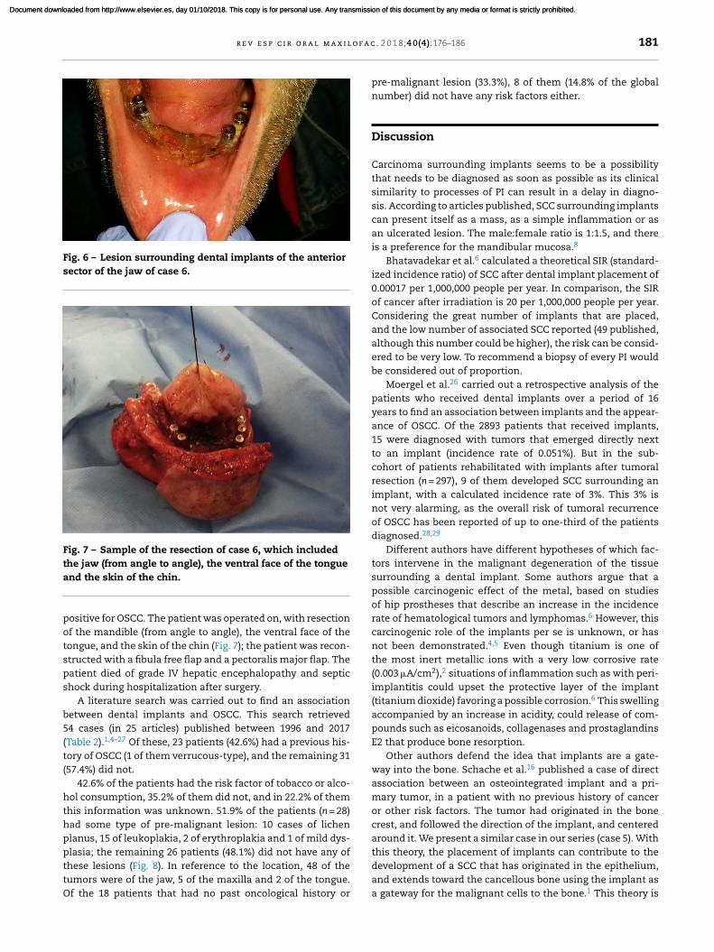
Case 3
Female patient, ex-smoker, had a past history of SCC of the
lower jaw. Two years after the placement of DI in the upper
maxilla, the patient was diagnosed with a verrucous-type SCC
(Fig. 3), and reconstructed with a buccal fat pad. Afterwards,
the patient presented tumoral recurrence and second primary
tumors of the mouth, which all required surgical treatment.
Case 4
Female patient, with a past history of leukoplakia, moderate-
to-intense dysplasias and SCC of the jaw, was treated with
surgery and reconstructed using a forearm flap. The patient
Document downloaded from http://www.elsevier.es, day 01/10/2018. This copy is for personal use. Any transmission of this document by any media or format is strictly prohibited.Document downloaded from http://www.elsevier.es, day 01/10/2018. This copy is for personal use. Any transmission of this document by any media or format is strictly prohibited.
180 r e v e s p c i r o r a l m a x i l o f a c . 2 0 1 8;4 0(4):176–186
Fig. 3 – Verrucous lesion in upper maxilla of case 3.
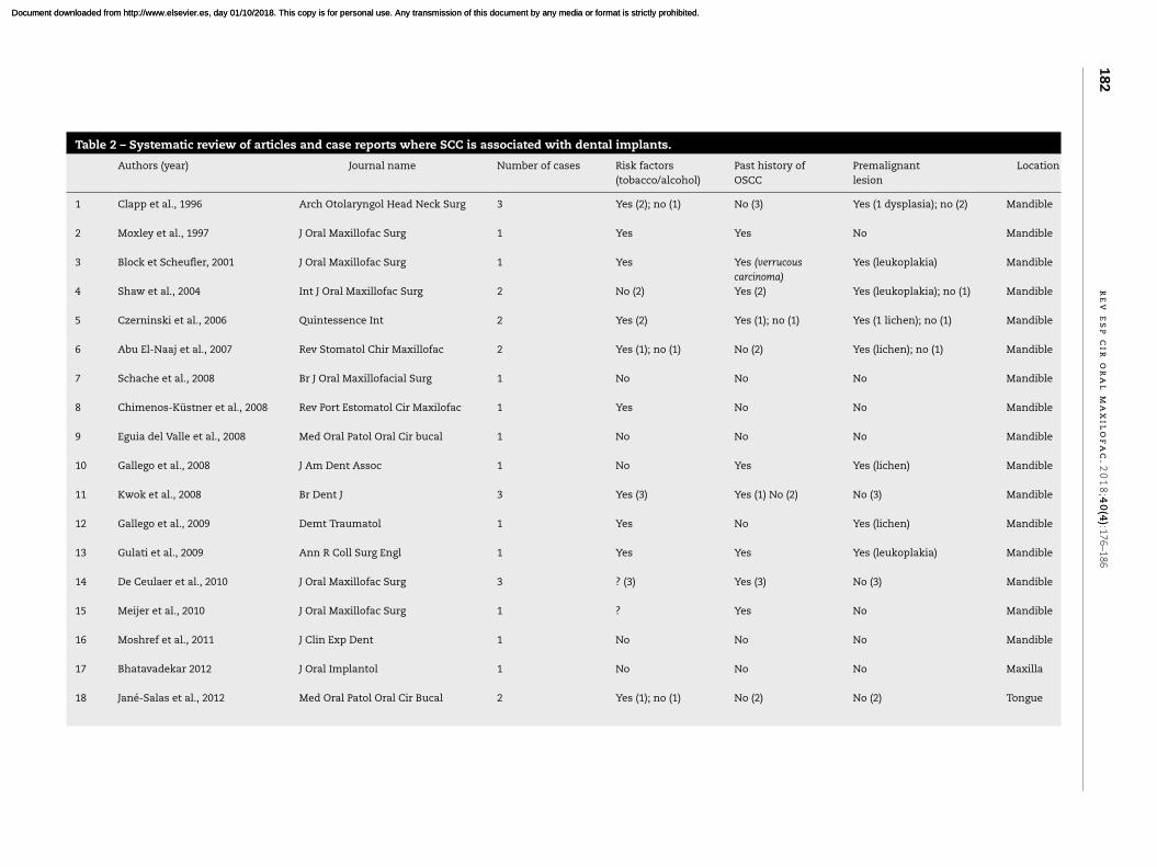
was rehabilitated with DI in the upper maxilla and the jaw.
6 years later, a leukoplasic-granulomatous lesion appeared in
the lower jaw (Fig. 4) adjacent to one of the implants (Fig. 5),
which turned out to be a moderately differentiated SCC. A
resection of the lesion and a marginal mandibulectomy was
done, reconstructed with a buccinator muscle-based myomu-
cosal flap. Years later, the patient has presented various
second primary tumors, which have required surgical treat-
ment and chemo and/or radiotherapy. Thirteen years after the
primary tumor, the patient was diagnosed with progression of
Fig. 4 – Leukoplasic-granulomatous lesion in the lower jaw
of case 4.
the disease, with local recurrence, extension to lymph nodes
and distant metastasis resulting in the death of the patient.
Case 5
Female patient, with a past history of carcinoma of the tongue,
treated with a partial glossectomy, ipsilateral neck dissection
and adjuvant radiotherapy, was rehabilitated with implants
in the jaw. Eight years after their placement, the patient pre-
sented cellulitis of the mental region with no bone lesions,
but with loss of dental implant integration. Removal of the
implant and curettage was done, obtaining the diagnosis of a
well-differentiated OSCC. A segmented resection of the lesion
was done, and reconstructed with a fibula free flap. The patient
currently is in follow-up.
Case 6
Male patient, with no previous history of cancer and tobacco
as a sole risk factor, presented a lesion surrounding implants
of the anterior sector of the mandible, with infiltration of
the floor of the oral cavity and of the lip (Fig. 6). Biopsy was
Fig. 5 – Panoramic X-ray of case 4 previous to tumoral resection of the third quadrant.
Document downloaded from http://www.elsevier.es, day 01/10/2018. This copy is for personal use. Any transmission of this document by any media or format is strictly prohibited.Document downloaded from http://www.elsevier.es, day 01/10/2018. This copy is for personal use. Any transmission of this document by any media or format is strictly prohibited.
r e v e s p c i r o r a l m a x i l o f a c . 2 0 1 8;4 0(4):176–186 181
Fig. 6 – Lesion surrounding dental implants of the anterior
sector of the jaw of case 6.
Fig. 7 – Sample of the resection of case 6, which included
the jaw (from angle to angle), the ventral face of the tongue
and the skin of the chin.
positive for OSCC. The patient was operated on, with resection
of the mandible (from angle to angle), the ventral face of the
tongue, and the skin of the chin (Fig. 7); the patient was recon-
structed with a fibula free flap and a pectoralis major flap. The
patient died of grade IV hepatic encephalopathy and septic
shock during hospitalization after surgery.
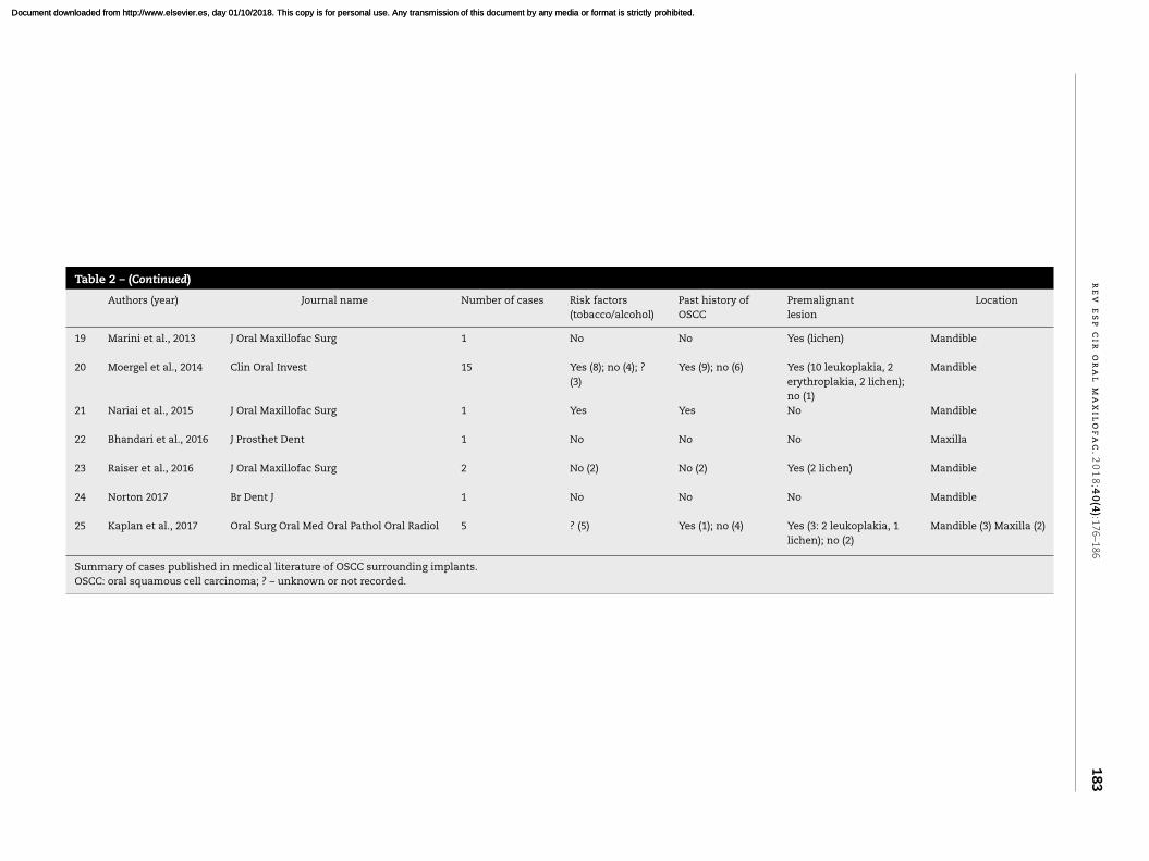
A literature search was carried out to find an association
between dental implants and OSCC. This search retrieved
54 cases (in 25 articles) published between 1996 and 2017
(Table 2).1,4–27 Of these, 23 patients (42.6%) had a previous his-
tory of OSCC (1 of them verrucous-type), and the remaining 31
(57.4%) did not.
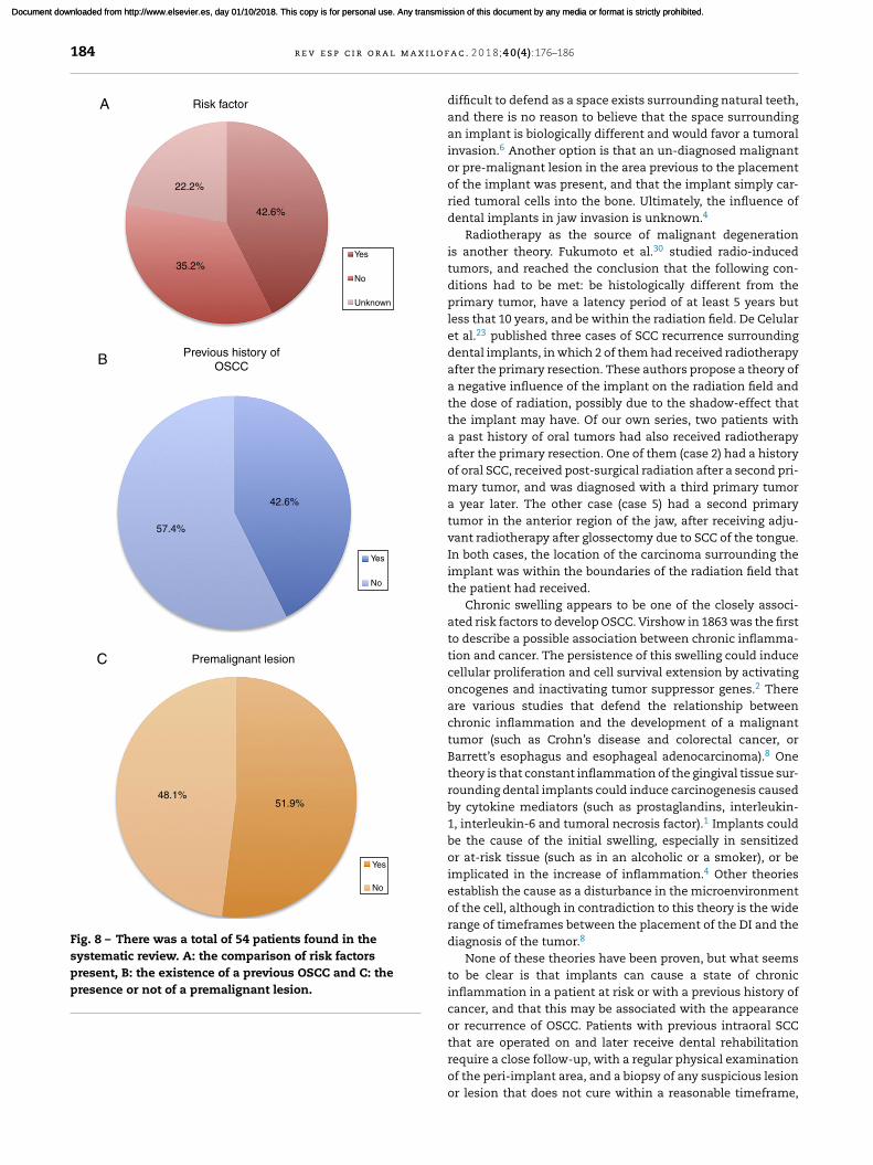
42.6% of the patients had the risk factor of tobacco or alco-
hol consumption, 35.2% of them did not, and in 22.2% of them
this information was unknown. 51.9% of the patients (n = 28)
had some type of pre-malignant lesion: 10 cases of lichen
planus, 15 of leukoplakia, 2 of erythroplakia and 1 of mild dys-
plasia; the remaining 26 patients (48.1%) did not have any of
these lesions (Fig. 8). In reference to the location, 48 of the
tumors were of the jaw, 5 of the maxilla and 2 of the tongue.
Of the 18 patients that had no past oncological history or
pre-malignant lesion (33.3%), 8 of them (14.8% of the global
number) did not have any risk factors either.
Discussion
Carcinoma surrounding implants seems to be a possibility
that needs to be diagnosed as soon as possible as its clinical
similarity to processes of PI can result in a delay in diagno-
sis. According to articles published, SCC surrounding implants
can present itself as a mass, as a simple inflammation or as
an ulcerated lesion. The male:female ratio is 1:1.5, and there
is a preference for the mandibular mucosa.8
Bhatavadekar et al.6 calculated a theoretical SIR (standard-
ized incidence ratio) of SCC after dental implant placement of
0.00017 per 1,000,000 people per year. In comparison, the SIR
of cancer after irradiation is 20 per 1,000,000 people per year.
Considering the great number of implants that are placed,
and the low number of associated SCC reported (49 published,
although this number could be higher), the risk can be consid-
ered to be very low. To recommend a biopsy of every PI would
be considered out of proportion.
Moergel et al.26 carried out a retrospective analysis of the
patients who received dental implants over a period of 16
years to find an association between implants and the appear-
ance of OSCC. Of the 2893 patients that received implants,
15 were diagnosed with tumors that emerged directly next
to an implant (incidence rate of 0.051%). But in the sub-
cohort of patients rehabilitated with implants after tumoral
resection (n = 297), 9 of them developed SCC surrounding an
implant, with a calculated incidence rate of 3%. This 3% is
not very alarming, as the overall risk of tumoral recurrence
of OSCC has been reported of up to one-third of the patients
diagnosed.28,29
Different authors have different hypotheses of which fac-
tors intervene in the malignant degeneration of the tissue
surrounding a dental implant. Some authors argue that a
possible carcinogenic effect of the metal, based on studies
of hip prostheses that describe an increase in the incidence
rate of hematological tumors and lymphomas.6 However, this
carcinogenic role of the implants per se is unknown, or has
not been demonstrated.4,5 Even though titanium is one of
the most inert metallic ions with a very low corrosive rate
(0.003 �A/cm2),2 situations of inflammation such as with peri-
implantitis could upset the protective layer of the implant
(titanium dioxide) favoring a possible corrosion.6 This swelling
accompanied by an increase in acidity, could release of com-
pounds such as eicosanoids, collagenases and prostaglandins
E2 that produce bone resorption.
Other authors defend the idea that implants are a gate-
way into the bone. Schache et al.16 published a case of direct
association between an osteointegrated implant and a pri-
mary tumor, in a patient with no previous history of cancer
or other risk factors. The tumor had originated in the bone
crest, and followed the direction of the implant, and centered
around it. We present a similar case in our series (case 5). With
this theory, the placement of implants can contribute to the
development of a SCC that has originated in the epithelium,
and extends toward the cancellous bone using the implant as
a gateway for the malignant cells to the bone.1 This theory is
Document downloaded from http://www.elsevier.es, day 01/10/2018. This copy is for personal use. Any transmission of this document by any media or format is strictly prohibited.Document downloaded from http://www.elsevier.es, day 01/10/2018. This copy is for personal use. Any transmission of this document by any media or format is strictly prohibited.
182
r
e
v
e
s
p
c
i
r
o
r
a
l
m
a
x
i
l
o
f
a
c
.
2
0
1
8;4
0(4
):17
6–1
86
Table 2 – Systematic review of articles and case reports where SCC is associated with dental implants.
Authors (year) Journal name Number of cases Risk factors
(tobacco/alcohol)
Past history of
OSCC
Premalignant
lesion
Location
1 Clapp et al., 1996 Arch Otolaryngol Head Neck Surg 3 Yes (2); no (1) No (3) Yes (1 dysplasia); no (2) Mandible
2 Moxley et al., 1997 J Oral Maxillofac Surg 1 Yes Yes No Mandible
3 Block et Scheufler, 2001 J Oral Maxillofac Surg 1 Yes Yes (verrucous
carcinoma)
Yes (leukoplakia) Mandible
4 Shaw et al., 2004 Int J Oral Maxillofac Surg 2 No (2) Yes (2) Yes (leukoplakia); no (1) Mandible
5 Czerninski et al., 2006 Quintessence Int 2 Yes (2) Yes (1); no (1) Yes (1 lichen); no (1) Mandible
6 Abu El-Naaj et al., 2007 Rev Stomatol Chir Maxillofac 2 Yes (1); no (1) No (2) Yes (lichen); no (1) Mandible
7 Schache et al., 2008 Br J Oral Maxillofacial Surg 1 No No No Mandible
8 Chimenos-Küstner et al., 2008 Rev Port Estomatol Cir Maxilofac 1 Yes No No Mandible
9 Eguia del Valle et al., 2008 Med Oral Patol Oral Cir bucal 1 No No No Mandible
10 Gallego et al., 2008 J Am Dent Assoc 1 No Yes Yes (lichen) Mandible
11 Kwok et al., 2008 Br Dent J 3 Yes (3) Yes (1) No (2) No (3) Mandible
12 Gallego et al., 2009 Demt Traumatol 1 Yes No Yes (lichen) Mandible
13 Gulati et al., 2009 Ann R Coll Surg Engl 1 Yes Yes Yes (leukoplakia) Mandible
14 De Ceulaer et al., 2010 J Oral Maxillofac Surg 3 ? (3) Yes (3) No (3) Mandible
15 Meijer et al., 2010 J Oral Maxillofac Surg 1 ? Yes No Mandible
16 Moshref et al., 2011 J Clin Exp Dent 1 No No No Mandible
17 Bhatavadekar 2012 J Oral Implantol 1 No No No Maxilla
18 Jané-Salas et al., 2012 Med Oral Patol Oral Cir Bucal 2 Yes (1); no (1) No (2) No (2) Tongue
Document downloaded from http://www.elsevier.es, day 01/10/2018. This copy is for personal use. Any transmission of this document by any media or format is strictly prohibited.Document downloaded from http://www.elsevier.es, day 01/10/2018. This copy is for personal use. Any transmission of this document by any media or format is strictly prohibited.
r
e
v
e
s
p
c
i
r
o
r
a
l
m
a
x
i
l
o
f
a
c
.
2
0
1
8;4
0(4
):17
6–1
86
183
Table 2 – (Continued)
Authors (year) Journal name Number of cases Risk factors
(tobacco/alcohol)
Past history of
OSCC
Premalignant
lesion
Location
19 Marini et al., 2013 J Oral Maxillofac Surg 1 No No Yes (lichen) Mandible
20 Moergel et al., 2014 Clin Oral Invest 15 Yes (8); no (4); ?
(3)
Yes (9); no (6) Yes (10 leukoplakia, 2
erythroplakia, 2 lichen);
no (1)
Mandible
21 Nariai et al., 2015 J Oral Maxillofac Surg 1 Yes Yes No Mandible
22 Bhandari et al., 2016 J Prosthet Dent 1 No No No Maxilla
23 Raiser et al., 2016 J Oral Maxillofac Surg 2 No (2) No (2) Yes (2 lichen) Mandible
24 Norton 2017 Br Dent J 1 No No No Mandible
25 Kaplan et al., 2017 Oral Surg Oral Med Oral Pathol Oral Radiol 5 ? (5) Yes (1); no (4) Yes (3: 2 leukoplakia, 1
lichen); no (2)
Mandible (3) Maxilla (2)
Summary of cases published in medical literature of OSCC surrounding implants.
OSCC: oral squamous cell carcinoma; ? – unknown or not recorded.
Document downloaded from http://www.elsevier.es, day 01/10/2018. This copy is for personal use. Any transmission of this document by any media or format is strictly prohibited.Document downloaded from http://www.elsevier.es, day 01/10/2018. This copy is for personal use. Any transmission of this document by any media or format is strictly prohibited.
184 r e v e s p c i r o r a l m a x i l o f a c . 2 0 1 8;4 0(4):176–186
Risk factorA
B
C
22.2%
35.2%
42.6%
42.6%
51.9%48.1%
57.4%
Yes
Yes
Yes
No
No
No
Unknown
Previous history of
OSCC
Premalignant lesion
Fig. 8 – There was a total of 54 patients found in the
systematic review. A: the comparison of risk factors
present, B: the existence of a previous OSCC and C: the
presence or not of a premalignant lesion.
difficult to defend as a space exists surrounding natural teeth,
and there is no reason to believe that the space surrounding
an implant is biologically different and would favor a tumoral
invasion.6 Another option is that an un-diagnosed malignant
or pre-malignant lesion in the area previous to the placement
of the implant was present, and that the implant simply car-
ried tumoral cells into the bone. Ultimately, the influence of
dental implants in jaw invasion is unknown.4
Radiotherapy as the source of malignant degeneration
is another theory. Fukumoto et al.30 studied radio-induced
tumors, and reached the conclusion that the following con-
ditions had to be met: be histologically different from the
primary tumor, have a latency period of at least 5 years but
less that 10 years, and be within the radiation field. De Celular
et al.23 published three cases of SCC recurrence surrounding
dental implants, in which 2 of them had received radiotherapy
after the primary resection. These authors propose a theory of
a negative influence of the implant on the radiation field and
the dose of radiation, possibly due to the shadow-effect that
the implant may have. Of our own series, two patients with
a past history of oral tumors had also received radiotherapy
after the primary resection. One of them (case 2) had a history
of oral SCC, received post-surgical radiation after a second pri-
mary tumor, and was diagnosed with a third primary tumor
a year later. The other case (case 5) had a second primary
tumor in the anterior region of the jaw, after receiving adju-
vant radiotherapy after glossectomy due to SCC of the tongue.
In both cases, the location of the carcinoma surrounding the
implant was within the boundaries of the radiation field that
the patient had received.
Chronic swelling appears to be one of the closely associ-
ated risk factors to develop OSCC. Virshow in 1863 was the first
to describe a possible association between chronic inflamma-
tion and cancer. The persistence of this swelling could induce
cellular proliferation and cell survival extension by activating
oncogenes and inactivating tumor suppressor genes.2 There
are various studies that defend the relationship between
chronic inflammation and the development of a malignant
tumor (such as Crohn’s disease and colorectal cancer, or
Barrett’s esophagus and esophageal adenocarcinoma).8 One
theory is that constant inflammation of the gingival tissue sur-
rounding dental implants could induce carcinogenesis caused
by cytokine mediators (such as prostaglandins, interleukin-
1, interleukin-6 and tumoral necrosis factor).1 Implants could
be the cause of the initial swelling, especially in sensitized
or at-risk tissue (such as in an alcoholic or a smoker), or be
implicated in the increase of inflammation.4 Other theories
establish the cause as a disturbance in the microenvironment
of the cell, although in contradiction to this theory is the wide
range of timeframes between the placement of the DI and the
diagnosis of the tumor.8
None of these theories have been proven, but what seems
to be clear is that implants can cause a state of chronic
inflammation in a patient at risk or with a previous history of
cancer, and that this may be associated with the appearance
or recurrence of OSCC. Patients with previous intraoral SCC
that are operated on and later receive dental rehabilitation
require a close follow-up, with a regular physical examination
of the peri-implant area, and a biopsy of any suspicious lesion
or lesion that does not cure within a reasonable timeframe,
Document downloaded from http://www.elsevier.es, day 01/10/2018. This copy is for personal use. Any transmission of this document by any media or format is strictly prohibited.Document downloaded from http://www.elsevier.es, day 01/10/2018. This copy is for personal use. Any transmission of this document by any media or format is strictly prohibited.
r e v e s p c i r o r a l m a x i l o f a c . 2 0 1 8;4 0(4):176–186 185
especially in patients in which curettage is used as part of the
treatment of PI.
A biopsy can also be useful for the diagnosis of other
pathologies surrounding implants. In the systematic review
of articles, other malignant lesions concurrent with DI were
also found: 5 cases of bone metastasis to the jaw (3 cases
of lung cancer and 3 of breast cancer). The literature review
also retrieved one case of osteosarcoma and another case of
type-B lymphoma (neither patient had any risk factor or past
oncological history). Another case of basal cell carcinoma was
also identified in a patient that had the same lesion in other
intraoral locations.
The presence of intraoral lesions should also be taken in
consideration when contemplating dental rehabilitation with
implants. Lichen planus is a pre-neoplastic condition with a
rate of malignant transformation risk of 1%5 (between 0 and
12.5%31). That dental implants favor this malignant transfor-
mation of premalignant lesions is unknown and should be
further studied in the future.
Conclusion
Although there are no studies that prove a direct relationship
between the presence of dental implants and the risk of SCC,
it appears that chronic inflammation of the tissue surround-
ing the implant could be an important factor. The incidence
rate of SCC surrounding DI seems to be high in patients with
previous oral tumors, and very low outside of this group. In
conclusion, a surveillance of patients with PI, especially those
with risk factors, a previous history of OSCC or presence of
leukoplakia or lichen planus, is highly recommended, and a
biopsy should be performed of lesions that are similar to PI
but do not respond well to regular treatment, have a sluggish
or rapid progression, or are accompanied by local anesthesia
or paresthesia.
Conflict of interests
The authors declare no conflict of interests.
r e f e r e n c e s
1. Jané-Salas E, López-López J, Roselló-Llabrés X,Rodriguez-Argueta OF, Chimeno-Küstner E. Relationshipbetween oral cancer and implants: clinical cases andsystematic literature review. Med Oral Patol Oral Cir Bucal.2012;17:e23.
2. Salgado-Peralvo AO, Arriba-Fuente L, Mateos-Moreno MV,Salgado-García A. Is there an association between dentalimplants and squamous cell carcinoma? Br Dent J. 2016;221.
3. Gonzalez-Garcia R, Naval-Gias L, Roman-Romero L,Sastre-Perez J, Rodriguez-Campo FJ. Local recurrences andsecond primary tumors from squamous cell carcinoma of theoral cavity: a retrospective analytic study of 500 patients.Head Neck. 2009;31:1168.
4. Nariai Y, Kanno T, Sekine J. Histopathological features ofsecondary squamous cell carcinoma around a dental implantin the mandible after chemoradiotherapy: a case report witha clinicopathological review. J Oral Maxillofac Surg.2016;74:982–90.
5. Marini E, Spink MJ, Messina AM. Peri-implant primarysquamous cell carcinoma: a case report with 5 years’follow-up. J Oral Maxillofac Surg. 2013;71:322–6.
6. Bhatavadekar NB. Squamous cell carcinoma in associationwith dental implants: an assessment of previouslyhypothesized carcinogenic mechanisms and a case report. JOral Implantol. 2012;37.
7. Raiser V, Abu-El Naaj I, Shlomi B, Fliss DM, Kaplan I. Primaryoral malignancy imitating peri-implantitis. J Oral MaxillofacSurg. 2016:1–8.
8. Kaplan I, Zeevi I, Tal H, Rosenfeld E, Chaushu G.Clinicopathologic evaluation of malignancy adjacent todental implants. Oral Surg Oral Med Oral Pathol Oral Radiol.2017;123:103–12.
9. Bhandari S, Rattan V, Panda N, Vaiphei K, Mittal BR. Oralcancer or periimplantitis: a clinical dilemma. J Prosthet Dent.2016;115:658–61.
10. Clapp C, Wheeler JC, Martof AB, Levine PA. Oral squamouscell carcinoma in association with dental osseointegratedimplants. An unusual occurrence. Arch Otolaryngol HeadNeck Surg. 1996;122:1402–3.
11. Moxley JE, Stoelinga PJ, Blijdorp PA. Squamous cell carcinomaassociated with a mandibular stable implant. J OralMaxillofac Surg. 1997;55:1020–2.
12. Block MS, Scheufler E. Squamous cell carcinoma appearing asperi-implant bone loss: a case report. J Oral Maxillofac Surg.2001;59:1349–52.
13. Shaw R, Sutton D, Brown J, Cawood J. Further malignancy infield change adjacent to osseointegrated implants. Int J OralMaxillofac Surg. 2004;33:353–5.
14. Czerninski R, Kaplan I, Almoznino G, Maly A, Regev E. Oralsquamous cell carcinoma around dental implants.Quintessence Int. 2006;37:707–11.
15. Abu El-Naaj I, Trost O, Tagger-Green N, Trouilloud P, Robe N,Malka G, et al. Peri-implantitis or squamous cell carcinoma?Rev Stomatol Chir Maxillofac. 2007;108:458–60.
16. Schache A, Thavaraj S, Kalavrezos N. Osseointegratedimplants: a potential route of entry for squamous cellcarcinoma of the mandible. Br J Oral Maxillofac Surg.2008;46:397–9.
17. Chimenos-Küstner E, López-López J, Finestres-Zubeldia F.Squamous carcinoma after dental implants: a clinical case.Rev Port Estomatol Cir Maxilofac. 2008;49:97–100.
18. Eguia del Valle A, Martínez-Conde L, Lamosas R, LópezVicente J, Uribarri Etxebarria A, Aguirre Urizar JM. Primaryoral squamous cell carcinoma arising around dentalosseointegrated implants mimicking peri-implantitis. MedOral Patol Oral Cir Bucal. 2008;13:489–91.
19. Gallego L, Junquera L, Baladrón J, Villarreal P. Oral squamouscell carcinoma associated with symphyseal dental implants:an unusual case report. J Am Dent Assoc. 2008;139:1061–5.
20. Kwok J, Eyeson J, Thompson I, McGurk M. Dental implantsand squamous cell carcinoma in the at risk patient-report ofthree cases. Br Dent J. 2008;205:543–5.
21. Gallego L, Junquera L, Llorente S. Oral carcinoma associatedwith implant-supported overdenture trauma: a case report.Dent Traumatol. 2009;25:3–4.
22. Gulati A, Puthussery FJ, Downie IP, Flood TR. Squamous cellcarcinoma presenting as peri-implantitis: a case report. AnnR Coll Surg Engl. 2009;91:8–10.
23. De Ceulaer J, Magremanne M, van Veen A, Scheerlinck J.Squamous cell carcinoma recurrence around dental implants.J Oral Maxillofac Surg. 2010;68:2507–12.
24. Meijer GJ, Dieleman FJ, Bergé SJ, Merkx MAW. Removal of anoral squamous cell carcinoma including parts ofosseointegrated implants in the marginal mandibulectomy: acase report. J Oral Maxillofac Surg. 2010;14:253–6.
Document downloaded from http://www.elsevier.es, day 01/10/2018. This copy is for personal use. Any transmission of this document by any media or format is strictly prohibited.Document downloaded from http://www.elsevier.es, day 01/10/2018. This copy is for personal use. Any transmission of this document by any media or format is strictly prohibited.

186 r e v e s p c i r o r a l m a x i l o f a c . 2 0 1 8;4 0(4):176–186
25. Moshref M, Jamilian A, Lotfi A, Showkatbakhsh R. Oralsquamous carcinoma associated with dental implant—a casereport and literature review. J Clin Exp Dent. 2011;3:166–8.
26. Moergel M, Karbach J, Kunkel M, Wagner W. Oral squamouscell carcinoma in the vicinity of dental implants. Clin OralInvest. 2014;18:277–84.
27. Norton MR. Dental implants. Potential relationship withcancer. Br Dent J. 2016;222 [Letter to the Editor].
28. Da Silva SD, Hier M, Mlynarek A, Kowalski LP, Alaoui-JamaliMA. Recurrent oral cancer: current and emerging therapeuticapproaches. Front Pharmacol. 2012;3:149.
29. Leemans CR, Tiwari R, Nauta JJ, van der Waal I, Snow GB.Recurrence at the primary site in head and neck cancer andthe significance of neck lymph node metastases as aprognostic factor. Cancer. 1994;73:187–90.
30. Fukumoto M, Matsumoto Y. Second malignancies afterradio-therapy: analysis of cases accumulated by aquestionnaire in Japan. Jpn J Cancer Clin. 2004;50:1081.
31. González-Moles MA, Scully C, Gil-Montoya JA. Oral lichenplanus: controversies surrounding malignant transformation.Oral Dis. 2008;14:229–43.
Document downloaded from http://www.elsevier.es, day 01/10/2018. This copy is for personal use. Any transmission of this document by any media or format is strictly prohibited.Document downloaded from http://www.elsevier.es, day 01/10/2018. This copy is for personal use. Any transmission of this document by any media or format is strictly prohibited.