Modeling Tumor
9
Introduction Cancer is a leading cause of death worldwide and, according to the World Health Organization, was responsible for the death of 7.9 million people in 2007 (www.who.int). This disease is characterized by the uncontrolled malignant growth of cells; however, the vast majority of human fatalities arise from secondary metastatic tumors. These secondary tumors generally spread from the original site via the blood or lymphatic system and are highly invasive and aggressive. The metastatic process involves several discrete biological steps: loss of cellular adhesion, increased motility and invasiveness, entry and survival of tumor cells in the circulation, their exit into new tissue, and their eventual colonization of a distant site (Chambers et al., 2002; Gupta and Massague, 2006). Understanding the mechanisms that promote tumor invasion and the role of the microenvironment is important for developing therapeutic strategies to treat metastatic cancers (Yang and Weinberg, 2008; Nguyen et al., 2009; Hanahan and Weinberg, 2011). Over the last decade, the fruit fly Drosophila melanogaster has become an important model system for cancer studies. Reduced redundancy in the Drosophila genome compared with that of humans, coupled with the ability to conduct large-scale genetic screens in this organism, has enabled its use to determine the molecular characterization of important signaling cascades, developmental processes and growth control. For example, our understanding of the Hippo, Notch, Dpp and JAK-STAT signaling pathways, all of which are involved in tumor formation, has been enhanced by research in Drosophila (for reviews, see Brumby and Richardson, 2005; Vidal and Cagan, 2006; Januschke and G onzalez, 2008). Drosophila genetics has revealed many genes that, when mutated or dysregulated, result in or contribute to tumorigenesis. Hyperplastic tumor suppressors, including components of the Hippo pathway, promote increased proliferation or survival, but do not disrupt tissue structure or differentiation. By contrast, Drosophila neoplastic tumor suppressors, such as the apical-basal cell polarity regulators ( e.g. Lgl), lead to loss of tissue architecture, defects in differentiation and failure to exit the cell cycle. Elegant genetic and cell biology techniques have enabled the effects of tumor suppressors and oncogenes to be examined in the context of the whole animal. The capacity to generate patches (clones) of mutant tissue for specified genes during fly development has facilitated investigations into the role of the microenvironmen t in tumor development. Similarly, studies using clonal analysis in Drosophila have begun to elucidate cell competition mechanisms, which could potentially confer malignant cells with a growth advantage over their neighbors. In conjunction with studies using other model organisms, flies have contributed greatly to our understanding of the mechanisms involved in cancer initiation and progression, revealing previously unknown molecular components and concepts. In turn, these have served to guide researchers who use mammalian systems to study cancer . This Perspective focuses on recent developments using fly tumor models that have been generated by tumor suppressor mutations or oncogene overexpression to induce neoplastic tumors of neuronal and epithelial origin as a means to probe the mechanisms involved in cellular invasion and metastasis. Insights from metastatic neuronal tumors in the developing larval brain Single gene mutations in a unique subset of genes { lethal (3) malignant brain tumor [l(3)mbt ], brain tumor (brat ), discs large PERSPECTIVE Disease Models & Mechanisms 1 Disease Models & Mechanisms 4, 000-000 (2011) doi:10.1242/dmm.006908 Modeling tumor invasion and metastasis in Drosophila Wayne O. Miles 1 , Nicholas J. Dyson 1 and James A. Walker 1,2, * 1 Massachusetts General Hospital Center for Cancer Research and Harvard Medical School, 149, 13th Street, Charlestown, MA 02129, USA 2 Center for Human Genetic Research, Massachusetts General Hospital, Boston, MA 02114, USA *Author for correspondenc e ([email protected] .edu) © 2011. Published by The Company of Biologists Ltd This is an Open Access article distributed under the terms of the Creative Commons Attribution Non-Commercial Share Alike License (http://creativecommons.org/licenses/by- nc-sa/3.0), which permits unrestricted non-commercial use, distribution and reproduction in any medium provided that the original work is properly cited and all further distributions of the work or adaptation are subject to the same Creative Commons License terms. Conservation of major signaling pathways between humans and flies has made Drosophila a useful model organism for cancer research. Our understanding of the mechanisms regulating cell growth, differentiation and development has been considerably advanced by studies in Drosophila. Several recent high profile studies have examined the processes constraining the metastatic growth of tumor cells in fruit fly models. Cell invasion can be studied in the context of an in vivo setting in flies, enabling the genetic requirements of the microenvironment of tumor cells undergoing metastasis to be analyzed. This Perspective discusses the strengths and limitations of Drosophila models of cancer invasion and the unique tools that have enabled these studies. It also highlights several recent reports that together make a strong case for Drosophila as a system with the potential for both testing novel concepts in tumor progression and cell invasion, and for uncovering players in metastasis.
-
Upload
fernando-lazzarotto-evaldt -
Category
Documents
-
view
218 -
download
0
Transcript of Modeling Tumor
8/3/2019 Modeling Tumor
http://slidepdf.com/reader/full/modeling-tumor 1/9
IntroductionCancer is a leading cause of death worldwide and, according to theWorld Health Organization, was responsible for the death of 7.9million people in 2007 (www.who.int). This disease is characterizedby the uncontrolled malignant growth of cells; however, the vastmajority of human fatalities arise from secondary metastatictumors. These secondary tumors generally spread from the originalsite via the blood or lymphatic system and are highly invasive andaggressive. The metastatic process involves several discretebiological steps: loss of cellular adhesion, increased motility andinvasiveness, entry and survival of tumor cells in the circulation,their exit into new tissue, and their eventual colonization of a distant
site (Chambers et al., 2002; Gupta and Massague, 2006).Understanding the mechanisms that promote tumor invasion andthe role of the microenvironment is important for developingtherapeutic strategies to treat metastatic cancers (Yang andWeinberg, 2008; Nguyen et al., 2009; Hanahan and Weinberg, 2011).
Over the last decade, the fruit fly Drosophila melanogaster hasbecome an important model system for cancer studies. Reducedredundancy in the Drosophila genome compared with that of humans, coupled with the ability to conduct large-scale geneticscreens in this organism, has enabled its use to determine themolecular characterization of important signaling cascades,developmental processes and growth control. For example, our
understanding of the Hippo, Notch, Dpp and JAK-STAT signalingpathways, all of which are involved in tumor formation, has beenenhanced by research in Drosophila (for reviews, see Brumby andRichardson, 2005; Vidal and Cagan, 2006; Januschke and Gonzalez,2008). Drosophila genetics has revealed many genes that, whenmutated or dysregulated, result in or contribute to tumorigenesis.Hyperplastic tumor suppressors, including components of theHippo pathway, promote increased proliferation or survival, butdo not disrupt tissue structure or differentiation. By contrast,
Drosophila neoplastic tumor suppressors, such as the apical-basalcell polarity regulators (e.g. Lgl), lead to loss of tissue architecture,defects in differentiation and failure to exit the cell cycle. Elegant
genetic and cell biology techniques have enabled the effects of tumor suppressors and oncogenes to be examined in the contextof the whole animal. The capacity to generate patches (clones) of mutant tissue for specified genes during fly development hasfacilitated investigations into the role of the microenvironment intumor development. Similarly, studies using clonal analysis in
Drosophila have begun to elucidate cell competition mechanisms,which could potentially confer malignant cells with a growthadvantage over their neighbors.
In conjunction with studies using other model organisms, flieshave contributed greatly to our understanding of the mechanismsinvolved in cancer initiation and progression, revealing previously unknown molecular components and concepts. In turn, these haveserved to guide researchers who use mammalian systems to study
cancer. This Perspective focuses on recent developments using fly tumor models that have been generated by tumor suppressormutations or oncogene overexpression to induce neoplastic tumorsof neuronal and epithelial origin as a means to probe themechanisms involved in cellular invasion and metastasis.
Insights from metastatic neuronal tumors in thedeveloping larval brainSingle gene mutations in a unique subset of genes {lethal (3)malignant brain tumor [l(3)mbt ], brain tumor (brat ), discs large
PERSPECTIVE
Disease Models & Mechanisms 1
Disease Models & Mechanisms 4, 000-000 (2011) doi:10.1242/dmm.006908
Modeling tumor invasion and metastasis inDrosophila
Wayne O. Miles1, Nicholas J. Dyson1 and James A. Walker1,2,*
1Massachusetts General Hospital Center for Cancer Research and Harvard MedicalSchool, 149, 13th Street, Charlestown, MA 02129, USA2Center for Human Genetic Research, Massachusetts General Hospital, Boston,MA 02114, USA*Author for correspondence ([email protected])
© 2011. Published by The Company of Biologists Ltd This is an Open Access article distributed under the terms of the Creative CommonsAttribution Non-Commercial Share Alike License (http://creativecommons.org/licenses/by-nc-sa/3.0), which permits unrestricted non-commercial use, distribution and reproductionin any medium provided that the original work is properly cited and all furtherdistributions of the work or adaptation are subject to the same Creative Commons Licenseterms.
Conservation of major signaling pathways between humans and flies has made Drosophila a useful model organism forcancer research. Our understanding of the mechanisms regulating cell growth, differentiation and development hasbeen considerably advanced by studies in Drosophila. Several recent high profile studies have examined the processesconstraining the metastatic growth of tumor cells in fruit fly models. Cell invasion can be studied in the context of an invivo setting in flies, enabling the genetic requirements of the microenvironment of tumor cells undergoing metastasisto be analyzed. This Perspective discusses the strengths and limitations of Drosophila models of cancer invasion andthe unique tools that have enabled these studies. It also highlights several recent reports that together make a strongcase for Drosophila as a system with the potential for both testing novel concepts in tumor progression and cellinvasion, and for uncovering players in metastasis.
8/3/2019 Modeling Tumor
http://slidepdf.com/reader/full/modeling-tumor 2/9
(dlg ), scribble ( scrib), lethal giant larvae (lgl ), miranda (mira), prospero ( pros), partner of inscuteable ( pins) (for reviews seeJanuschke and Gonzalez, 2008; Froldi et al., 2008)} cause malignantneoplastic tumors in the Drosophila larval brain. These genes havecrucial roles in regulating proliferation and development [l(3)mbt ](Gateff et al., 1993) and apical-basal cell polarity (dlg and scrib)(Woods et al., 1989; Bilder et al., 2000).
The human homologs of Dlg, Scrib and Lgl are importantregulators of cell polarity, and mutations or splice variants havebeen linked to poor prognosis for colorectal (Schimanski et al.,2005) and hepatocarcinoma (Lu et al., 2009) patients. Importantly,all three complex components are targets for E6 papillomavirus-mediated degradation (Nakagawa and Huibregtse, 2000; Humbertet al., 2003; Handa et al., 2007). L(3)mbt has also been linked to various myeloid haemopoietic disorders (Boccuni et al., 2003) andis important for DNA replication and genomic stability (Gurvichet al., 2010).
Of these fly neoplastic tumor suppressors, lgl and brat are thebest characterized. Lgl is localized to the cellular cortex, andfunctions in the same genetic pathway as dlg and scrib to maintaincellular polarity. Neuroblasts from lgl mutant larvae undergo
aberrant symmetrical cell divisions, rather than the normalasymmetric divisions that are required for neuroblast and ganglionmother cell production (Mechler et al., 1985). Brat regulatesribosomal RNA (rRNA) synthesis and is a translational repressor.Brat is also asymmetrically localized to the ganglion mother cellafter neuroblast division and is necessary for neuronaldifferentiation and proliferation control (Arama et al., 2000).Malignant tumors resulting from mutations in these fly neoplastictumor suppressors cause late larval lethality but can be propagatedand will metastasize when allografted into a recipient adult host(Gateff, 1978; Januschke and Gonzalez, 2008).
Metastasis of lgl and brat tumors
The Shearn group has utilized allograft experiments to investigatethe metastatic behavior of lgl and brat mutant cells (Fig. 1A).Neoplastic brain tumors labeled with -galactosidase (encodedby lacZ ) from mutant larvae were dissected and allografted intothe abdomens of wild-type adult flies. These tumors grew rapidly and resulted in host lethality within 12 days. Ovaries fromallograft recipients were dissected and assayed for cells expressinglacZ . Because the ovary is contained in a non-porous epithelialsheet and muscle layers surround the ovarioles, only cells thatare capable of metastasizing can invade into this organ (Fig. 1A).lacZ -positive cells were detected in recipient ovarioles from lgl and brat mutant tumors but not from control brains. These lacZ -positive cells in the ovary were shown to retain their neuronaland glial cell markers. This work provided the first evidence that
Drosophila cells can metastasize through epithelial tissue andcolonize new sites distant from the initial tumor (Beaucher et al.,2007a).
To understand the processes regulating this metastasis, acandidate approach was used to identify genes that are requiredfor invasion by lgl and brat cells. Matrix metalloproteases (MMPs)have long been linked to metastasis in human cancer and weretherefore excellent candidates. The Drosophila genome encodestwo MMPs (MMP1 and MMP2). MMP1 expression is strongly upregulated in lgl mutant cells and is necessary for their metastatic
behavior (Beaucher et al., 2007b). By contrast, MMP1 expressionis not required in brat mutant cells but is required in the host tissue,suggesting that MMP1 co-operates non-cell-autonomously withbrat mutant cells to enable their metastasis.
Molecular profiling of l(3)mbt neuronal tumorsRecent studies from the Gonzalez group have taken a fresh
approach to identifying genes required for l(3)mbt tumor growth(Janic et al., 2010). l(3)mbt is a substoichiometric member of the
Drosophila dREAM-Myb complex, which is required for genesilencing, and functions in establishing and maintainingdifferentiation states (Lewis et al., 2004). Similar to lgl and brat tumors, those arising from l(3)mbt mutations result in larvallethality and can be allografted (Fig. 1B).
By conducting extensive expression arrays on larval neuronal andallograft cultured tumors, gene expression profiles were identifiedfor tumors that result from single gene mutations [in l(3)mbt , brat ,lgl , mira, pros and pins] (Janic et al., 2010). By comparing profiles,Janic and colleagues characterized an l(3)mbt -specific tumorsignature. The concatenate sequencing of Piwi-interacting RNAs(piRNAs) and microRNAs from these tumors identified small RNA
changes that are characteristic of the l(3)mbt tumors. Together withimmunohistochemical analyses, these approaches provide a basisfor understanding the gene expression changes that are specific tol(3)mbt tumors. The l(3)mbt tumor signature surprisingly containsa disproportionately high number of germline-specific genes andmicroRNAs. This result suggests that a germline fate is associatedwith l(3)mbt tumor growth, and is in agreement with studies fromCaenorhabditis elegans that linked soma-to-germline transitionwith increased fitness and longevity (Curran et al., 2009; Wang etal., 2005).
Genetic studies then tested the capacity of mutations inupregulated germline-specific genes to modify l(3)mbt tumorformation and allograft metastasis ( Janic et al., 2010). Interestingly,
only mutations in a subset of germline-specific genes ( piwi, vasa,aub and nos) could suppress l(3)mbt tumor formation. By contrast,other upregulated germline-specific transcripts (zpg , Pxt and AGO)were unable to prevent tumor formation or metastatic growth. Thisresult suggests that only a limited number of germline-specificgenes are capable of restricting tumor growth. These findingsprovide a detailed understanding of the gene expression and smallRNA changes in l(3)mbt neoplastic tumors, and highlight thepossibility that a soma-to-germline transition accompaniesmetastasis. However, this approach has not yet identified genes thatare specifically required for invasion, because all metastaticsuppressors also prevented l(3)mbt tumor growth. Further analysisof the l(3)mbt tumor signature will hopefully identify othermetastasis-promoting factors.
Using mutations to generate neoplastic tumors has significantadvantages. First, only a single mutational event is required toinitiate tumor growth in an isogenic background. Second, thel(3)mbt alleles are particularly advantageous because they aretemperature sensitive, permitting gene expression arrays to beconducted from a single stock at both permissive and restrictivetemperatures. Finally, these tumors develop rapidly andmetastasize when allografted. These experiments permitexpansion of the tumor mass for biochemical analysis andmetastasis modeling.
dmm.biologists.org2
Drosophila tumor invasion and metastasisPERSPECTIVE
8/3/2019 Modeling Tumor
http://slidepdf.com/reader/full/modeling-tumor 3/9
Insights from neuronal metastatic tumors induced byoncogene overexpressionModeling RasV12 tumors in the larval eye neuralepitheliumOverexpression of oncogenic Ras protein (RasV12) causes benigntumor growth in Drosophila. This model has been used to identify pathways that are required for tumor development and metastasis.The mosaic analysis with a repressible cell marker (MARCM)technique can be used to generate clones expressing activatedRasV12 by FLP-FRT-mediated mitotic recombination; clones areconcomitantly labeled with a visible marker (UAS-GFP )(Theodosiou and Xu, 1998; Lee and Luo, 2001; Elliott and Brand,
2008). Using an eye-specific flippase (ey-FLP ) to convert an inactiveGAL4 driver ( Act>y+>GAL4) to an active conformation
( Act>GAL4) enables the ectopic expression of UAS transgenes tobe stimulated in the larval neural tissue, in an otherwise wild-typeanimal (Pagliarini and Xu, 2003). Expression of activated Ras (UAS-
RasV12) causes benign tumor growth in the larval midbrain, whichcan be visualized through imaging living larvae or pupae directly.These benign tumors can then be used to identify genetic modifiersthat are required for either initial tumor development and/ormetastasis (Fig. 2A).
This system identified the cell polarity genes scrib, lgl and dlg asbeing crucial for constraining metastatic growth (Pagliarini and Xu,
Disease Models & Mechanisms 3
Drosophila tumor invasion and metastasis PERSPECTIVE
l(3)mbt temperature-sensitive mutantAllograft of tumors
in adult flies
Tumorsignature
B
Sequencing ofmicroRNAsand piRNAs
Larval CNS
Permissivetemperature
Restrictive
A
(lgl or brat mutant)
lacZ -labeled tumors
Larval CNS Cells dissected andtransplanted into
abdomen of adult fly
VNC
OPC
and CB
lacZ -labeledcells metastasize
to ovary
Ovaries within
peritoneal sheath
c
Ovariole
OPC
and CB
VNC
temperature
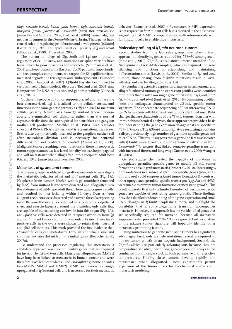
Fig. 1. Drosophila models of tumor metastasis caused by loss-of-function mutations in lgl or brat and l(3)mbt . The Drosophila larval brain is composed of
two hemispheres and the ventral nerve cord (VNC). Mutant tumorous tissue in these models is restricted to the outer proliferative center (OPC) and central brain
(CB) regions. (A)Neoplastic brain tumors caused by mutations in either lgl or brat can be engineered to express a reporter gene (lacZ ; orange). Larval brains from
these lgl or brat mutant animals are quartered and transplanted into the abdomens of adult female flies. The transplanted tissue continues to proliferate in the
abdomen and, after several days, the ovaries of the host can be dissected and examined by immunofluorescence to detect labeled tumor cells that have
metastasized. Drosophila ovaries are encased within a peritoneal sheath. Each ovary consists of 15-20 ovarioles, which are surrounded by an epithelial sheath of
cells in addition to a muscle layer (blue) between two layers of extracellular matrix (green). Any lacZ -expressing cells found within the ovarioles must have
traversed these layers and are therefore considered to have undergone metastasis. (B) Modeling metastasis using tumors caused by loss-of-function mutations in
l(3)mbt . Temperature-sensitive mutations in l(3)mbt result in tissue overgrowth in the developing larval CNS when grown at restrictive temperatures (29 °C).
Tumor tissue (engineered to express GFP) can be dissected and transplanted into the abdomens of wild-type adult flies (allografts). These can be maintained and
multiple rounds of allografts can be performed by transplantation into other flies, thereby generating sufficient material for biochemical analysis (e.g. microarray
analysis, sequencing and western blotting). Comparison of the expression profiles of l(3)mbt and brat mutant tumors enabled an l(3)mbt -specific tumor
expression signature to be obtained (Janic et al., 2010).

8/3/2019 Modeling Tumor
http://slidepdf.com/reader/full/modeling-tumor 4/9
2003). All three of them genetically interact and cause overgrowthof RasV12 tissue when mutated. These studies also identifiedbazooka, stardust and cdc42 as factors that do not induceovergrowth when mutated singly but can strongly stimulateoncogenic Ras-mediated tumor growth and metastasis (Pagliariniand Xu, 2003). Each of these genes can also regulate cell polarity and E-cadherin expression. Crucially, although downregulation of E-cadherin is necessary for RasV12 ;scrib–/–-induced metastasis, itis not sufficient, implicating other contributing factors in metastaticgrowth.
The relationship between oncogenic Ras and JNK signalingSeveral Drosophila laboratories have highlighted the importanceof the oncogenic cooperation between Ras and JNK signaling.Constitutive activation of the Ras signaling pathway prevents fly cells from undergoing JNK-mediated apoptosis in response tocellular stresses (Brumby and Richardson, 2003; Brumby andRichardson, 2005). The Xu group has linked JNK upregulation tocell polarity and changes in E-cadherin expression in RasV12 ;scrib–/–
clones (Igaki et al., 2006). They also demonstrated thatoverexpression of negative regulators of the JNK pathway [using atransgene to express a dominant-negative form of Drosophila JNK
(encoded by the bsk gene)] prevents tumor formation andmetastasis (Igaki et al., 2006). The Bohmann laboratory has shownthat the invasion potential of RasV12 ;scrib–/– clones is dependenton the Fos-mediated transcriptional activation of mmp1downstream of JNK (Uhlirova and Bohmann, 2006). Expression of the MMP inhibitor, TIMP, or mmp1 RNA interference (RNAi)knockdown, was able to suppress cell invasiveness (Uhlirova andBohmann, 2006).
Recent studies of the sds22 gene in Drosophila have strengthenedthe idea that epithelial integrity and JNK signaling cooperate todrive the metastatic growth of RasV12 ;scrib–/– clones (Jiang et al.,2011). Sds22 is a conserved regulatory subunit of proteinphosphatase 1 (PP1), and acts as a regulator of epithelial polarity
and as a neoplastic tumor suppressor in Drosophila (Grusche etal., 2009). Loss of sds22 in RasV12 clones results in reduced epithelialintegrity, and the clones become invasive. Overexpression of Sds22in RasV12 ;scrib–/– cells largely suppresses their tumorigenic growth,and mechanistic studies suggest that Sds22-PP1 inhibits non-muscle myosin II and JNK activity ( Jiang et al., 2011). The authorsof this study also showed that human SDS22 is deleted ordownregulated in multiple types of carcinoma.
A recent study by the Richardson group identified key regulatorsof the actin cytoskeleton and cell morphology, including Rho1-
dmm.biologists.org4
Drosophila tumor invasion and metastasisPERSPECTIVE
A
Wild type Ras V12 clonesGFP positive
(benign overgrowth)
+ suppressingmutation
+ enhancing mutation + metastasis-promotingmutation
Ras V12 ;scrib –/ – clones
Genetic
screen
Invasion of GFP-positive cells into
VNC
Eye-
antennaldiscs
vergrowth andinvasion of VNC
B
Cepahliccomplex
Ras V12 scrib –/ –
scrib –/ –
Ras V12
FRT-FLP
clones
Interclonalcooperation
scrib –/ – clones eliminated
Overgrowth andinvasion
Benign overgrowth
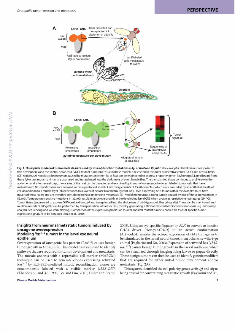
Fig. 2. Modeling tumor invasion and metastasis in
the Drosophila larval brain. (A)RasV12
overexpression clones generated by the MARCM
system in eye-antennal discs. These clones (marked
with GFP, and shown in green) produce a benign
overgrowth throughout the cephalic complex. When
RasV12 clones are also mutant for scrib, they become
tumorigenic and metastasize, as shown by invasion
of GFP-labeled cells into the ventral nerve cord(VNC). Genetic screens have identified modifiers of
the RasV12 phenotype [suppressor, JNK; enhancer,
large giant lethal (lgl ); metastatic enhancer, deep
orange (dor )] (Chi et al., 2010; Wu et al., 2010).
(B)Interclonal cooperation of RasV12 and scrib–/–
clones. As described in A, RasV12 overexpression
produces benign overgrowth. scrib–/– clones grow at
a reduced rate and are excluded via cell competition.
Adjacent RasV12 and scrib–/– clones cooperate to
overproliferate and undergo metastasis. The large
tumor and metastatic growth consists almost
exclusively of RasV12 cells, suggesting that scrib–/– cells
actively cooperate with RasV12 cells during the early
stages of tumor growth and metastasis (Wu et al.,
2010).
8/3/2019 Modeling Tumor
http://slidepdf.com/reader/full/modeling-tumor 5/9
family GTPases and RhoGEFs, as Ras-cooperating proteins(Brumby et al., 2011). The hyperplastic eye phenotype producedby driving UAS-RasV12 with ey-GAL4 was screened for modifiersusing a collection of P-element enhancer insertion lines bearingUAS promoter sequences. These lines result in overexpression of the gene adjacent to the insertion element in RasV12 cells. JNKpathway activation is crucial for the cooperation of these actin
cytoskeletal regulators with RasV12
. The relevance to human cancerof the collaboration between oncogenic Ras and JNK wasdemonstrated in this study by correlating JNK signaling with theupregulation of Ras in breast cancers (Brumby et al., 2011).
The role of JNK signaling in promoting metastatic behaviorseems to be context specific. In contrast to the promotion of Ras-induced metastasis by JNK, analysis of lgl –/– clones suggests thatthe activity of Diap1 (a caspase inhibitor) and JNK loss are essentialfor invasion of mutant cells out of the clone (Grzeschik et al., 2010a;Grzeschik et al., 2010b). Although Diap1 expression is sufficientto inhibit widespread apoptosis, cells at the boundary of lgl –/– clonesundergo cell death in a JNK-dependent manner. By blocking theJNK signaling cascade, apoptosis of these cells is specifically inhibited, stimulating de-differentiation and cellular invasion.
Lysosome dysfunction in RasV12 tumor growth andinvasionRecent studies from the Xu laboratory have used genetic screensto identify mutations that promote RasV12 cell metastasis (Fig. 2A).The authors tested 3119 ethyl-methanesulfonate-mutagenized lines(on the X chromosome) for their capacity to modify RasV12 larvaltumors. This screen identified 516 suppressors and 351 enhancersof tumor growth, of which 23 enhanced tumor growth as well asmetastasis (Chi et al., 2010). Two of these metastasis-promotingmutations occurred within the class C vacuolar protein sorting(VPS) complex member deep orange (dor ). By testing mutations ingenes encoding other components of this lysosome complex,
namely carnation and vps16A, they confirmed that loss of lysosomeactivity stimulates metastatic growth of the RasV12 tissue (Chi etal., 2010). Characterization of other modifiers from this screenmight provide a clearer understanding of the mechanismsunderlying Ras-mediated tumor growth in Drosophila. This study also confirmed their genetic findings: feeding chemical inhibitorsof lysosomal function to larvae with RasV12 overgrowths promotedtumor development and metastasis. This work illustrates thepotential of Drosophila to screen for therapeutic cancer drugs in vivo, a system that might one day provide a cheap and rapid meansfor drug discovery.
Interclonal cooperation between RasV12 and scrib mutantcellsExciting recent work using the RasV12- scrib–/– system hasdemonstrated a non-cell-autonomous effect between neighboringclones (called interclonal cooperation; Fig. 2B). Adjacent RasV12
clones and scrib–/– clones actively cooperate to form tumors andcan metastasize (Wu et al., 2010). Interclonal tumors and metastaticgrowths are smaller than those produced from a single
RasV12 ;scrib–/– clone but still represent a significant tumor burdento the hosts (Fig. 2B). During later stages of tumor development,these interclonal tumors are made up almost exclusively of RasV12-expressing cells, suggesting that the scrib–/– mutant cells are only
required for the initial stages of tumor formation and metastasis(Wu et al., 2010). These studies also identified the JAK-STATsignaling pathway as being a crucial downstream target of JNKactivity, implicating JNK and JAK-STAT as important oncogenicdrivers for Ras-mediated tumor growth (Wu et al., 2010). Inagreement with these findings, the authors went on to demonstratethat JNK activation after wounding is also able to promote the over-
growth of RasV12
cells. This study potentially forges a link betweentissue damage and Ras-stimulated JNK activity – a situationresembling the chronic inflammation that has been reported tocontribute to tumorigenesis in humans (Mantovani, 2010). Otherrecent Drosophila studies have also highlighted the role of theimmune response in tumor growth (Box 1).
Screening for regulators of invasive growth caused byactivation of the Notch pathwayThe Notch signaling cascade was originally identified as animportant regulator of proliferation and differentiation in flies.Extensive genetic and biochemical studies have identified andcharacterized the components and regulators of this pathway (Artavanis-Tsakonas and Muskavitch, 2010). Drosophila studies
have revealed that, similar to oncogenic Ras, Notch signalingcooperates with JNK to promote dysregulation of epithelial integrity (Brumby and Richardson, 2003; Leong et al., 2009). Aberrant Notchsignaling is associated with several human cancers, including skin,breast, lung and ovarian cancer (for a review, see Allenspach et al.,2002). Tumors containing amplifications of genes encoding Notchsignaling components (e.g. Notch and Jagged) tend to be highly aggressive and metastatic.
Overexpression of the Notch activator Delta (Dl) using theeyeless-GAL4 driver (ey-GAL4>UAS-Dl ) stimulates a non-metastatic overproliferation of eye tissue. Flies treated with a -secretase inhibitor to prevent Notch receptor proteolysis showedcomplete rescue of the ey-GAL4>UAS-Dl phenotype (Palomero et
al., 2007). This model has been employed by the Dominguez groupto screen for modifiers of this phenotype using a library of P-element UAS insertion lines that result in gene overexpression
Disease Models & Mechanisms 5
Drosophila tumor invasion and metastasis PERSPECTIVE
Box 1. The role of the immune system in tumorgrowth inDrosophilaSeveral recent reports suggest that the immune system plays a crucial role in
Drosophila tumor models, as is the case in mammalian tumors. Circulating
blood cells, known in Drosophila as hemocytes, are part of the fly immune
system and have been found to associate with RasV12;scrib–/– tumors and to
reduce tumor growth in scrib–/– animals (Pastor-Pareja et al., 2008). The
Drosophila genome encodes a single member of the tumor necrosis factor
(TNF) family, named Eiger (Egr). Egr has been shown to be required for the
JNK-dependent cell death of scrib or dlg clonal tissue (Igaki et al., 2009). In the
absence of egr , these mutant clones grow aggressively and develop intotumors (Igaki et al., 2009). By contrast, another study showed that loss of egr in
RasV12 ;scrib–/– tumors prevented invasive overgrowth, which was correlated
with reduced JNK activation and a failure to express MMP1 (Cordero et al.,
2010). Therefore, in the presence of oncogenic Ras, Egr seems to play a role as
a tumor promoter. This study also revealed that Egr is produced in the
hemocytes associated with RasV12 ;scrib–/– tumors (Cordero et al., 2010).
Together, these Drosophila models might provide an excellent parallel to
mammalian tumors, in which TNF is produced in both tumor cells and
associated immune cells, where it has been shown to have both oncogenic
and tumor-suppressive roles (Balkwill, 2009).
8/3/2019 Modeling Tumor
http://slidepdf.com/reader/full/modeling-tumor 6/9
(Ferres-Marco et al., 2006). One of these lines, eyeful , strongly promoted the metastatic growth of ey-GAL4>UAS-Dl eye tissue,resulting in secondary eye growths throughout the body. Using various tissue-specificGAL4 drivers, the authors demonstrated thatco-expression of Dl and eyeful could stimulate massive overgrowthand metastatic invasion in multiple tissue settings. The eyeful construct was mapped within the divergently transcribed genes of
longitudinals lacking (lola) and pipsqueak ( psq ), which produce amyriad of alternatively spliced BTB (BR-C, ttk and bab) proteins,which are required for the recruitment of histone deacetylases andPolycomb complexes to promoter regions. Further biochemicalstudies demonstrated that silencing of Retinoblastoma (Rbf)expression via promoter hypermethylation strongly contributed tothe metastatic phenotype (Ferres-Marco et al., 2006).
This screen also identified the P-element insertion GS1D233Cas an enhancer of the ey-GAL4>UAS-Dl phenotype. This insertionwas mapped to the Akt1 locus, which encodes an importantserine/tyrosine kinase linked to phosphatase and tensin homolog(PTEN) and Notch signaling (Palomero et al., 2007). These resultssuggest that flies will provide a useful system to test new pharmacological reagents targeted against the Notch pathway.
The Hassan group used the ey-GAL4>UAS-Dl/eyeful phenotypeto screen for mutations that could further enhance metastaticgrowth (Bossuyt et al., 2009). This screen identified atonal (ato),a transcription factor required for retinal terminal differentiation,as a crucial tumor suppressor. Mutations affecting ato dramatically enhanced tumor burden and metastasis rates, and tumors displayedelevated levels of proliferation markers (such as phosphorylatedhistone H3). Conversely, overexpression of atonal upregulatedboth Decapo (also known as p21 cell-cycle inhibitor) andphosphorylated-JNK levels, inhibiting proliferation and inducingapoptosis. Furthermore, overexpressing dominant-negative JNK(bsk ) partially mimics ato downregulation in the eyeful (ey-GAL4>UAS-Dl/eyeful ) model, indicating that JNK signaling is
downstream of atonal and that atonal requires active JNK signalingto inhibit overgrowth (Bossuyt et al., 2009).
Modeling glioma in DrosophilaDeveloping Drosophila models of specific human tumor types islimited in many cases owing to the lack of directly homologousorgans (e.g. pancreas, liver and lung). However, recent studies havecapitalized on the similarity of mammalian and Drosophila glialcells to model glioblastoma in flies (Read et al., 2009; Witte et al.,2009). Glioblastomas are the most common tumors of the centralnervous system (CNS), and their rapid proliferation and malignancy result in poor patient prognosis.
Mutation or amplification of the gene encoding epidermalgrowth factor receptor (EGFR) tyrosine kinase, loss of PTEN [which
antagonizes the growth promoting effects of phosphoinositide 3-kinase (PI3K) signaling] or activating mutations in PI3K (Furnariet al., 2007) are genetic lesions that are commonly associated withgliomas. Consequently, the glial-specific Repo-GAL4 driver(incorporating the promoter reversed polarity) was used tosimultaneously express transgenes encoding constitutively activeEGFR and PI3K in larval glia. Co-activation of EGFR (or Ras) andPI3K in larval glia results in neoplasia, neurological defects andlethality (Read et al., 2009; Witte et al., 2009). The overproliferationand neoplastic transformation seems to be specific for glia, because
overexpression of EGFR and PI3K in neurons, neuroblasts or otherglial cells (such as oligodendrocyte-like neuropil glia and astrocyte-like cortex glia) failed to transform them.
The neoplastic glia induced in this Drosophila model mimic thehighly proliferative anaplastic glia from high-grade human gliomas.They ectopically express Cyclin-B (CycB), Cyclin-E (CycE) andMMP1, promoting cell cycle entry and invasive growth. These
studies demonstrated that glia expressing activated EGFR or Rasand PI3K invade into inappropriate areas of the brain, such as alongBolwig nerves, which are not normally accompanied by glial cells(Read et al., 2009; Witte et al., 2009).
Genetic experiments have revealed that the malignant neoplastictransformation of larval glia induced by EGFR and PI3K occurs viaa complex network of genetic factors that are commonly mutatedor activated in human gliomas. These downstream effectors includeTor, Myc, G1 cyclin-Cdk complexes (such as those including CyclinB or E) and the Rb-E2F pathway. Interestingly, pharmacologicalinhibitors, including compounds that are used to treat patients,rescued these phenotypes in larvae. An EGFR inhibitor (gefitinib)partially reduced the migration of EGFR- and PI3K-transformedglia, whereas a PI3K inhibitor (wortmannin) and an Akt inhibitor
(triciribine) completely prevented invasion (Witte et al., 2009).Together, these studies suggest that this Drosophila model is usefulfor deciphering the signaling cascades underlying the abnormalbehavior of glioma cells, including their metastatic potential.
Modeling tumor invasion in the epithelia of theDrosophila wing discRoles for both receptor and non-receptor tyrosine kinases in celltransformation and progression towards malignant phenotypes arewell established (Blume-Jensen and Hunter, 2001). The SRC family kinases (SFKs) are membrane-linked non-receptor tyrosine kinasesthat are required for regulating adhesion and cytoskeletonreorganization, cell cycle progression, and migration (Guarino,
2010; Thomas and Brugge, 1997). Elevated SRC levels have beenreported in a wide variety of human cancers, including those of the colon, liver, lung, breast and pancreas (Ishizawar and Parsons,2004; Summy and Gallick, 2003). SFK activity is inhibited by theC-terminal SRC kinase (CSK) family of tyrosine kinases [CSK andCSK homologous kinase (CHK)] (Chong et al., 2005); both CSKand CHK phosphorylate and inactivate SFKs, and mutationsdisrupting this activity have been implicated in a plethora of cancers. Elevated SRC levels (either by amplification of SRC or lossof CSK function) promote anchorage-independent cell growth,tumor cell invasion and metastasis (Guarino, 2010). Although SRCis a crucial regulator of epithelial-mesenchymal transition (EMT),the exact mechanism of how SRC promotes metastatic growthremains elusive.
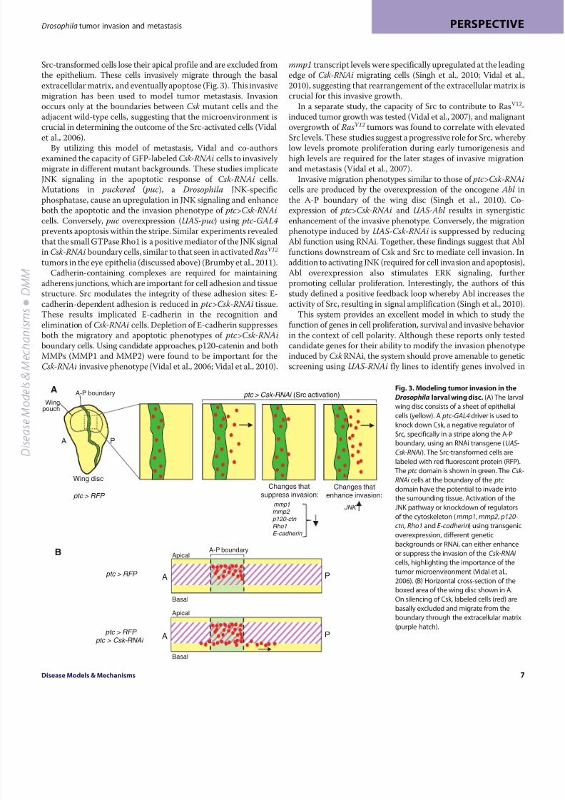
Recent studies from the Cagan group have utilized thepseudostratified epithelia of the Drosophila larval wing imaginaldisc to model invasive cell growth (Vidal et al., 2006). Globaldepletion of the sole Drosophila CSK/CHK ortholog, Csk, usingeither RNAi or Csk mutations, elevates active levels of Src, leadingto significant overgrowth in developing larvae. By contrast,targeting Csk depletion to a discrete stripe along the anterior-posterior (A-P) boundary of the larval wing disc using a patched-GAL4 driver ( ptc-GAL4) to express a Csk RNAi transgene (UAS-Csk-RNAi) produces a metastatic phenotype (Vidal et al., 2006).
dmm.biologists.org6
Drosophila tumor invasion and metastasisPERSPECTIVE
8/3/2019 Modeling Tumor
http://slidepdf.com/reader/full/modeling-tumor 7/9
Src-transformed cells lose their apical profile and are excluded fromthe epithelium. These cells invasively migrate through the basalextracellular matrix, and eventually apoptose (Fig. 3). This invasivemigration has been used to model tumor metastasis. Invasionoccurs only at the boundaries between Csk mutant cells and theadjacent wild-type cells, suggesting that the microenvironment iscrucial in determining the outcome of the Src-activated cells (Vidal
et al., 2006).By utilizing this model of metastasis, Vidal and co-authors
examined the capacity of GFP-labeled Csk-RNAi cells to invasively migrate in different mutant backgrounds. These studies implicateJNK signaling in the apoptotic response of Csk - RNAi cells.Mutations in puckered ( puc), a Drosophila JNK-specificphosphatase, cause an upregulation in JNK signaling and enhanceboth the apoptotic and the invasion phenotype of ptc>Csk-RNAicells. Conversely, puc overexpression (UAS-puc) using ptc-GAL4prevents apoptosis within the stripe. Similar experiments revealedthat the small GTPase Rho1 is a positive mediator of the JNK signalin Csk-RNAi boundary cells, similar to that seen in activated RasV12
tumors in the eye epithelia (discussed above) (Brumby et al., 2011).Cadherin-containing complexes are required for maintaining
adherens junctions, which are important for cell adhesion and tissuestructure. Src modulates the integrity of these adhesion sites: E-cadherin-dependent adhesion is reduced in ptc>Csk-RNAi tissue.These results implicated E-cadherin in the recognition andelimination of Csk-RNAi cells. Depletion of E-cadherin suppressesboth the migratory and apoptotic phenotypes of ptc>Csk-RNAiboundary cells. Using candidate approaches, p120-catenin and bothMMPs (MMP1 and MMP2) were found to be important for theCsk-RNAi invasive phenotype (Vidal et al., 2006; Vidal et al., 2010).
mmp1 transcript levels were specifically upregulated at the leadingedge of Csk-RNAi migrating cells (Singh et al., 2010; Vidal et al.,2010), suggesting that rearrangement of the extracellular matrix iscrucial for this invasive growth.
In a separate study, the capacity of Src to contribute to RasV12-induced tumor growth was tested (Vidal et al., 2007), and malignantovergrowth of RasV12 tumors was found to correlate with elevated
Src levels. These studies suggest a progressive role for Src, whereby low levels promote proliferation during early tumorigenesis andhigh levels are required for the later stages of invasive migrationand metastasis (Vidal et al., 2007).
Invasive migration phenotypes similar to those of ptc>Csk-RNAicells are produced by the overexpression of the oncogene Abl inthe A-P boundary of the wing disc (Singh et al., 2010). Co-expression of ptc>Csk-RNAi and UAS-Abl results in synergisticenhancement of the invasive phenotype. Conversely, the migrationphenotype induced by UAS-Csk-RNAi is suppressed by reducingAbl function using RNAi. Together, these findings suggest that Ablfunctions downstream of Csk and Src to mediate cell invasion. Inaddition to activating JNK (required for cell invasion and apoptosis),Abl overexpression also stimulates ERK signaling, further
promoting cellular proliferation. Interestingly, the authors of thisstudy defined a positive feedback loop whereby Abl increases theactivity of Src, resulting in signal amplification (Singh et al., 2010).
This system provides an excellent model in which to study thefunction of genes in cell proliferation, survival and invasive behaviorin the context of cell polarity. Although these reports only testedcandidate genes for their ability to modify the invasion phenotypeinduced by Csk RNAi, the system should prove amenable to geneticscreening using UAS-RNAi fly lines to identify genes involved in
Disease Models & Mechanisms 7
Drosophila tumor invasion and metastasis PERSPECTIVE
A
ptc > RFP
Wing discChanges that
suppress invasion:
mmp1mmp2
p120-ctn Rho1E-cadherin
Changes thatenhance invasion:
JNK
B
ptc > RFP
ptc > RFP ptc > Csk-RNAi
A P
A P
A P
Apical
Basal
Apical
Basal
A-P boundary
Wingpouch
ptc > Csk-RNAi (Src activation)
A-P boundary
Fig. 3. Modeling tumor invasion in the
Drosophila larval wing disc. (A) The larval
wing disc consists of a sheet of epithelial
cells (yellow). A ptc-GAL4 driver is used to
knock down Csk, a negative regulator of
Src, specifically in a stripe along the A-P
boundary, using an RNAi transgene (UAS-
Csk-RNAi ). The Src-transformed cells are
labeled with red fluorescent protein (RFP).
The ptc domain is shown in green. The Csk-
RNAi cells at the boundary of the ptc
domain have the potential to invade into
the surrounding tissue. Activation of the
JNK pathway or knockdown of regulators
of the cytoskeleton (mmp1, mmp2, p120-
ctn, Rho1 and E-cadherin) using transgenic
overexpression, different genetic
backgrounds or RNAi, can either enhance
or suppress the invasion of the Csk-RNAi
cells, highlighting the importance of thetumor microenvironment (Vidal et al.,
2006). (B) Horizontal cross-section of the
boxed area of the wing disc shown in A.
On silencing of Csk, labeled cells (red) are
basally excluded and migrate from the
boundary through the extracellular matrix
(purple hatch).

8/3/2019 Modeling Tumor
http://slidepdf.com/reader/full/modeling-tumor 8/9
metastasis. In addition, the identification of the factors thatcollaborate to control cell migration suggests possible approachesfor dual therapeutic targeting in combating metastasis in variouscancers.
Conclusions, limitations and perspectivesThis review has highlighted the many advantages of Drosophila as
a model for studying tumor progression. A streamlined genome,coupled with powerful genetic tools, provides a unique system inwhich to explore the mechanisms regulating the metastatic growthof tumor cells. However, as with any model organism, it has severallimitations that should be considered. In mammals, malignant cellsundergoing metastasis enter a local blood or lymph vessel beforecolonizing a distant tissue and forming secondary tumors. This isdifficult to model in Drosophila because flies have rudimentary hematopoietic systems and a dramatically different lymphaticsystem compared with mammals. In addition, the metastaticpotential of tumors induced in Drosophila is greatly reducedcompared with their mammalian counterparts; tumor cells tend toinvade the local surrounding tissue distally (e.g. in the cephaliccomplex in the case of RasV12 overexpression).
Tumor formation is generally stimulated by a single mutationor activated oncogene expression in Drosophila. However, as theexamples outlined here demonstrate, Drosophila has also provedto be a powerful experimental system for examining ‘two-hit’models of tumor overgrowth and invasion. Although the Drosophilamodels discussed here are largely limited to tumors that developin the larval stage, adult flies can be employed to examine themetastatic potential of these tumors using allografts, providing anideal way to propagate tumors for extended studies.
The systems of modeling tumor development in Drosophilacapitalize on the unique advantages of this model organism coupledwith recent innovative technologies (see Box 2). Undoubtedly, themajor strength of Drosophila is the ease of conducting large-scale
genetic screens, which can now make use of the large publicly available collections of isogenic deficiencies and RNAi lines thatcover the entire genome (Dietzl et al., 2007; Parks et al., 2004).Elegant targeting techniques – including the UAS/GAL4 system,FLP-FRT-mediated recombination and MARCM clonal analysis –enable gene knockdown in specific tissues or patches of cells,bypassing issues of organism lethality. Because these cancer models
involve an in vivo setting, they are also ideally suited for studies
that examine the role of the microenvironment. In addition,
Drosophila offers the potential for drug screening. Together, these
approaches make Drosophila an excellent model organism to
elucidate the basic mechanisms governing tumorigenesis and
tumor progression. Validation of this research in mammalian
cancer models and human cancer cell lines could lead to new
insights into tumor invasion. Metastasis modeling in Drosophilais in its infancy but, as better tools and models of specific tumor
types are developed, it offers the potential to probe the basic
mechanisms regulating cancer cell invasion.
FUNDING
Work in the authors’ laboratories is supported by the National Institutes of Health
(NIH) [R01GM53202] to N.J.D.; and the Department of Defense (DOD) [W81XWH-09-1-0487] to J.A.W.
COMPETING INTERESTS
The authors declare that they do not have any competing or financial interests.
REFERENCES
Allenspach, E. J., Maillard, I., Aster, J. C. and Pear, W. S. (2002). Notch signaling incancer. Cancer Biol. Ther. 1, 466-476.
Arama, E., Dickman, D., Kimchie, Z., Shearn, A. and Lev, Z. (2000). Mutations in the
beta-propeller domain of the Drosophila brain tumor (brat) protein induce neoplasmin the larval brain. Oncogene 19, 3706-3716.
Artavanis-Tsakonas, S. and Muskavitch, M. A. (2010). Notch: the past, the present,
and the future. Curr. Top. Dev. Biol. 92, 1-29.
Balkwill, F. (2009). Tumour necrosis factor and cancer. Nat. Rev. Cancer 9, 361-371.Beaucher, M., Goodliffe, J., Hersperger, E., Trunova, S., Frydman, H. and Shearn, A.
(2007a). Drosophila brain tumor metastases express both neuronal and glial cell typemarkers. Dev. Biol. 301, 287-297.
Beaucher, M., Hersperger, E., Page-McCaw, A. and Shearn, A. (2007b). Metastaticability of Drosophila tumors depends on MMP activity. Dev. Biol. 303, 625-634.
Bilder, D., Li, M. and Perrimon, N. (2000). Cooperative regulation of cell polarity andgrowth by Drosophila tumor suppressors. Science 289, 113-116.
Blume-Jensen, P. and Hunter, T. (2001). Oncogenic kinase signalling. Nature 411, 355-365.
Boccuni, P., MacGrogan, D., Scandura, J. M. and Nimer, S. D. (2003). The human
L(3)MBT polycomb group protein is a transcriptional repressor and interactsphysically and functionally with TEL (ETV6). J. Biol. Chem. 278, 5412-5420.
Bossuyt, W., De Geest, N., Aerts, S., Leenaerts, I., Marynen, P. and Hassan, B. A.(2009). The atonal proneural transcription factor links differentiation and tumor
formation in Drosophila . PLoS Biol. 7, e40.Brumby, A. M. and Richardson, H. E. (2003). scribble mutants cooperate with
oncogenic Ras or Notch to cause neoplastic overgrowth in Drosophila . EMBO J. 22,5769-5779.
Brumby, A. M. and Richardson, H. E. (2005). Using Drosophila melanogaster to maphuman cancer pathways. Nat. Rev. Cancer 5, 626-639.
Brumby, A. M., Goulding, K. R., Schlosser, T., Loi, S., Galea, R., Khoo, P., Bolden, J.E., Aigaki, T., Humbert, P. O. and Richardson, H. E. (2011). Identification of novel
ras-cooperating oncogenes in Drosophila melanogaster : a RhoGEF/Rho-family/JNK
pathway is a central driver of tumorigenesis. Genetics 188, 105-125.Chambers, A. F., Groom, A. C. and MacDonald, I. C. (2002). Dissemination and
growth of cancer cells in metastatic sites. Nat. Rev. Cancer 2, 563-572.Chi, C., Zhu, H., Han, M., Zhuang, Y., Wu, X. and Xu, T. (2010). Disruption of lysosome
function promotes tumor growth and metastasis in Drosophila. J. Biol. Chem. 285,
21817-21823.
Chong, Y. P., Mulhern, T. D. and Cheng, H. C. (2005). C-terminal Src kinase (CSK) andCSK-homologous kinase (CHK)-endogenous negative regulators of Src-family proteinkinases. Growth Factors 23, 233-244.
Cordero, J. B., Macagno, J. P., Stefanatos, R. K., Strathdee, K. E., Cagan, R. L. and
Vidal, M. (2010). Oncogenic Ras diverts a host TNF tumor suppressor activity intotumor promoter. Dev. Cell 18, 999-1011.
Curran, S. P., Wu, X., Riedel, C. G. and Ruvkun, G. (2009). A soma-to-germlinetransformation in long-lived Caenorhabditis elegans mutants. Nature 459, 1079-1084.
Dietzl, G., Chen, D., Schnorrer, F., Su, K. C., Barinova, Y., Fellner, M., Gasser, B.,Kinsey, K., Oppel, S., Scheiblauer, S. et al. (2007). A genome-wide transgenic RNAi
library for conditional gene inactivation in Drosophila . Nature 448, 151-156.Elliott, D. A. and Brand, A. H. (2008). The GAL4 system: a versatile system for the
expression of genes. Methods Mol. Biol. 420, 79-95.
dmm.biologists.org8
Drosophila tumor invasion and metastasisPERSPECTIVE
Box 2. Advantages of Drosophila for modeling tumorinvasion and metastasis• The signaling pathways controlling growth, differentiation and development
that are involved in tumorigenesis and tumor progression are largely
conserved between Drosophila and humans.
• Models of tumor formation and cell invasion have been created in
Drosophila using a wide variety of gene targeting strategies, such as loss-of-function mutations and tissue-specific RNAi knockdown, as well as transgenic
overexpression of activated oncogenes found in human cancers.
• The ability to generate clones that are mutant for specific genes juxtaposed
with wild-type cells using the FLP-FRT and MARCM systems allows the
genetics of the tumor microenvironment required for invasion and metastasis
to be examined.
• Genome-wide screens using either de novo mutagenesis or tissue-specific
knockdown by RNAi in Drosophila can identify genes with previously
unidentified roles in cancer progression.
• Drosophila tumor models can be used for pharmacological screening.
8/3/2019 Modeling Tumor
http://slidepdf.com/reader/full/modeling-tumor 9/9
Ferres-Marco, D., Gutierrez-Garcia, I., Vallejo, D. M., Bolivar, J., Gutierrez-Avino, F. J. and Dominguez, M. (2006). Epigenetic silencers and Notch collaborate topromote malignant tumours by Rb silencing. Nature 439, 430-436.
Froldi, F., Ziosi, M., Tomba, G., Parisi, F., Garoia, F., Pession, A. and Grifoni, D.(2008). Drosophila lethal giant larvae neoplastic mutant as a genetic tool for cancermodeling. Curr. Genomics 9, 147-154.
Furnari, F. B., Fenton, T., Bachoo, R. M., Mukasa, A., Stommel, J. M., Stegh, A.,Hahn, W. C., Ligon, K. L., Louis, D. N., Brennan, C. et al. (2007). Malignantastrocytic glioma: genetics, biology, and paths to treatment. Genes Dev. 21, 2683-
2710.Gateff, E. (1978). Malignant neoplasms of genetic origin in Drosophila melanogaster .Science 200, 1448-1459.
Gateff, E., Loffler, T. and Wismar, J. (1993). A temperature-sensitive brain tumorsuppressor mutation of Drosophila melanogaster : developmental studies andmolecular localization of the gene. Mech. Dev. 41, 15-31.
Grusche, F. A., Hidalgo, C., Fletcher, G., Sung, H. H., Sahai, E. and Thompson, B. J.(2009). Sds22, a PP1 phosphatase regulatory subunit, regulates epithelial cell polarityand shape [Sds22 in epithelial morphology]. BMC Dev. Biol. 9, 14.
Grzeschik, N. A., Parsons, L. M., Allott, M. L., Harvey, K. F. and Richardson, H. E.(2010a). Lgl, aPKC, and Crumbs regulate the Salvador/Warts/Hippo pathway throughtwo distinct mechanisms. Curr. Biol. 20, 573-581.
Grzeschik, N. A., Parsons, L. M. and Richardson, H. E. (2010b). Lgl, the SWH pathwayand tumorigenesis: it’s a matter of context & competition! Cell Cycle 9, 3202-3212.
Guarino, M. (2010). Src signaling in cancer invasion. J. Cell. Physiol. 223, 14-26.Gupta, G. P. and Massague, J. (2006). Cancer metastasis: building a framework. Cell
127, 679-695.Gurvich, N., Perna, F., Farina, A., Voza, F., Menendez, S., Hurwitz, J. and Nimer, S.
D. (2010). L3MBTL1 polycomb protein, a candidate tumor suppressor in del(20q12)myeloid disorders, is essential for genome stability. Proc. Natl. Acad. Sci. USA 107,22552-22557.
Hanahan, D. and Weinberg, R. A. (2011). Hallmarks of cancer: the next generation.Cell 144, 646-674.
Handa, K., Yugawa, T., Narisawa-Saito, M., Ohno, S., Fujita, M. and Kiyono, T.(2007). E6AP-dependent degradation of DLG4/PSD95 by high-risk humanpapillomavirus type 18 E6 protein. J. Virol. 81, 1379-1389.
Humbert, P., Russell, S. and Richardson, H. (2003). Dlg, Scribble and Lgl in cellpolarity, cell proliferation and cancer. BioEssays 25, 542-553.
Igaki, T., Pagliarini, R. A. and Xu, T. (2006). Loss of cell polarity drives tumor growthand invasion through JNK activation in Drosophila. Curr. Biol. 16, 1139-1146.
Igaki, T., Pastor-Pareja, J. C., Aonuma, H., Miura, M. and Xu, T. (2009). Intrinsictumor suppression and epithelial maintenance by endocytic activation of Eiger/TNFsignaling in Drosophila . Dev. Cell 16, 458-465.
Ishizawar, R. and Parsons, S. J. (2004). c-Src and cooperating partners in humancancer. Cancer Cell 6, 209-214.
Janic, A., Mendizabal, L., Llamazares, S., Rossell, D. and Gonzalez, C. (2010). Ectopicexpression of germline genes drives malignant brain tumor growth in Drosophila.Science 330, 1824-1827.
Januschke, J. and Gonzalez, C. (2008). Drosophila asymmetric division, polarity andcancer. Oncogene 27, 6994-7002.
Jiang, Y., Scott, K. L., Kwak, S. J., Chen, R. and Mardon, G. (2011). Sds22/PP1 linksepithelial integrity and tumor suppression via regulation of myosin II and JNK signaling. Oncogene 30, 3248-3260.
Lee, T. and Luo, L. (2001). Mosaic analysis with a repressible cell marker (MARCM) forDrosophila neural development. Trends Neurosci. 24, 251-254.
Leong, G. R., Goulding, K. R., Amin, N., Richardson, H. E. and Brumby, A. M. (2009).Scribble mutants promote aPKC and JNK-dependent epithelial neoplasiaindependently of Crumbs. BMC Biol. 7, 62.
Lewis, P. W., Beall, E. L., Fleischer, T. C., Georlette, D., Link, A. J. and Botchan, M. R.(2004). Identification of a Drosophila Myb-E2F2/RBF transcriptional repressorcomplex. Genes Dev. 18, 2929-2940.
Lu, X., Feng, X., Man, X., Yang, G., Tang, L., Du, D., Zhang, F., Yuan, H., Huang, Q.,
Zhang, Z. et al. (2009). Aberrant splicing of Hugl-1 is associated with hepatocellularcarcinoma progression. Clin. Cancer Res. 15, 3287-3296.
Mantovani, A. (2010). Molecular pathways linking inflammation and cancer. Curr. Mol.
Med. 10, 369-373.
Mechler, B. M., McGinnis, W. and Gehring, W. J. (1985). Molecular cloning of
lethal(2)giant larvae, a recessive oncogene of Drosophila melanogaster . EMBO J. 4,
1551-1557.
Nakagawa, S. and Huibregtse, J. M. (2000). Human scribble (Vartul) is targeted for
ubiquitin-mediated degradation by the high-risk papillomavirus E6 proteins and the
E6AP ubiquitin-protein ligase. Mol. Cell. Biol. 20, 8244-8253.
Nguyen, D. X., Bos, P. D. and Massague, J. (2009). Metastasis: from dissemination to
organ-specific colonization. Nat. Rev. Cancer 9, 274-284.Pagliarini, R. A. and Xu, T. (2003). A genetic screen in Drosophila for metastatic
behavior. Science 302, 1227-1231.
Palomero, T., Sulis, M. L., Cortina, M., Real, P. J., Barnes, K., Ciofani, M., Caparros,
E., Buteau, J., Brown, K., Perkins, S. L. et al. (2007). Mutational loss of PTEN
induces resistance to NOTCH1 inhibition in T-cell leukemia. Nat. Med. 13, 1203-1210.
Parks, A. L., Cook, K. R., Belvin, M., Dompe, N. A., Fawcett, R., Huppert, K., Tan, L.
R., Winter, C. G., Bogart, K. P., Deal, J. E. et al. (2004). Systematic generation of
high-resolution deletion coverage of the Drosophila melanogaster genome. Nat.
Genet. 36, 288-292.
Pastor-Pareja, J. C., Wu, M. and Xu. T. (2008). An innate immune response of blood
cells to tumors and tissue damage in Drosophila. Dis. Model Mech. 1, 144-154.
Read, R. D., Cavenee, W. K., Furnari, F. B. and Thomas, J. B. (2009). A Drosophila
model for EGFR-Ras and PI3K-dependent human glioma. PLoS Genet. 5, e1000374.
Schimanski, C. C., Schmitz, G., Kashyap, A., Bosserhoff, A. K., Bataille, F., Schafer,
S. C., Lehr, H. A., Berger, M. R., Galle, P. R., Strand, S. et al. (2005). Reduced
expression of Hugl-1, the human homologue of Drosophila tumour suppressor gene
lgl, contributes to progression of colorectal cancer. Oncogene 24, 3100-3109.Singh, J., Aaronson, S. A. and Mlodzik, M. (2010). Drosophila Abelson kinase
mediates cell invasion and proliferation through two distinct MAPK pathways.
Oncogene 29, 4033-4045.
Summy, J. M. and Gallick, G. E. (2003). Src family kinases in tumor progression and
metastasis. Cancer Metastasis Rev. 22, 337-358.
Theodosiou, N. A. and Xu, T. (1998). Use of FLP/FRT system to study Drosophila
development. Methods 14, 355-365.
Thomas, S. M. and Brugge, J. S. (1997). Cellular functions regulated by Src family
kinases. Annu. Rev. Cell Dev. Biol. 13, 513-609.
Uhlirova, M. and Bohmann, D. (2006). JNK- and Fos-regulated Mmp1 expression
cooperates with Ras to induce invasive tumors in Drosophila . EMBO J. 25, 5294-5304.
Vidal, M. and Cagan, R. L. (2006). Drosophila models for cancer research. Curr. Opin.
Genet. Dev. 16, 10-16.
Vidal, M., Larson, D. E. and Cagan, R. L. (2006). Csk-deficient boundary cells are
eliminated from normal Drosophila epithelia by exclusion, migration, and apoptosis.Dev. Cell 10, 33-44.
Vidal, M., Warner, S., Read, R. and Cagan, R. L. (2007). Differing Src signaling levels
have distinct outcomes in Drosophila . Cancer Res. 67, 10278-10285.
Vidal, M., Salavaggione, L., Ylagan, L., Wilkins, M., Watson, M., Weilbaecher, K.
and Cagan, R. (2010). A role for the epithelial microenvironment at tumor
boundaries: evidence from Drosophila and human squamous cell carcinomas. Am. J.
Pathol. 176, 3007-3014.
Wang, D., Kennedy, S., Conte, D., Jr, Kim, J. K., Gabel, H. W., Kamath, R. S., Mello, C.
C. and Ruvkun, G. (2005). Somatic misexpression of germline P granules and
enhanced RNA interference in retinoblastoma pathway mutants. Nature 436, 593-
597.
Witte, H. T., Jeibmann, A., Klambt, C. and Paulus, W. (2009). Modeling glioma
growth and invasion in Drosophila melanogaster . Neoplasia 11, 882-888.
Woods, D. F. and Bryant, P. J. (1989). Molecular cloning of the lethal(1)discs large-1
oncogene of Drosophila . Dev. Biol. 134, 222-235.
Wu, M., Pastor-Pareja, J. C. and Xu, T. (2010). Interaction between Ras(V12) and
scribbled clones induces tumour growth and invasion. Nature 463, 545-548.
Yang, J. and Weinberg, R. A. (2008). Epithelial-mesenchymal transition: at thecrossroads of development and tumor metastasis. Dev. Cell 14, 818-829.
Disease Models & Mechanisms 9
Drosophila tumor invasion and metastasis PERSPECTIVE