Metabolites of the Nitric Oxide (NO) Pathway Are Altered...
14
Research Article Metabolites of the Nitric Oxide (NO) Pathway Are Altered and Indicative of Reduced NO and Arginine Bioavailability in Patients with Cardiometabolic Diseases Complicated with Chronic Wounds of Lower Extremities: Targeted Metabolomics Approach (LC-MS/MS) Małgorzata Krzystek-Korpacka , 1 Jerzy Wiśniewski, 1 Mariusz G. Fleszar , 1,2 Iwona Bednarz-Misa , 1 Agnieszka Bronowicka-Szydełko, 1 Małgorzata Gacka, 3 Leszek Masłowski, 4 Krzysztof Kędzior, 1 Wojciech Witkiewicz, 5,6 and Andrzej Gamian 1 1 Department of Medical Biochemistry, Wroclaw Medical University, Wroclaw 50-368, Poland 2 PORT Polski Ośrodek Rozwoju Technologii sp, ZOO, Wroclaw 54-066, Poland 3 Department of Angiology, Hypertension and Diabetes, Wroclaw Medical University, Wroclaw 50-556, Poland 4 Department of Angiology, Regional Specialist Hospital, Wroclaw 51-124, Poland 5 Department of Vascular Surgery, Regional Specialist Hospital, Wroclaw 51-124, Poland 6 Research and Development Centre, Regional Specialist Hospital, Wroclaw 51-124, Poland Correspondence should be addressed to Małgorzata Krzystek-Korpacka; [email protected] Received 3 May 2019; Accepted 26 June 2019; Published 14 July 2019 Guest Editor: Aneta Radziwon-Balicka Copyright © 2019 Małgorzata Krzystek-Korpacka et al. This is an open access article distributed under the Creative Commons Attribution License, which permits unrestricted use, distribution, and reproduction in any medium, provided the original work is properly cited. Objective. The status of metabolites of the nitric oxide (NO) pathway in patients with chronic wounds in the course of cardiometabolic diseases is largely unknown. Yet arginine supplementation and citrulline supplementation as novel therapeutic modalities aimed at increasing NO are tested. Material and Methods. Targeted metabolomics approach (LC-MS/MS) was applied to determine the concentrations of L-arginine, L-citrulline, asymmetric and symmetric dimethylarginines (ADMA and SDMA), and arginine/ADMA and arginine/SDMA ratios as surrogate markers of NO and arginine availability in ulnar and femoral veins, representing systemic and local levels of metabolites, in patients with chronic wounds in the course of cardiometabolic diseases (n = 59) as compared to patients without chronic wounds but with similar cardiometabolic burden (n = 55) and healthy individuals (n = 88). Results. Patients with chronic wounds had significantly lower systemic L-citrulline and higher ADMA and SDMA concentrations and lower L-arginine/ADMA and L-arginine/SDMA as compared to healthy controls. The presence of chronic wounds in patients with cardiometabolic diseases was associated with decreased L-arginine but with increased L-citrulline, ADMA, and SDMA concentrations and decreased L-arginine/ADMA and L-arginine/SDMA. Serum obtained from the ulnar and femoral veins of patients with chronic wounds differed by L-arginine concentrations and L-arginine/SDMA ratio, both lower in the femoral vein. Wound etiology affected L-citrulline and SDMA concentrations, lower and higher, respectively, in patients with venous stasis, and the L-arginine/SDMA ratio—lower in venous stasis. The wound type affected L-arginine/ADMA and citrulline—lower in patients with ulcerations or gangrene. IL-6 was an independent predictor of L-arginine/ADMA, VEGF-A of ADMA, G-CSF of L-arginine/SDMA, and GM-CSF of L-citrulline and SDMA. Conclusion. Chronic wounds in the course of cardiometabolic diseases are associated with reduced NO and arginine availability due to ADMA and SDMA accumulation rather than arginine deficiency, not supporting its supplementation. Wound character seems to affect NO bioavailability and wound etiology—arginine bioavailability. Arginine concentration and its availability are more markedly reduced at the local level than the systemic level. Hindawi Oxidative Medicine and Cellular Longevity Volume 2019, Article ID 5965721, 13 pages https://doi.org/10.1155/2019/5965721
Transcript of Metabolites of the Nitric Oxide (NO) Pathway Are Altered...
Research ArticleMetabolites of the Nitric Oxide (NO) Pathway Are Altered andIndicative of Reduced NO and Arginine Bioavailability inPatients with Cardiometabolic Diseases Complicated withChronic Wounds of Lower Extremities: Targeted MetabolomicsApproach (LC-MS/MS)
Małgorzata Krzystek-Korpacka ,1 Jerzy Wiśniewski,1 Mariusz G. Fleszar ,1,2
Iwona Bednarz-Misa ,1 Agnieszka Bronowicka-Szydełko,1 Małgorzata Gacka,3
Leszek Masłowski,4 Krzysztof Kędzior,1 Wojciech Witkiewicz,5,6 and Andrzej Gamian1
1Department of Medical Biochemistry, Wroclaw Medical University, Wroclaw 50-368, Poland2PORT Polski Ośrodek Rozwoju Technologii sp, ZOO, Wroclaw 54-066, Poland3Department of Angiology, Hypertension and Diabetes, Wroclaw Medical University, Wroclaw 50-556, Poland4Department of Angiology, Regional Specialist Hospital, Wroclaw 51-124, Poland5Department of Vascular Surgery, Regional Specialist Hospital, Wroclaw 51-124, Poland6Research and Development Centre, Regional Specialist Hospital, Wroclaw 51-124, Poland
Correspondence should be addressed to Małgorzata Krzystek-Korpacka; [email protected]
Received 3 May 2019; Accepted 26 June 2019; Published 14 July 2019
Guest Editor: Aneta Radziwon-Balicka
Copyright © 2019 Małgorzata Krzystek-Korpacka et al. This is an open access article distributed under the Creative CommonsAttribution License, which permits unrestricted use, distribution, and reproduction in any medium, provided the original workis properly cited.
Objective. The status of metabolites of the nitric oxide (NO) pathway in patients with chronic wounds in the course ofcardiometabolic diseases is largely unknown. Yet arginine supplementation and citrulline supplementation as novel therapeuticmodalities aimed at increasing NO are tested. Material and Methods. Targeted metabolomics approach (LC-MS/MS) wasapplied to determine the concentrations of L-arginine, L-citrulline, asymmetric and symmetric dimethylarginines (ADMA andSDMA), and arginine/ADMA and arginine/SDMA ratios as surrogate markers of NO and arginine availability in ulnar andfemoral veins, representing systemic and local levels of metabolites, in patients with chronic wounds in the course ofcardiometabolic diseases (n = 59) as compared to patients without chronic wounds but with similar cardiometabolic burden(n = 55) and healthy individuals (n = 88). Results. Patients with chronic wounds had significantly lower systemic L-citrulline andhigher ADMA and SDMA concentrations and lower L-arginine/ADMA and L-arginine/SDMA as compared to healthy controls.The presence of chronic wounds in patients with cardiometabolic diseases was associated with decreased L-arginine butwith increased L-citrulline, ADMA, and SDMA concentrations and decreased L-arginine/ADMA and L-arginine/SDMA.Serum obtained from the ulnar and femoral veins of patients with chronic wounds differed by L-arginine concentrations andL-arginine/SDMA ratio, both lower in the femoral vein. Wound etiology affected L-citrulline and SDMA concentrations, lowerand higher, respectively, in patients with venous stasis, and the L-arginine/SDMA ratio—lower in venous stasis. The woundtype affected L-arginine/ADMA and citrulline—lower in patients with ulcerations or gangrene. IL-6 was an independentpredictor of L-arginine/ADMA, VEGF-A of ADMA, G-CSF of L-arginine/SDMA, and GM-CSF of L-citrulline and SDMA.Conclusion. Chronic wounds in the course of cardiometabolic diseases are associated with reduced NO and arginineavailability due to ADMA and SDMA accumulation rather than arginine deficiency, not supporting its supplementation.Wound character seems to affect NO bioavailability and wound etiology—arginine bioavailability. Arginine concentration andits availability are more markedly reduced at the local level than the systemic level.
HindawiOxidative Medicine and Cellular LongevityVolume 2019, Article ID 5965721, 13 pageshttps://doi.org/10.1155/2019/5965721
1. Introduction
Sufficient synthesis and bioavailability of nitric oxide(NO)—a free radical and a key vasodilator—are crucialfor proper functioning of the vascular endothelium. Conse-quently, NO deficiency is a prerequisite for and a hallmarkof endothelial dysfunction, a pathology preceding thedevelopment of cardiovascular diseases (CVD) [1]. CVDand their main risk factors, such as obesity, hypertension,and type 2 diabetes mellitus (T2DM), are, in turn, amongkey factors negatively affecting proper wound healing [2].Nonhealing wounds constitute a serious problem foraffected people and a growing burden for public healthcare [3]. Currently, they affect 4.5 million people in theUnited States alone [4] but the incidence of chronicwounds is likely to increase along with estimated rise inthe incidence of CVD, obesity, and T2DM [5–8]. The over-all prevalence of peripheral artery disease (PAD) in Europeis estimated to be 5.3% but differs by country [9]. The dis-turbed blood flow and blood vessel damage accompanyingCVD and specifically PAD may result in ulcerations organgrene located in the lower extremities [9–11]. NO-releasing wound dressings as well as diet supplementationwith L-arginine, a NO substrate, are currently being evalu-ated as novel modalities in the treatment of chronicwounds [12, 13].
Successful healing requires spacial and temporal cooper-ation of a myriad of players, mediating three key phases ofthe process: inflammatory, proliferative, and remodelling[5]. Nonhealing wounds are believed to be locked in theinflammatory phase [14]. While most mediators are proteins,other molecules, such as NO, are recently gaining attention aspotentially relevant for all phases of the healing [15–19]. Theimportance of NO has been demonstrated by the delayedhealing of animals with genetically impaired NO synthesis[20, 21]. Moreover, it has been shown that NO therapy iseffective in healing ischemic [22] and diabetic ulcers [23] inexperimental animals by inducing reepithelization, angio-genesis, and collagen synthesis.
NO is synthesized by nitric oxide synthases (NOS) fromL-arginine, and L-citrulline is the other reaction product.There are three isoforms of the enzyme: constitutivelyexpressed endothelial (eNOS; NOS1) and neuronal (nNOS;NOS3) isoforms and the inducible isoform (iNOS; NOS2)[24]. The activity of NOS enzymes is regulated by methylatedderivatives of arginine, of which asymmetric dimethylargi-nine (ADMA) is believed to be a strong and symmetricdimethylarginine (SDMA) a weak enzyme inhibitor. BothADMA and SDMA compete with L-arginine for its trans-porters, and therefore, their accumulation decreases NOproduction by diminishing L-arginine availability for theNOS enzymes. The ADMA and SDMA pool is regulatedat the level of their synthesis, conducted by the protein argi-nine methyltransferases (PRMTs), and degradation. WhileADMA is mostly catabolized to L-citrulline and dimethyla-mine (DMA), by dimethylarginine dimethylaminohydro-lases (DDAHs), SDMA is preferentially excreted withurine. L-Citrulline may be used in the arginine-citrullinecycle to satisfy the body demand for L-arginine [24].
The gaseous nature, high diffusion capacity, and shorthalf-life make NO the ideal signalling molecule but causeits quantification to be a challenge. Therefore, relatively morestable products of NO oxidation, nitrites and nitrates, aremeasured instead of NO. Nitrate has even been proposed asa “wound healing biomarker and surrogate end point” fortreatment of diabetic foot ulcers [18]. Another approach forthe assessment of NO and its bioavailability is the evaluationof intermediates in the NO synthesis pathway and inhibitorsof NOS enzymes. Of those, the measurement of L-arginineand/or ADMA is the most popular.
Despite the relevance of NO for wound healing, the statusof its pathway metabolites and surrogate markers is largelyunknown. Recently, an elevation in serum ADMA [25] hasbeen reported in patients with chronic wounds while ourown preliminary research showed a decrease in serum L-arginine [26]. However, there seem to be a paucity of dataon SDMA and citrulline or regarding the possible associationbetween NO metabolites and wound etiology and the type ortheir interplay with inflammatory and immune mediators.Therefore, this study was designed to simultaneously evalu-ate a wider panel of L-arginine/NO pathway metabolitesusing targeted metabolomics approach and a novel assayrecently developed by our group [27]. We measured L-argi-nine, L-citrulline (referred to hereafter simply as arginineand citrulline), ADMA, and SDMA to determine their statusand clinical relevance in patients with chronic wounds of var-ious etiologies and types, at systemic and local levels.
2. Materials and Methods
2.1. Ethical Approval. The study protocol was approved bythe Medical Ethics Committees of Wroclaw Medical Univer-sity (#KB-384/2012) from April 12, 2012, and the study wasconducted in accordance with the Helsinki Declaration of1975, as revised in 1983, and an informed consent has beenobtained from all patients.
2.2. Patients. The study population consisted of 202 individ-uals: 114 patients with cardiovascular diseases and/or diabe-tes, of whom 59 had chronic wounds of lower extremities,and 88 healthy individuals. Patients were recruited fromamong patients of the Dept. of Angiology of the RegionalSpecialist Hospital and the Dept. of Angiology, Hypertensionand Diabetes of the Wroclaw Medical University. Concern-ing patients with nonhealing wounds, only those withwounds in the course of vascular disease and diabetes wereincluded while others, with wounds due to autoimmune dis-eases, malignancy, infections, or drugs, were not enrolled.Wound etiology was determined by the evaluation of woundcharacteristics (location and an appearance of the wound, itsborders and those of the surrounding skin, pain, and thepresence of bleeding on manipulation) in addition to thepatient’s history and clinical assessment, which was basedon the ankle-brachial pressure index, ultrasound, angiogra-phy, and computer tomography. The wound etiology wasas follows: venous stasis (n = 25), ischemic (arterial) (n = 17),and neurotrophic (n = 17). Patients’ wounds were mechani-cally cleaned of necrotic tissue and excess wound exudate,
2 Oxidative Medicine and Cellular Longevity
photographs were taken, and wound material was collectedfor bacteriological examination. Subsequently, wounds werewashed with antiseptic octenidine dihydrochloride anddressed using a sterile gauze. None of the patients used argi-nine supplements.
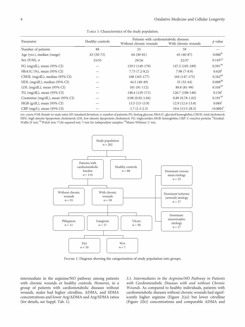
The control group consisted of healthy volunteersrecruited from hospital staff and blood donors from LowerSilesian Center of Blood Donation and Therapy, Wroclaw,Poland (recruited on the basis of standard eligibility criteriafor blood donation with an age > 50 yrs and an insignificantmedical history as additional inclusion criteria for the currentstudy). Table 1 presents detailed characteristics of the studypopulation, and the categorization into examined groups isdepicted in Figure 1.
In a subgroup of patients (n = 7), blood was sampledfrom the ulnar and femoral veins in order to compare sys-temic metabolite concentrations with those more local andcloser to the wound.
2.3. Analytical Methods. Blood (7.5mL), after an overnightfasting, was drawn into serum separator tubes from ulnarveins and, additionally, from femoral veins. Blood wasclotted for 30min and subsequently centrifuged (15min,10°C, 720×g). Collected serum was aliquoted and kept frozenat -80° until examination.
2.3.1. Materials. LC-MS-grade acetonitrile, water, andmetha-nol were purchased fromMerckMillipore (Warsaw, Poland).L-Arginine, SDMA, ADMA, L-citrulline, sodium tetraborate,benzoyl chloride (BCl), and HPLC-grade formic acid (FA)were obtained from Sigma-Aldrich (Poznan, Poland).Isotope-labeled asymmetric dimethylarginine (2,3,3,4,4,5,5-D7-ADMA, 98%) and L-arginine:HCl (D7-arginine, 98%)were acquired from Cambridge Isotope Laboratories (MA,USA). Leucine-enkephalin was purchased from Waters(Warsaw, Poland).
2.3.2. Quantitative Analysis of Metabolites Involved in NOSynthesis. Serum concentrations of metabolites involved inNO synthesis were measured by stable isotope dilution liquidchromatography tandemmass spectrometry using a Xevo G2quadrupole-TOF instrument (Waters, Milford, MA, USA) asdescribed in detail by Fleszar et al. [27]. Briefly, aliquots of100 μL of sera or the calibration sample, 10μL of internalstandard solution in water (100 μM D7-arginine and20 μM D7-ADMA), and 50μL of borate buffer (0.025MNa2B4O7·10H2O, 1.77mM NaOH, pH = 9 2) were trans-ferred into polypropylene microtubes and vortexed (1min,1100 rpm, 25°C). Then, 400 μL of acetonitrile and 10μL of10% BCl in acetonitrile were added and vortexed (5min,1100 rpm, 25°C). After derivatization, samples were centri-fuged (7min, 4°C, 15000 × g) and 100 μL of supernatantswas transferred into a chromatographic glass vial with300μL of water for LC-MS analysis.
2.3.3. LC-ESI-MS Analysis. The LC analysis was carried outon a nanoACQUITY UPLC System equipped with anACQUITY HSS T3 column (50 × 1 0mm, 1.75 μm) with a0.22μm membrane inline filter (Waters). The total run timeof the method was 10min with a flow rate of 80μL/min.
Mobile phase A consisted of 0.1% FA in water, while mobilephase B consisted of 0.1% FA in methanol. For ADMAand SDMA isomer separation, the following gradient wasapplied: 11% B for 0–1min, 11%–13% B for 1–2min,13%–60% B for 2–5min, 60%–90% B for 5–5.5min, 90%B for 5.5–6min, and 90%–11% B for 6–6.05min. Thesample injection volume was 2 μL.
Mass spectra for the compounds were acquired in a XevoG2 Q-TOF mass spectrometer (Waters) in positive ion modeelectrospray ionization (ESI). The MS operating conditionswere as follows: capillary voltage, 3000V; cone voltage,40V; source temperature, 120°C; cone gas flow, 85L/hour;desolvation temperature, 350°C; and desolvation gas flow,800 L/hour. Data acquisition was carried on MassLynx Soft-ware (Waters) using the following ions: 279.1457 m/z,286.1897 m/z, 307.1770 m/z, 314.2076 m/z, and 280.1136m/z for L-arginine, D7-arginine, ADMA and SDMA, D7-ADMA, and L-citrulline, respectively.
As previously described [27], the method is character-ized by intra- and interassay coefficients of variation of1.6% and 3.3% for arginine, 3.2% and 3.1% for citrulline,7.5% and 9.4% for ADMA, and 6.4% and 7.1% forSDMA determination.
2.4. Statistical Analysis.Data were tested for normality of dis-tribution using the Kolmogorov-Smirnoff test and for homo-geneity of variances using Levene’s test and presented asmeans or medians with 95% confidence interval (CI) aroundthem. Between-group differences in means or medians weretested using a t-test for independent samples withWelch cor-rection if appropriate or with the Mann-Whitney U test(two-group comparisons) and with a one-way ANOVA withthe Tukey-Kramer post hoc test or with the Kruskal-WallisHtest with the Conover post hoc test (multigroup compari-sons). Log-transformation was used if necessary to obtainnormality of distribution and/or homogeneity of variances.Additionally, a t-test for paired samples was used to analyzedifferences in metabolite concentrations between femoraland ulnar veins. Frequency analysis was conducted usingthe chi-squared test. Univariate correlations were examinedusing Pearson tests. Multivariate linear regression wasconducted to discern the independent predictor of NO-associated metabolites. Regression models were built witha stepwise method using the following criteria: enter vari-able if p < 0 05 and remove variable if p > 0 1. All calculatedprobabilities were two-tailed, and p values ≤ 0.05 were con-sidered statistically significant. The analyses were per-formed using MedCalc Statistical Software version 18.11.6(MedCalc Software bvba, Ostend, Belgium; https://www.medcalc.org; 2019).
3. Results
Both patients’ cohorts were well matched with respect to sexdistribution, age, and concentrations of biochemical indices,indicative of similar disease burden. There were, however,significant differences in HDL cholesterol and CRP lowerand higher, respectively, in patients with chronic wounds(Table 1). There were no sex-related differences in any
3Oxidative Medicine and Cellular Longevity
intermediate in the arginine/NO pathway among patientswith chronic wounds or healthy controls. However, in agroup of patients with cardiometabolic diseases withoutwounds, males had higher citrulline, ADMA, and SDMAconcentrations and lower Arg/ADMA and Arg/SDMA ratios(for details, see Suppl. Tab. 1).
3.1. Intermediates in the Arginine/NO Pathway in Patientswith Cardiometabolic Diseases with and without ChronicWounds. As compared to healthy individuals, patients withcardiometabolic diseases without chronic wounds had signif-icantly higher arginine (Figure 2(a)) but lower citrulline(Figure 2(b)) concentrations and comparable ADMA and
Table 1: Characteristics of the study population.
Parameter Healthy controlsPatients with cardiometabolic diseases
p valueWithout chronic wounds With chronic wounds
Number of patients 88 55 59 —
Age (yrs.), median (range) 63 (50-73) 64 (49-81) 65 (40-87) 0.086K
Sex (F/M), n 33/55 29/26 22/37 0.145χ2
FG (mg/dL), mean (95% CI) — 159.5 (149-179) 147.2 (105-189) 0.591W
HbA1C (%), mean (95% CI) — 7.73 (7.2-8.2) 7.98 (7-8.9) 0.620t
CHOL (mg/dL), median (95% CI) — 168 (163-177) 164 (147-175) 0.162M
HDL (mg/dL), median (95% CI) — 44.5 (40-49) 35 (32-44) 0.008M
LDL (mg/dL), mean (95% CI) — 101 (91-112) 89.8 (81-99) 0.104W
TG (mg/dL), mean (95% CI) — 148.4 (129-171) 126.7 (108-148) 0.156t
Creatinine (mg/dL), mean (95% CI) — 0.98 (0.92-1.04) 0.89 (0.78-1.02) 0.191W
HGB (g/dL), mean (95% CI) — 13.5 (13-13.9) 12.9 (12.4-13.8) 0.084t
CRP (mg/L), mean (95% CI) — 1.7 (1.3-2.3) 19.6 (13.5-28.5) <0.0001t
yrs.: years; F/M: female-to-male ratio; SD: standard deviation; n: number of patients; FG: fasting glucose; HbA1C: glycated hemoglobin; CHOL: total cholesterol;HDL: high-density lipoprotein cholesterol; LDL: low-density lipoprotein cholesterol; TG: triglycerides; HGB: hemoglobin; CRP: C-reactive protein; KKruskal-Wallis H test; WWelch test; χ2chi-squared test; tt-test for independent samples; MMann-Whitney U test.
Study populationn = 202
Patients withcardiometabolic
burdenn = 114
Healthy controlsn = 88
Without chronicwoundsn = 55
With chronicwoundsn = 59
Phlegmonn = 11
Gangrenen = 17
Ulcersn = 50
Dominant ischemic(arterial) etiology
n = 17
Dominant neurotrophic
etiologyn = 17
Dryn = 10
Wetn = 7
Dominant venousstasis etiology
n = 25
Figure 1: Diagram showing the categorization of study population into groups.
4 Oxidative Medicine and Cellular Longevity
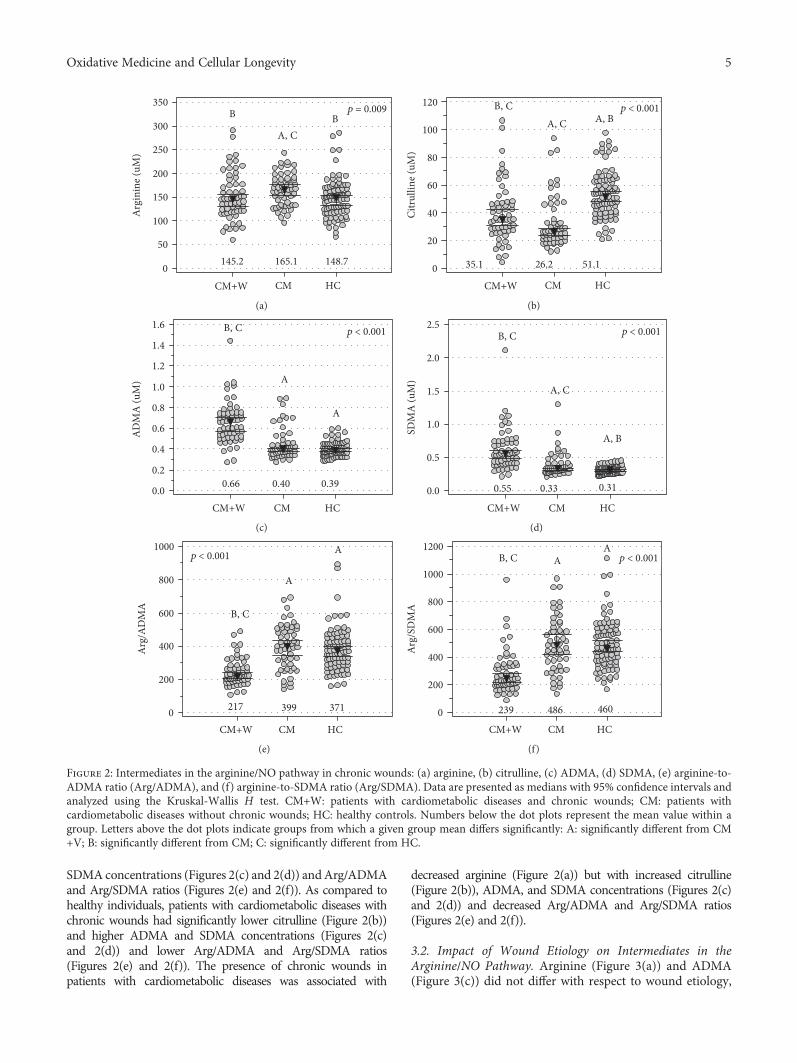
SDMA concentrations (Figures 2(c) and 2(d)) andArg/ADMAand Arg/SDMA ratios (Figures 2(e) and 2(f)). As compared tohealthy individuals, patients with cardiometabolic diseases withchronic wounds had significantly lower citrulline (Figure 2(b))and higher ADMA and SDMA concentrations (Figures 2(c)and 2(d)) and lower Arg/ADMA and Arg/SDMA ratios(Figures 2(e) and 2(f)). The presence of chronic wounds inpatients with cardiometabolic diseases was associated with
decreased arginine (Figure 2(a)) but with increased citrulline(Figure 2(b)), ADMA, and SDMA concentrations (Figures 2(c)and 2(d)) and decreased Arg/ADMA and Arg/SDMA ratios(Figures 2(e) and 2(f)).
3.2. Impact of Wound Etiology on Intermediates in theArginine/NO Pathway. Arginine (Figure 3(a)) and ADMA(Figure 3(c)) did not differ with respect to wound etiology,
0
50
100
150
200
250
300
350
Arg
inin
e (uM
)
CM+W CM HC
145.2 165.1 148.7
B
A, CB
p = 0.009
(a)
0
20
40
60
80
100
120
Citr
ullin
e (uM
)
p < 0.001
CM+W CM HC
35.1 26.2 51.1
B, C A, C A, B
(b)
0.0
0.2
0.4
0.6
0.8
1.0
1.2
1.4
1.6
AD
MA
(uM
)
CM+W CM HC
0.66 0.40 0.39
p < 0.001B, C
A
A
(c)
0.0
0.5
1.0
1.5
2.0
2.5
SDM
A (u
M)
CM+W CM HC
0.55 0.33 0.31
p < 0.001B, C
A, C
A, B
(d)
0
200
400
600
800
1000
Arg
/AD
MA
CM+W CM HC
217 399 371
p < 0.001
B, C
A
A
(e)
0
200
400
600
800
1000
1200
Arg
/SD
MA
CM+W CM HC
239 486 460
p < 0.001B, C AA
(f)
Figure 2: Intermediates in the arginine/NO pathway in chronic wounds: (a) arginine, (b) citrulline, (c) ADMA, (d) SDMA, (e) arginine-to-ADMA ratio (Arg/ADMA), and (f) arginine-to-SDMA ratio (Arg/SDMA). Data are presented as medians with 95% confidence intervals andanalyzed using the Kruskal-Wallis H test. CM+W: patients with cardiometabolic diseases and chronic wounds; CM: patients withcardiometabolic diseases without chronic wounds; HC: healthy controls. Numbers below the dot plots represent the mean value within agroup. Letters above the dot plots indicate groups from which a given group mean differs significantly: A: significantly different from CM+V; B: significantly different from CM; C: significantly different from HC.
5Oxidative Medicine and Cellular Longevity
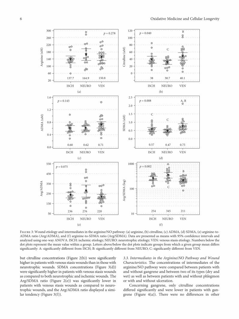
but citrulline concentrations (Figure 2(b)) were significantlyhigher in patients with venous stasis wounds than in those withneurotrophic wounds. SDMA concentrations (Figure 3(d))were significantly higher in patients with venous stasis woundsas compared to both neurotrophic and ischemic wounds. TheArg/SDMA ratio (Figure 2(e)) was significantly lower inpatients with venous stasis wounds as compared to neuro-trophic wounds, and the Arg/ADMA ratio displayed a simi-lar tendency (Figure 3(f)).
3.3. Intermediates in the Arginine/NO Pathway and WoundCharacteristics. The concentrations of intermediates of thearginine/NO pathway were compared between patients withand without gangrene and between two of its types (dry andwet) as well as between patients with and without phlegmonor with and without ulceration.
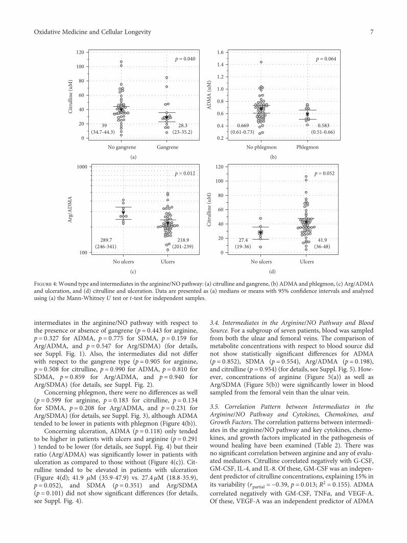
Concerning gangrene, only citrulline concentrationsdiffered significantly and were lower in patients with gan-grene (Figure 4(a)). There were no differences in other
20
60
100
140
180
220
260
300
Arg
inin
e (uM
)
ISCH NEURO VEN
p = 0.278
137.7 164.9 150.8
(a)
0
20
40
60
80
100
120
Citr
ullin
e (uM
)
ISCH NEURO VEN
Bp = 0.040
C
38 30.7 40.1
(b)
0.0
0.4
0.8
1.2
1.6
AD
MA
(uM
)
ISCH NEURO VEN
0.60 0.62 0.71
p = 0.143
(c)
0.0
0.5
1.0
1.5
2.0
2.5
SDM
A (u
M)
ISCH NEURO VEN
0.57
A, Bp = 0.008
CC
0.47 0.75
(d)
50
150
250
350
450
550
Arg
/AD
MA
ISCH NEURO VEN
p = 0.075
236 276 220
(e)
10
100
1000
Arg
/SD
MA
(log
)
ISCH NEURO VEN
p = 0.002
254 345 211
B
C
(f)
Figure 3: Wound etiology and intermediates in the arginine/NO pathway: (a) arginine, (b) citrulline, (c) ADMA, (d) SDMA, (e) arginine-to-ADMA ratio (Arg/ADMA), and (f) arginine-to-SDMA ratio (Arg/SDMA). Data are presented as means with 95% confidence intervals andanalyzed using one-way ANOVA. ISCH: ischemic etiology; NEURO: neurotrophic etiology; VEN: venous stasis etiology. Numbers below thedot plots represent the mean value within a group. Letters above/below the dot plots indicate groups from which a given group mean differssignificantly: A: significantly different from ISCH; B: significantly different from NEURO; C: significantly different from VEN.
6 Oxidative Medicine and Cellular Longevity
intermediates in the arginine/NO pathway with respect tothe presence or absence of gangrene (p = 0 443 for arginine,p = 0 327 for ADMA, p = 0 775 for SDMA, p = 0 159 forArg/ADMA, and p = 0 547 for Arg/SDMA) (for details,see Suppl. Fig. 1). Also, the intermediates did not differwith respect to the gangrene type (p = 0 905 for arginine,p = 0 508 for citrulline, p = 0 990 for ADMA, p = 0 810 forSDMA, p = 0 859 for Arg/ADMA, and p = 0 940 forArg/SDMA) (for details, see Suppl. Fig. 2).
Concerning phlegmon, there were no differences as well(p = 0 599 for arginine, p = 0 183 for citrulline, p = 0 134for SDMA, p = 0 208 for Arg/ADMA, and p = 0 231 forArg/SDMA) (for details, see Suppl. Fig. 3), although ADMAtended to be lower in patients with phlegmon (Figure 4(b)).
Concerning ulceration, ADMA (p = 0 118) only tendedto be higher in patients with ulcers and arginine (p = 0 291) tended to be lower (for details, see Suppl. Fig. 4) but theirratio (Arg/ADMA) was significantly lower in patients withulceration as compared to those without (Figure 4(c)). Cit-rulline tended to be elevated in patients with ulceration(Figure 4(d); 41.9 μM (35.9-47.9) vs. 27.4μM (18.8-35.9),p = 0 052), and SDMA (p = 0 351) and Arg/SDMA(p = 0 101) did not show significant differences (for details,see Suppl. Fig. 4).
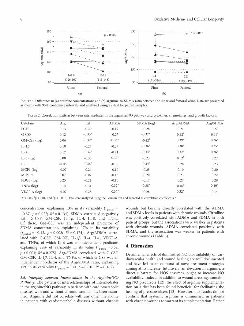
3.4. Intermediates in the Arginine/NO Pathway and BloodSource. For a subgroup of seven patients, blood was sampledfrom both the ulnar and femoral veins. The comparison ofmetabolite concentrations with respect to blood source didnot show statistically significant differences for ADMA(p = 0 852), SDMA (p = 0 554), Arg/ADMA (p = 0 198),and citrulline (p = 0 954) (for details, see Suppl. Fig. 5). How-ever, concentrations of arginine (Figure 5(a)) as well asArg/SDMA (Figure 5(b)) were significantly lower in bloodsampled from the femoral vein than the ulnar vein.
3.5. Correlation Pattern between Intermediates in theArginine/NO Pathway and Cytokines, Chemokines, andGrowth Factors. The correlation patterns between intermedi-ates in the arginine/NO pathway and key cytokines, chemo-kines, and growth factors implicated in the pathogenesis ofwound healing have been examined (Table 2). There wasno significant correlation between arginine and any of evalu-ated mediators. Citrulline correlated negatively with G-CSF,GM-CSF, IL-4, and IL-8. Of these, GM-CSF was an indepen-dent predictor of citrulline concentrations, explaining 15% inits variability (rpartial = −0 39, p = 0 013; R2 = 0 155). ADMAcorrelated negatively with GM-CSF, TNFα, and VEGF-A.Of these, VEGF-A was an independent predictor of ADMA
0
20
40
60
80
100
120
Citr
ullin
e (uM
)
No gangrene Gangrene
p = 0.040
39(34.7-44.3)
28.3(23-35.2)
(a)
0.2
0.4
0.6
0.8
1.0
1.2
1.4
1.6
AD
MA
(uM
)
No phlegmon Phlegmon
p = 0.064
0.669(0.61-0.73)
0.583(0.51-0.66)
(b)
100
1000
Arg
/AD
MA
No ulcers Ulcers
p = 0.012
289.7(246-341)
218.9(201-239)
(c)
0
20
40
60
80
100
120
Citr
ullin
e (uM
)
No ulcers Ulcers
p = 0.052
27.4(19-36)
41.9(36-48)
(d)
Figure 4: Wound type and intermediates in the arginine/NO pathway: (a) citrulline and gangrene, (b) ADMA and phlegmon, (c) Arg/ADMAand ulceration, and (d) citrulline and ulceration. Data are presented as (a) medians or means with 95% confidence intervals and analyzedusing (a) the Mann-Whitney U test or t-test for independent samples.
7Oxidative Medicine and Cellular Longevity
concentrations, explaining 13% in its variability (rpartial =−0 37, p = 0 022; R2 = 0 134). SDMA correlated negativelywith G-CSF, GM-CSF, IL-1β, IL-4, IL-8, and TNFα.Of these, GM-CSF was an independent predictor ofSDMA concentrations, explaining 17% in its variability(rpartial = −0 42, p = 0 008; R2 = 0 174). Arg/ADMA corre-lated with G-CSF, GM-CSF, IL-1β, IL-4, IL-6, VEGF-A,and TNFα, of which IL-6 was an independent predictor,explaining 28% of variability in its value (rpartial = 0 52,p < 0 001; R2 = 0 275). Arg/SDMA correlated with G-CSF,GM-CSF, IL-1β, IL-4, and TNFα, of which G-CSF was anindependent predictor of the Arg/SDMA ratio, explaining17% in its variability (rpartial = 0 41, p = 0 010; R2 = 0 167).
3.6. Interplay between Intermediates in the Arginine/NOPathway. The pattern of interrelationships of intermediatesin the arginine/NO pathway in patients with cardiometabolicdiseases with and without chronic wounds has been exam-ined. Arginine did not correlate with any other metabolitein patients with cardiometabolic diseases without chronic
wounds but became directly correlated with the ADMAand SDMA levels in patients with chronic wounds. Citrullinewas positively correlated with ADMA and SDMA in bothpatient groups, but the associations were weaker in patientswith chronic wounds. ADMA correlated positively withSDMA, and the association was weaker in patients withchronic wounds (Table 3).
4. Discussion
Detrimental effects of diminished NO bioavailability on car-diovascular health and wound healing are well documentedand have led to an outburst of novel treatment strategiesaiming at its increase. Intuitively, an elevation in arginine, adirect substrate for NOS enzymes, ought to increase NOavailability. Indeed, in addition to wound dressings contain-ing NO precursors [12], the effect of arginine supplementa-tion on a diet has been found beneficial for facilitating thehealing of pressure ulcers [13]. However, our results do notconfirm that systemic arginine is diminished in patientswith chronic wounds to warrant its supplementation. Rather
80
100
120
140
160
180
Arg
inin
e (uM
)
Ulnar Femoral
142.8(126-160)
130.9(113-148)
p = 0.005
(a)
50
150
250
350
450
Arg
/SD
MA
Ulnar Femoral
243(171-344)
226(160-319)
p = 0.037
(b)
Figure 5: Difference in (a) arginine concentrations and (b) arginine-to-SDMA ratio between the ulnar and femoral veins. Data are presentedas means with 95% confidence intervals and analyzed using a t-test for paired samples.
Table 2: Correlation pattern between intermediates in the arginine/NO pathway and cytokines, chemokines, and growth factors.
Cytokine Arg Cit ADMA SDMA (log) Arg/ADMA Arg/SDMA
FGF2 0.13 -0.29 -0.17 -0.28 0.21 0.27
G-CSF 0.12 -0.35∗ -0.27 -0.37∗ 0.42‡ 0.41‡
GM-CSF (log) 0.06 -0.39∗ -0.36∗ -0.42‡ 0.39∗ 0.36∗
IL-1β 0.10 -0.27 -0.27 -0.36∗ 0.38∗ 0.35∗
IL-4 0.17 -0.32∗ -0.21 -0.34∗ 0.32∗ 0.36∗
IL-6 (log) 0.08 -0.30 -0.39∗ -0.23 0.52† 0.27
IL-8 -0.06 -0.36∗ -0.30 -0.34∗ 0.28 0.23
MCP1 (log) -0.07 -0.24 -0.10 -0.25 0.10 0.20
MIP-1α 0.07 -0.07 -0.16 -0.20 0.23 0.22
PDGF (log) 0.25 -0.21 -0.10 -0.17 0.27 0.28
TNFα (log) 0.14 -0.31 -0.32∗ -0.38∗ 0.46‡ 0.40∗
VEGF-A (log) -0.07 -0.28 -0.37∗ -0.28 0.32∗ 0.14∗p ≤ 0 05, ‡p < 0 01, and †p < 0 001. Data were analyzed using the Pearson test and reported as correlation coefficients r.
8 Oxidative Medicine and Cellular Longevity
unexpectedly, they were increased in patients with cardio-metabolic burden but without chronic wounds. This observa-tion, however, agrees well with previously reported increasedrisk for CVD in patients with elevated arginine, independentfrom traditional risk factors [28]. Still, we also demonstratedthat arginine concentrations in the blood from the femoralvein, draining the wounded leg, are significantly lower imply-ing that arginine might indeed be depleted locally. Moreover,also the Arg/SDMA ratio was markedly reduced in the fem-oral vein than in the ulnar vein, indicative of more severelocal arginine depletion.
Nonetheless, it has been suggested that an elevation insystemic or local arginine may not directly translate intoincreased NO concentrations, a phenomenon explained byimpaired arginine availability for NO synthesis by NOSenzymes. At least several mechanisms are in operation, thatis, concomitant upregulation of NOS inhibitors, NOSuncoupling due to oxidative stress and tetrahydrobiopterindeficiency, and arginine utilization by an upregulated argi-nase [29]. Indeed, corroborating the first mechanism, weshowed that patients with chronic wounds had increasedboth ADMA and SDMA concentrations as compared tohealthy individuals and patients with cardiometabolic dis-eases without chronic wounds. Elevated ADMA increasescardiovascular risk [30] and is associated with every diseasewithin the CVD spectrum as well as with CVD risk factors[31]. Mechanistically, ADMA interferes with NO synthesisby inhibiting NOS enzymes and by reducing arginine avail-ability by competing for its membrane transporters. Addi-tionally, it impairs NO signalling by inhibiting eNOSphosphorylation [32]. Also, El-Mesallamy et al. [25] demon-strated that ADMA was elevated in patients with leg ulcerssignificantly more so than in T2DM patients without neu-ropathy. SDMA negatively affects arginine availability byinhibiting its membrane transport as well but has not gainedas much attention as ADMA as it is only a weak NOS inhib-itor. There seems to be a paucity of information on SDMA inchronic wounds in the course of cardiometabolic diseases. Inturn, data linking SDMA with a risk of CVD and CVD-caused mortality derived from meta-analyses are contradic-tory [30, 33]. However, individual studies indicate SDMAelevation to be an independent predictor of CVD-relatedmortality [34, 35] and to predict renal and cardiovascularoutcomes in patients with chronic kidney disease [36]. Fur-thermore, functional studies have shown SDMA to abolish
anti-inflammatory and antiatherogenic properties of HDL.Consequently, SDMA has been claimed to be a marker ofHDL dysfunction [34].
We also observed that due to ADMA and SDMA accu-mulation, both Arg/ADMA and Arg/SDMA ratios weresignificantly decreased in patients with chronic wounds, indi-cating reduced availability of NO and arginine, respectively.Wound character seemed to have an impact on NO bioavail-ability since the Arg/ADMA ratio was markedly reduced inpatients with ulcerations. In turn, wound etiology affectedarginine bioavailability as patients with venous stasis hadmarkedly elevated SDMA and decreased Arg/SDMA. TheADMA and SDMA pool is regulated mainly by the rates oftheir synthesis by type I (ADMA) and type II (SDMA)PRMTs and the rate of their degradation by DDAH enzymes(ADMA) and renal excretion [24]. Impaired renal secretiondoes not seem to explain the phenomenon of their accumu-lation in full as cardiometabolic burden, including chronickidney disease, was similar in patients with and withoutchronic wounds. There is a paucity of data regarding PRMTand DDAH activity and expression in metabolic disordersand in chronic wounds. However, limited animal studieshave linked PRMT overexpression with obesity, nonalcoholicfatty liver disease, and diabetic retinopathy [37, 38] andDDAH downregulation with diabetic retinopathy [38] andimpaired vascular homeostasis [39]. PRMT and DDAHmight be altered more strongly among patients with chronicwounds than without as they are positively (PRMTs) andnegatively (DDAHs) affected by inflammatory mediators[31, 40] and, as we have previously demonstrated, chronicwounds are accompanied by systemic elevation in proinflam-matory cytokines [41].
An interesting observation is the difference betweenSDMA, which was elevated in patients with cardiometabolicburden as compared to controls, and ADMA, which was not.It might result from differences in sensitivity of distinctPRMT enzymes. It has been shown that ADMA-yieldingPRMT2 expression is inhibited by high glucose concentra-tion, a common occurrence among our patients, which, inturn, upregulates SDMA synthesis owing to substrate scav-enging by type II PRMTs [37].
A direct link between arginine and NO concentrations isfurther disturbed by NOS uncoupling [29]. The lack of argi-nine [42], high ADMA concentrations [43], inflammationand oxidative stress [44], and tetrahydrobiopterin deficiency
Table 3: Correlation pattern between intermediates in the arginine/NO pathway in patients with cardiometabolic diseases with and withoutchronic wounds.
Metabolite Arginine Citrulline ADMA SDMA (log) Arg/ADMA Arg/SDMA
Arginine — -0.14 -0.15 0.23 — —
Citrulline 0.20 — 0.81† 0.85† -0.62† -0.63†
ADMA 0.45† 0.69† — 0.84† — —
SDMA (log) 0.28∗ 0.63† 0.68† — — —
Arg/ADMA — -0.35‡ — — —
Arg/SDMA — -0.36‡ — — — —
The right side of the table (in italics) presents correlations in patients without wounds; the left side (in a straight script) presents correlations in patients withchronic wounds. ∗p ≤ 0 05, ‡p < 0 01, and †p < 0 001. Data were analyzed using the Pearson test and reported as correlation coefficients r.
9Oxidative Medicine and Cellular Longevity
[45] are considered main culprits of switching enzymeactivity from NO synthesis to the production of superoxideanion. As such, the uncoupling of NOS leads not only tothe concomitant decrease in NO availability but also to theexacerbation of oxidative stress. Furthermore, NOS enzymescompete for arginine with arginases, the enzymes convertingarginine to ornithine. Correspondingly, the upregulatedARG1 expression has been reported in patients with diabeticfoot and venous ulcers [46] as well as in chronic wounds inrecessive dystrophic epidermolysis bullosa [47].
Data regarding citrulline association with chronicwounds in the course of cardiometabolic diseases are miss-ing, and those on citrulline in underlying conditions seemto be contradictory. Elevated citrulline has been associatedwith an increased CVD risk [28] and has a negative impacton arginine bioavailability in obesity and T2DM [48]. How-ever, it is also an effective antioxidant [49], argued to be moreeffective in restoring cardiometabolic health via increasingNO availability than arginine [49]. In addition to improvingendothelial function via NO-associated mechanisms, citrul-line supplementation in patients with vasospastic anginahas been shown to reduce the concentrations of oxidizedLDL thus alleviating oxidative stress [50]. Citrulline in ourstudy was diminished in patients with cardiometabolic bur-den as compared to healthy individuals and strongly andpositively correlated with NOS inhibitors—ADMA andSDMA. Considering that citrulline is a second reaction prod-uct and an effective arginine precursor, these findings seem toconfirm reduced rates of NO synthesis among patients withcardiometabolic burden and especially among patients withgangrene. Still, citrulline was less diminished in patients withchronic wounds than those without, particularly in case ofwounds of venous stasis etiology. Also, in line with its nega-tive impact on arginine availability, it negatively and stronglycorrelated with the Arg/SDMA ratio. Debats et al. [51], inturn, found citrulline to be elevated exclusively in patientswith infected chronic wounds but not with noninfected oracute wounds.
Inflammatory cytokines are among the initiators of endo-thelial dysfunction [44] and key players in sustaining inflam-mation in chronic wounds. Among others, they induce theexpression of iNOS [16] but inhibit that of eNOS [52, 53]and contribute to ADMA accumulation [31, 40]. Recently,we have demonstrated that chronic wounds are accompaniedby systemic elevation of IL-1β, IL-4, IL-6, IL-8, FGF-2, MIP-1α, PDGF-BB, and VEGF-A [41]. Also, markedly elevatedCRP and reduced HDL in patients with chronic woundscompared to those with similar cardiometabolic burden areindicative of a higher grade of inflammation in the former.Among our patients, ADMA concentrations were indepen-dently and inversely associated with VEGF-A, indicative ofthe negative impact of ADMA accumulation and resultingdiminished NO synthesis on angiogenesis in patients withchronic wounds. We also observed that the higher inflamma-tory response, indicated by IL-6 concentration, the lower theNO availability, indicated by the reduced Arg/ADMA ratio.We found that GM-CSF was an independent predictor forcitrulline and SDMA and G-CSF for Arg/SDMA. G-CSFand GM-CSF are hematopoietic cytokines displaying immu-
nomodulatory and antibiotic-enhancing activities with aproven beneficial effect on wound healing [54–56]. Theirconcentrations are reduced in patients with chronic wounds[41]. Park et al. [57] demonstrated that G-CSF exerts a pro-tective effect on endothelial cells via stimulating eNOSexpression and phosphorylation and thus enhancing NOsynthesis and signalling. The close relation between G-CSFand Arg/SDMA observed here may indicate an additionalmechanism, that is, increased arginine availability.
5. Conclusions
Taken together, our results demonstrate that patients withchronic wounds in the course of cardiometabolic diseaseshave reduced bioavailability of NO and its substrate, argi-nine, resulting from ADMA and SDMA accumulationrather than from arginine deficiency. Citrulline, in turn, isdecreased in patients with cardiometabolic diseases in gen-eral, but the presence of chronic wounds is associated withits elevation, reflecting degree of ADMA, and SDMA accu-mulation and inversely related to NO and arginine bioavail-ability. As such, our findings do not support arginine orcitrulline supplementation in patients with chronic woundsand rather suggest the need for treatment aiming at decreas-ing ADMA and SDMA concentrations.
Data Availability
The raw data used to support the findings of this study areavailable from the corresponding author upon reasonablerequest.
Conflicts of Interest
The authors declare that there is no conflict of interestregarding the publication of this paper.
Authors’ Contributions
Małgorzata Krzystek-Korpacka and Jerzy Wiśniewski con-tributed equally.
Acknowledgments
The research was supported by statutory funding fromWroclaw Medical University (#ST-911).
Supplementary Materials
Supplementary 1. Supplementary Table 1. Effect of sex onintermediates in the arginine/NO pathway among controlsand patients with cardiometabolic burden without or withchronic wounds.
Supplementary 2. Supplementary Figure 1. The association ofthe gangrene presence with intermediates in the arginine/NOpathway.
Supplementary 3. Supplementary Figure 2. The associationof the gangrene type with intermediates in the arginine/NOpathway.
10 Oxidative Medicine and Cellular Longevity
Supplementary 4. Supplementary Figure 3. The association ofthe phlegmon presence with intermediates in the argini-ne/NO pathway.
Supplementary 5. Supplementary Figure 4. The association ofulcers with intermediates in the arginine/NO pathway.
Supplementary 6. Supplementary Figure 5. Difference inintermediates in the arginine/NO pathway between ulnarand femoral veins.
References
[1] J. Barthelmes, M. P. Nägele, V. Ludovici, F. Ruschitzka,I. Sudano, and A. J. Flammer, “Endothelial dysfunction in car-diovascular disease and Flammer syndrome-similarities anddifferences,” EPMA Journal, vol. 8, no. 2, pp. 99–109, 2017.
[2] E. Avishai, K. Yeghiazaryan, and O. Golubnitschaja, “Impairedwound healing: facts and hypotheses for multi-professionalconsiderations in predictive, preventive and personalised med-icine,” EPMA Journal, vol. 8, no. 1, pp. 23–33, 2017.
[3] M. Olsson, K. Järbrink, U. Divakar et al., “The humanistic andeconomic burden of chronic wounds: a systematic review,”Wound Repair and Regeneration, vol. 27, no. 1, pp. 114–125,2019.
[4] R. E. Jones, D. S. Foster, and M. T. Longaker, “Management ofchronic wounds-2018,” JAMA, vol. 320, no. 14, pp. 1481-1482,2018.
[5] G. Han and R. Ceilley, “Chronic wound healing: a review ofcurrent management and treatments,” Advances in Therapy,vol. 34, no. 3, pp. 599–610, 2017.
[6] C.M.Hales,M. D. Carroll, C. D. Fryar, and C. L. Ogden, ““Prev-alence of obesity among adults and youth: United States, 2015–2016,”NCHS Data Brief,” no. 288, pp. 1–8, 2017, October 2017,https://www.cdc.gov/nchs/data/databriefs/db288.pdf.
[7] Y. Zheng, S. H. Ley, and F. B. Hu, “Global aetiology andepidemiology of type 2 diabetes mellitus and its complica-tions,” Nature Reviews Endocrinology, vol. 14, no. 2, pp. 88–98, 2018.
[8] T. R. Einarson, A. Acs, C. Ludwig, and U. H. Panton, “Preva-lence of cardiovascular disease in type 2 diabetes: a systematicliterature review of scientific evidence from across the world in2007–2017,” Cardiovascular Diabetology, vol. 17, no. 1, pp. 83–101, 2018.
[9] D.-M. Olinic, M. Spinu, M. Olinic et al., “Epidemiology ofperipheral artery disease in Europe: VAS Educational Paper,”International Angiology, vol. 37, no. 4, pp. 327–334, 2018.
[10] R. Rayner, K. Carville, J. Keaton, J. Prentice, andN. Santamaria, “Leg ulcers: atypical presentations and associ-ated comorbidities,” Wound Practice and Research, vol. 17,pp. 168–185, 2017.
[11] J. Green, R. Jester, R. McKinley, and A. Pooler, “The impact ofchronic venous leg ulcers: a systematic review,” Journal ofWound Care, vol. 23, no. 12, pp. 601–612, 2014.
[12] J. Durão, N. Vale, S. Gomes, P. Gomes, C. C. Barrias, andL. Gales, “Nitric oxide release from antimicrobial peptidehydrogels for wound healing,” Biomolecules, vol. 9, no. 1,p. 4, 2019.
[13] J. C. L. Neyens, E. Cereda, E. P. Meijer, C. Lindholm, and J. M.G. A. Schols, “Arginine-enriched oral nutritional supplemen-tation in the treatment of pressure ulcers: a literature review,”Wound Medicine, vol. 16, pp. 46–51, 2017.
[14] S. Guo and L. A. DiPietro, “Factors affecting wound heal-ing,” Journal of Dental Research, vol. 89, no. 3, pp. 219–229, 2010.
[15] J. D. Luo and A. F. Chen, “Nitric oxide: a newly discoveredfunction on wound healing,” Acta Pharmacologica Sinica,vol. 26, no. 3, pp. 259–264, 2005.
[16] P. Tripathi, P. Tripathi, L. Kashyap, and V. Singh, “The roleof nitric oxide in inflammatory reactions,” FEMS Immunol-ogy & Medical Microbiology, vol. 51, no. 3, pp. 443–452,2007.
[17] C. Napoli, G. Paolisso, A. Casamassimi et al., “Effects of nitricoxide on cell proliferation: novel insights,” Journal of theAmerican College of Cardiology, vol. 62, no. 2, pp. 89–95,2013.
[18] J. V. Boykin Jr., “Wound nitric oxide bioactivity,” Journal ofWound, Ostomy and Continence Nursing, vol. 37, no. 1,pp. 25–32, 2010.
[19] W. T. Weng, C. S. Wu, F. S. Wang et al., “α-Melanocyte-stim-ulating hormone attenuates neovascularization by inducingnitric oxide deficiency via MC-Rs/PKA/NF-κB signaling,”International Journal of Molecular Sciences, vol. 19, no. 12,p. 3823, 2018.
[20] K. Yamasaki, H. D. Edington, C. McClosky et al., “Reversal ofimpaired wound repair in iNOS-deficient mice by topicaladenoviral-mediated iNOS gene transfer,” Journal of ClinicalInvestigation, vol. 101, no. 5, pp. 967–971, 1998.
[21] P. C. Lee, A. N. Salyapongse, G. A. Bragdon et al.,“Impaired wound healing and angiogenesis in eNOS-deficient mice,” American Journal of Physiology-Heart andCirculatory Physiology, vol. 277, no. 4, pp. H1600–H1608,1999.
[22] M. Jones, J. G. Ganopolsky, A. Labbé et al., “Novel nitric oxideproducing probiotic wound healing patch: preparation andin vivo analysis in a New Zealand white rabbit model of isch-aemic and infected wounds,” International Wound Journal,vol. 9, no. 3, pp. 330–343, 2012.
[23] K. S. B. Masters, S. J. Leibovich, P. Belem, J. L. West, and L. A.Poole-Warren, “Effects of nitric oxide releasing poly(vinylalcohol) hydrogel dressings on dermal wound healing in dia-betic mice,” Wound Repair and Regeneration, vol. 10, no. 5,pp. 286–294, 2002.
[24] R. Keshet and A. Erez, “Arginine and the metabolic regula-tion of nitric oxide synthesis in cancer,” Disease Models &Mechanisms, vol. 11, no. 8, pp. dmm033332–dmm033341,2018.
[25] H. O. El-Mesallamy, N. M. Hamdy, O. A. Ezzat, and A. M.Reda, “Levels of soluble advanced glycation end product-receptors and other soluble serum markers as indicators ofdiabetic neuropathy in the foot,” Journal of Investigative Med-icine, vol. 59, no. 8, pp. 1233–1238, 2011.
[26] J. Wiśniewski, M. G. Fleszar, J. Piechowicz et al., “A novel massspectrometry-based method for simultaneous determinationof asymmetric and symmetric dimethylarginine, l-arginineand l-citrulline optimized for LC-MS-TOF and LC-MS/MS,”Biomedical Chromatography, vol. 31, no. 11, article e3994,2017.
[27] M. G. Fleszar, J. Wiśniewski, M. Krzystek-Korpacka et al.,“Quantitative analysis of L-arginine, dimethylated argininederivatives, L-citrulline, and dimethylamine in human serumusing liquid chromatography-mass spectrometric method,”Chromatographia, vol. 81, no. 6, pp. 911–921, 2018.
11Oxidative Medicine and Cellular Longevity
[28] M. Magnusson, T. Wang, B. Hedblad et al., “High levels ofarginine, citrulline and ADMA are independent predictorsof cardiovascular disease,” European Heart Journal, vol. 34,article P5687, Supplement 1, 2013.
[29] Y. C. Luiking, G. A. M. Ten Have, R. R. Wolfe, and N. E. P.Deutz, “Arginine de novo and nitric oxide production indisease states,” American Journal of Physiology Endocrinol-ogy and Metabolism, vol. 303, no. 10, pp. E1177–E1189,2012.
[30] P. Willeit, D. F. Freitag, J. A. Laukkanen et al., “Asymmetricdimethylarginine and cardiovascular risk: systematic reviewand meta-analysis of 22 prospective studies,” Journal of theAmerican Heart Association, vol. 4, no. 6, article e001833,2015.
[31] G. Bouras, S. Deftereos, D. Tousoulis et al., “Asymmetricdimethylarginine (ADMA): a promising biomarker for cardio-vascular disease?,” Current Topics in Medicinal Chemistry,vol. 13, no. 2, pp. 180–200, 2013.
[32] H. Kajimoto, H. Kai, H. Aoki et al., “Inhibition of eNOS phos-phorylation mediates endothelial dysfunction in renal failure:new effect of asymmetric dimethylarginine,” Kidney Interna-tional, vol. 81, no. 8, pp. 762–768, 2012.
[33] S. Schlesinger, S. R. Sonntag, W. Lieb, and R. Maas, “Asym-metric and symmetric dimethylarginine as risk markers fortotal mortality and cardiovascular outcomes: a systematicreview and meta-analysis of prospective studies,” PLoS ONE,vol. 11, no. 11, article e0165811, 2016.
[34] S. Zewinger, M. E. Kleber, L. Rohrer et al., “Symmetricdimethylarginine, high-density lipoproteins and cardiovascu-lar disease,” European Heart Journal, vol. 38, no. 20,pp. 1597–1607, 2017.
[35] P. Jud, F. Hafner, N. Verheyen et al., “Homoarginine/ADMAratio and homoarginine/SDMA ratio as independent predic-tors of cardiovascular mortality and cardiovascular events inlower extremity arterial disease,” Scientific Reports, vol. 8,no. 1, article 14197, 2018.
[36] I. E. Emrich, A. M. Zawada, J. Martens-Lobenhoffer et al.,“Symmetric dimethylarginine (SDMA) outperforms asym-metric dimethylarginine (ADMA) and other methylargininesas predictor of renal and cardiovascular outcome in non-dialysis chronic kidney disease,” Clinical Research in Cardiol-ogy, vol. 107, no. 3, pp. 201–213, 2018.
[37] R. S. Blanc and S. Richard, “Arginine methylation: thecoming of age,” Molecular Cell, vol. 65, no. 1, pp. 8–24,2017.
[38] Y. Chen, X. Xu, M. Sheng, X. Zhang, Q. Gu, and Z. Zheng,“PRMT-1 and DDAHs-induced ADMA upregulation isinvolved in ROS- and RAS-mediated diabetic retinopathy,”Experimental Eye Research, vol. 89, no. 6, pp. 1028–1034,2009.
[39] J. Leiper, M. Nandi, B. Torondel et al., “Disruption of methy-larginine metabolism impairs vascular homeostasis,” NatureMedicine, vol. 13, no. 2, pp. 198–203, 2007.
[40] J. H. Kim, B. C. Yoo, W. S. Yang, E. Kim, S. Hong, and J. Y.Cho, “The role of protein arginine methyltransferases ininflammatory responses,” Mediators of Inflammation,vol. 2016, Article ID 4028353, 11 pages, 2016.
[41] M. Krzystek-Korpacka, K. Kędzior, L. Masłowski et al.,“Impact of chronic wounds of various etiology on systemicprofiles of key inflammatory cytokines, chemokines, andgrowth factors and their interplay,” Advances in Clinical andExperimental Medicine, vol. 28, no. 10, 2019.
[42] S. Luo, H. Lei, H. Qin, and Y. Xia, “Molecular mechanisms ofendothelial NO synthase uncoupling,” Current Pharmaceuti-cal Design, vol. 20, no. 22, pp. 3548–3553, 2014.
[43] C. Xuan, F. J. Chang, X. C. Liu et al., “Endothelial nitricoxide synthase enhancer for protection of endothelial func-tion from asymmetric dimethylarginine–induced injury inhuman internal thoracic artery,” The Journal of Thoracicand Cardiovascular Surgery, vol. 144, no. 3, pp. 697–703,2012.
[44] S. Karbach, P. Wenzel, A. Waisman, T. Munzel, and A. Daiber,“eNOS uncoupling in cardiovascular diseases–the role of oxi-dative stress and inflammation,” Current PharmaceuticalDesign, vol. 20, no. 22, pp. 3579–3594, 2014.
[45] A. Daiber, N. Xia, S. Steven et al., “New therapeutic implica-tions of endothelial nitric oxide synthase (eNOS) function/-dysfunction in cardiovascular disease,” International Journalof Molecular Sciences, vol. 20, no. 1, p. 187, 2019.
[46] S. A. Abd-El-Aleem, M. W. J. Ferguson, I. Appleton et al.,“Expression of nitric oxide synthase isoforms and arginase innormal human skin and chronic venous leg ulcers,” The Jour-nal of Pathology, vol. 191, no. 4, pp. 434–442, 2000.
[47] E. B. Jude, A. J. M. Boulton, M. W. J. Ferguson, andI. Appleton, “The role of nitric oxide synthase isoforms andarginase in the pathogenesis of diabetic foot ulcers: possiblemodulatory effects by transforming growth factor beta 1,”Dia-betologia, vol. 42, no. 6, pp. 748–757, 1999.
[48] M. Sailer, C. Dahlhoff, P. Giesbertz et al., “Increased plasmacitrulline in mice marks diet-induced obesity and may predictthe development of the metabolic syndrome,” PLoS ONE,vol. 8, no. 5, article e63950, 2013.
[49] T. D. Allerton, D. N. Proctor, J. M. Stephens, T. R. Dugas,G. Spielmann, and B. A. Irving, “L-Citrulline supplementation:impact on cardiometabolic health,” Nutrients, vol. 10, no. 7,p. 921, 2018.
[50] M. Morita, M. Sakurada, F. Watanabe et al., “Effects of oralL-citrulline supplementation on lipoprotein oxidation andendothelial dysfunction in humans with vasospastic angina,”Immunology, Endocrine & Metabolic Agents in MedicinalChemistry, vol. 13, no. 3, pp. 214–220, 2013.
[51] I. B. J. G. Debats, D. Booi, N. E. P. Deutz, W. A. Buurman,W. D. Boeckx, and R. R. W. J. van der Hulst, “Infected chronicwounds show different local and systemic arginine conversioncompared with acute wounds,” Journal of Surgical Research,vol. 134, no. 2, pp. 205–214, 2006.
[52] K. P. Koh, Y. Wang, T. Yi et al., “T cell–mediated vascular dys-function of human allografts results from IFN-γ dysregulationof NO synthase,” Journal of Clinical Investigation, vol. 114,no. 6, pp. 846–856, 2004.
[53] P. F. Lai, F. Mohamed, J. C. Monge, and D. J. Stewart, “Down-regulation of eNOS mRNA expression by TNFalpha: identifi-cation and functional characterization of RNA-proteininteractions in the 3′UTR,” Cardiovascular Research, vol. 59,no. 1, pp. 160–168, 2003.
[54] F. de Lalla, G. Pellizzer, M. Strazzabosco et al., “Randomizedprospective controlled trial of recombinant granulocytecolony-stimulating factor as adjunctive therapy for limb-threatening diabetic foot infection,” Antimicrobial Agentsand Chemotherapy, vol. 45, no. 4, pp. 1094–1098, 2001.
[55] S. Dogra and R. Sarangal, “Summary of recommendations forleg ulcers,” Indian Dermatology Online Journal, vol. 5, no. 3,pp. 400–407, 2014.
12 Oxidative Medicine and Cellular Longevity

[56] S. K. Beidler, C. D. Douillet, D. F. Berndt, B. A. Keagy, P. B.Rich, and W. A. Marston, “Inflammatory cytokine levels inchronic venous insufficiency ulcer tissue before and after com-pression therapy,” Journal of Vascular Surgery, vol. 49, no. 4,pp. 1013–1020, 2009.
[57] K. W. Park, Y. W. Kwon, H. J. Cho et al., “G-CSF exerts dualeffects on endothelial cells–opposing actions of direct eNOSinduction versus indirect CRP elevation,” Journal of Molecularand Cellular Cardiology, vol. 45, no. 5, pp. 670–678, 2008.
13Oxidative Medicine and Cellular Longevity
Stem Cells International
Hindawiwww.hindawi.com Volume 2018
Hindawiwww.hindawi.com Volume 2018
MEDIATORSINFLAMMATION
of
EndocrinologyInternational Journal of
Hindawiwww.hindawi.com Volume 2018
Hindawiwww.hindawi.com Volume 2018
Disease Markers
Hindawiwww.hindawi.com Volume 2018
BioMed Research International
OncologyJournal of
Hindawiwww.hindawi.com Volume 2013
Hindawiwww.hindawi.com Volume 2018
Oxidative Medicine and Cellular Longevity
Hindawiwww.hindawi.com Volume 2018
PPAR Research
Hindawi Publishing Corporation http://www.hindawi.com Volume 2013Hindawiwww.hindawi.com
The Scientific World Journal
Volume 2018
Immunology ResearchHindawiwww.hindawi.com Volume 2018
Journal of
ObesityJournal of
Hindawiwww.hindawi.com Volume 2018
Hindawiwww.hindawi.com Volume 2018
Computational and Mathematical Methods in Medicine
Hindawiwww.hindawi.com Volume 2018
Behavioural Neurology
OphthalmologyJournal of
Hindawiwww.hindawi.com Volume 2018
Diabetes ResearchJournal of
Hindawiwww.hindawi.com Volume 2018
Hindawiwww.hindawi.com Volume 2018
Research and TreatmentAIDS
Hindawiwww.hindawi.com Volume 2018
Gastroenterology Research and Practice
Hindawiwww.hindawi.com Volume 2018
Parkinson’s Disease
Evidence-Based Complementary andAlternative Medicine
Volume 2018Hindawiwww.hindawi.com
Submit your manuscripts atwww.hindawi.com
![Research into oxide inclusions in silicon bronze ...journals.bg.agh.edu.pl/METALLURGY/2016.42.1/mafe.2016.42.1.41.pdf · 42 hydraulic valve bodies (especially), and valve safety [1].](https://static.fdocuments.pl/doc/165x107/5c78474d09d3f268558b8938/research-into-oxide-inclusions-in-silicon-bronze-42-hydraulic-valve-bodies.jpg)