
Long-term in vivo monitoring of mouse and human ... in vivo monitoring of mouse and human...
6
Long-term in vivo monitoring of mouse and human hematopoietic stem cell engraftment with a human positron emission tomography reporter gene Melissa N. McCracken a , Eric H. Gschweng b , Evan Nair-Gill a , Jami McLaughlin b , Aaron R. Cooper c , Mireille Riedinger a , Donghui Cheng d , Christopher Nosala e , Donald B. Kohn b,e,f , and Owen N. Witte a,b,c,d,e,1 a Department of Molecular and Medical Pharmacology, David Geffen School of Medicine, University of California, Los Angeles, CA 90095; b Department of Microbiology, Immunology and Molecular Genetics, University of California, Los Angeles, CA 90095; c Molecular Biology Institute, University of California, Los Angeles, CA 90095; d Howard Hughes Medical Institute, University of California, Los Angeles, CA 90095; e Eli and Edythe Broad Center of Regenerative Medicine and Stem Cell Research, University of California, Los Angeles, CA 90095; and f Department of Pediatrics, University of California, Los Angeles, CA 90095 Contributed by Owen N. Witte, December 14, 2012 (sent for review October 29, 2012) Positron emission tomography (PET) reporter genes allow nonin- vasive whole-body imaging of transplanted cells by detection with radiolabeled probes. We used a human deoxycytidine kinase con- taining three amino acid substitutions within the active site (hdCK3mut) as a reporter gene in combination with the PET probe [ 18 F]-L-FMAU (1-(2-deoxy-2- 18 fluoro-β-L-arabinofuranosyl)-5-meth- yluracil) to monitor models of mouse and human hematopoietic stem cell (HSC) transplantation. These mutations in hdCK3mut ex- panded the substrate capacity allowing for reporter-specific de- tection with a thymidine analog probe. Measurements of long- term engrafted cells (up to 32 wk) demonstrated that hdCK3mut expression is maintained in vivo with no counter selection against reporter-labeled cells. Reporter cells retained equivalent engraft- ment and differentiation capacity being detected in all major he- matopoietic lineages and tissues. This reporter gene and probe should be applicable to noninvasively monitor therapeutic cell transplants in multiple tissues. gene therapy | molecular imaging G enetically modifying cells can offer novel therapeutic strate- gies for currently untreatable diseases (1). Standard methods for monitoring the long-term viability of transplanted cells are inadequate. Improved methods to serially detect transplanted cells in several tissues throughout the body simultaneously and non- invasively are critical to measure therapeutic efficacy (2, 3). Hematopoietic stem cell transplants (HSCT) from both autol- ogous and allogeneic sources have been successfully used in re- generative medicine (4). Genetic engineering through viral vector integration repairs defects in HSCs expanding clinical applica- tions (5). Effective transplantation requires the engraftment of HSCs followed by an expansion into mature hematopoietic line- ages repopulating multiple organs and peripheral blood. Mea- surement of mature hematopoietic cells in the peripheral blood is the primary diagnostic method for evaluating transplant efficacy. The major limitation of this approach is the lack of information about the engraftment within hematopoietic tissues. Cells engineered to express a positron emission tomography (PET) reporter gene can be serially imaged in vivo with a re- porter-specific probe (2). Most studies have used variants of the herpes simplex virus type 1 thymidine kinase (HSV1-TK or HSV1-sr39TK) and a radiolabeled penciclovir analog (9-(4- [ 18 F]-fluoro-3-hydroxymethylbutyl)guanine, [ 18 F]-FHBG) to de- tect reporter-labeled cells (6, 7). However, HSV1-TK is immu- nogenic and cells expressing this enzyme are selectively cleared over time potentially causing therapeutic failure (8–10). This immunogenicity has prevented the routine use of PET reporter genes clinically (11, 12). Alternative potentially nonimmunogenic PET reporter genes have been investigated (3). Human nucleoside kinases deoxy- cytidine kinase (dCK) and thymidine kinase 2 (TK2) have similar substrate specificity to HSV1-TK. Several studies demonstrated the specific detection of reporter-labeled cells in mouse models with these human nucleoside kinases as PET reporters. Two studies developed xenografts expressing truncated TK2 or a mutant TK2 demonstrating reporter-specific imaging when probed with [ 18 F]- thymidine analogs (13, 14). Infiltrating tumor-specific human T cells expressing a mutant dCK (dCKDM: R104M, D133A) re- porter were detected within lung lesions of mice after trans- plantation by 2′-[ 18 F]fluoro-5-ethyl-1-beta-D-arabinofuranosyluracil ([ 18 F]-FEAU) PET (15). These reporter-labeled T cells were tested for cytolytic activity in vitro against target cells demonstrating that expression of dCKDM did not alter their short-term function (15). Further investigation of human dCK as a PET reporter was selected based on multiple factors. mRNA encoding DCK is ∼800 bp, smaller than HSV1-TK, causing a minimal size increase when inserted into therapeutic vectors. The biological function of dCK has been described in genetic knockout mice (16, 17). The enzyme structure and kinetics of dCK are well characterized (18) with known point mutations that shift substrate specificity (18–20). A previous study successfully demonstrated the use of an alternate mutant dCK reporter and probe (15). Endogenous dCK activity can be monitored with PET using an alternate radiolabeled nucleoside analog (21). How human nucleoside kinase reporters affect long-term cell- based therapies remains uncertain. Specifically it is unknown if con- stitutive expression in reporter-labeled cells is maintained within the recipient with no perturbation on cell function. Knockout dCK mice have a significant reduction in the total quantity of T and B lymphocytes caused by cellular stress from imbalanced nucleotide pools (22). Ectopic expression of nucleoside kinases could cause similar imbalances. Potential complications may include growth defect, disadvantage, or counterselection resulting in the loss of engrafted cells over time. Our study demonstrates that a hdCK PET reporter can success- fully monitor transplanted cells long term with no toxicity or survival disadvantage in modified cells. Models of mouse and human he- matopoietic reconstitution were used to compare our reporter tracking for monitoring engraftment to peripheral blood sam- pling. Reporter-labeled cells exhibited identical behavior to non- labeled cells with no differences detected regarding cell cycle, lineage, or tissue location. Our data provide evidence that hdCK3mut is an optimal reporter gene for hematopoietic cell tracking with future applications in a broad range of therapeutic cell transplants. Author contributions: M.N.M., D.B.K., and O.N.W. designed research; M.N.M., E.H.G., E.N.-G., J.M., A.R.C., M.R., D.C., C.N., and O.N.W. performed research; M.R. contributed new reagents/analytic tools; M.N.M., E.H.G., E.N.-G., J.M., A.R.C., D.C., C.N., and O.N.W. analyzed data; and M.N.M. and O.N.W. wrote the paper. The authors declare no conflict of interest. Freely available online through the PNAS open access option. 1 To whom correspondence should be addressed. E-mail: [email protected]. This article contains supporting information online at www.pnas.org/lookup/suppl/doi:10. 1073/pnas.1221840110/-/DCSupplemental. www.pnas.org/cgi/doi/10.1073/pnas.1221840110 PNAS Early Edition | 1 of 6 MEDICAL SCIENCES
Transcript of Long-term in vivo monitoring of mouse and human ... in vivo monitoring of mouse and human...
Long-term in vivo monitoring of mouse and humanhematopoietic stem cell engraftment with a humanpositron emission tomography reporter geneMelissa N. McCrackena, Eric H. Gschwengb, Evan Nair-Gilla, Jami McLaughlinb, Aaron R. Cooperc, Mireille Riedingera,Donghui Chengd, Christopher Nosalae, Donald B. Kohnb,e,f, and Owen N. Wittea,b,c,d,e,1
aDepartment of Molecular and Medical Pharmacology, David Geffen School of Medicine, University of California, Los Angeles, CA 90095; bDepartment ofMicrobiology, Immunology and Molecular Genetics, University of California, Los Angeles, CA 90095; cMolecular Biology Institute, University of California, LosAngeles, CA 90095; dHoward Hughes Medical Institute, University of California, Los Angeles, CA 90095; eEli and Edythe Broad Center of RegenerativeMedicine and Stem Cell Research, University of California, Los Angeles, CA 90095; and fDepartment of Pediatrics, University of California, Los Angeles,CA 90095
Contributed by Owen N. Witte, December 14, 2012 (sent for review October 29, 2012)
Positron emission tomography (PET) reporter genes allow nonin-vasive whole-body imaging of transplanted cells by detection withradiolabeled probes. We used a human deoxycytidine kinase con-taining three amino acid substitutions within the active site(hdCK3mut) as a reporter gene in combination with the PET probe[18F]-L-FMAU (1-(2-deoxy-2-18fluoro-β-L-arabinofuranosyl)-5-meth-yluracil) to monitor models of mouse and human hematopoieticstem cell (HSC) transplantation. These mutations in hdCK3mut ex-panded the substrate capacity allowing for reporter-specific de-tection with a thymidine analog probe. Measurements of long-term engrafted cells (up to 32 wk) demonstrated that hdCK3mutexpression is maintained in vivo with no counter selection againstreporter-labeled cells. Reporter cells retained equivalent engraft-ment and differentiation capacity being detected in all major he-matopoietic lineages and tissues. This reporter gene and probeshould be applicable to noninvasively monitor therapeutic celltransplants in multiple tissues.
gene therapy | molecular imaging
Genetically modifying cells can offer novel therapeutic strate-gies for currently untreatable diseases (1). Standard methods
for monitoring the long-term viability of transplanted cells areinadequate. Improvedmethods to serially detect transplanted cellsin several tissues throughout the body simultaneously and non-invasively are critical to measure therapeutic efficacy (2, 3).Hematopoietic stem cell transplants (HSCT) from both autol-
ogous and allogeneic sources have been successfully used in re-generative medicine (4). Genetic engineering through viral vectorintegration repairs defects in HSCs expanding clinical applica-tions (5). Effective transplantation requires the engraftment ofHSCs followed by an expansion into mature hematopoietic line-ages repopulating multiple organs and peripheral blood. Mea-surement of mature hematopoietic cells in the peripheral blood isthe primary diagnostic method for evaluating transplant efficacy.The major limitation of this approach is the lack of informationabout the engraftment within hematopoietic tissues.Cells engineered to express a positron emission tomography
(PET) reporter gene can be serially imaged in vivo with a re-porter-specific probe (2). Most studies have used variants of theherpes simplex virus type 1 thymidine kinase (HSV1-TK orHSV1-sr39TK) and a radiolabeled penciclovir analog (9-(4-[18F]-fluoro-3-hydroxymethylbutyl)guanine, [18F]-FHBG) to de-tect reporter-labeled cells (6, 7). However, HSV1-TK is immu-nogenic and cells expressing this enzyme are selectively clearedover time potentially causing therapeutic failure (8–10). Thisimmunogenicity has prevented the routine use of PET reportergenes clinically (11, 12).Alternative potentially nonimmunogenic PET reporter genes
have been investigated (3). Human nucleoside kinases deoxy-cytidine kinase (dCK) and thymidine kinase 2 (TK2) have similarsubstrate specificity to HSV1-TK. Several studies demonstrated
the specific detection of reporter-labeled cells in mouse modelswith these human nucleoside kinases as PET reporters. Two studiesdeveloped xenografts expressing truncated TK2 or a mutant TK2demonstrating reporter-specific imaging when probed with [18F]-thymidine analogs (13, 14). Infiltrating tumor-specific human Tcells expressing a mutant dCK (dCKDM: R104M, D133A) re-porter were detected within lung lesions of mice after trans-plantation by 2′-[18F]fluoro-5-ethyl-1-beta-D-arabinofuranosyluracil([18F]-FEAU) PET (15). These reporter-labeled T cells were testedfor cytolytic activity in vitro against target cells demonstrating thatexpression of dCKDM did not alter their short-term function (15).Further investigation of human dCK as a PET reporter was
selected based on multiple factors. mRNA encoding DCK is∼800 bp, smaller than HSV1-TK, causing a minimal size increasewhen inserted into therapeutic vectors. The biological functionof dCK has been described in genetic knockout mice (16, 17).The enzyme structure and kinetics of dCK are well characterized(18) with known point mutations that shift substrate specificity(18–20). A previous study successfully demonstrated the use ofan alternate mutant dCK reporter and probe (15). EndogenousdCK activity can be monitored with PET using an alternateradiolabeled nucleoside analog (21).How human nucleoside kinase reporters affect long-term cell-
based therapies remains uncertain. Specifically it is unknown if con-stitutive expression in reporter-labeled cells is maintained withinthe recipient with no perturbation on cell function. Knockout dCKmice have a significant reduction in the total quantity of T and Blymphocytes caused by cellular stress from imbalanced nucleotidepools (22). Ectopic expression of nucleoside kinases could causesimilar imbalances. Potential complications may include growthdefect, disadvantage, or counterselection resulting in the loss ofengrafted cells over time.Our study demonstrates that a hdCK PET reporter can success-
fullymonitor transplanted cells long termwith no toxicity or survivaldisadvantage in modified cells. Models of mouse and human he-matopoietic reconstitution were used to compare our reportertracking for monitoring engraftment to peripheral blood sam-pling. Reporter-labeled cells exhibited identical behavior to non-labeled cells with no differences detected regarding cell cycle,lineage, or tissue location. Our data provide evidence that hdCK3mutis an optimal reporter gene for hematopoietic cell tracking withfuture applications in a broad range of therapeutic cell transplants.
Author contributions: M.N.M., D.B.K., and O.N.W. designed research; M.N.M., E.H.G.,E.N.-G., J.M., A.R.C., M.R., D.C., C.N., and O.N.W. performed research; M.R. contributednew reagents/analytic tools; M.N.M., E.H.G., E.N.-G., J.M., A.R.C., D.C., C.N., and O.N.W.analyzed data; and M.N.M. and O.N.W. wrote the paper.
The authors declare no conflict of interest.
Freely available online through the PNAS open access option.1To whom correspondence should be addressed. E-mail: [email protected].
This article contains supporting information online at www.pnas.org/lookup/suppl/doi:10.1073/pnas.1221840110/-/DCSupplemental.
www.pnas.org/cgi/doi/10.1073/pnas.1221840110 PNAS Early Edition | 1 of 6
MED
ICALSC
IENCE
S
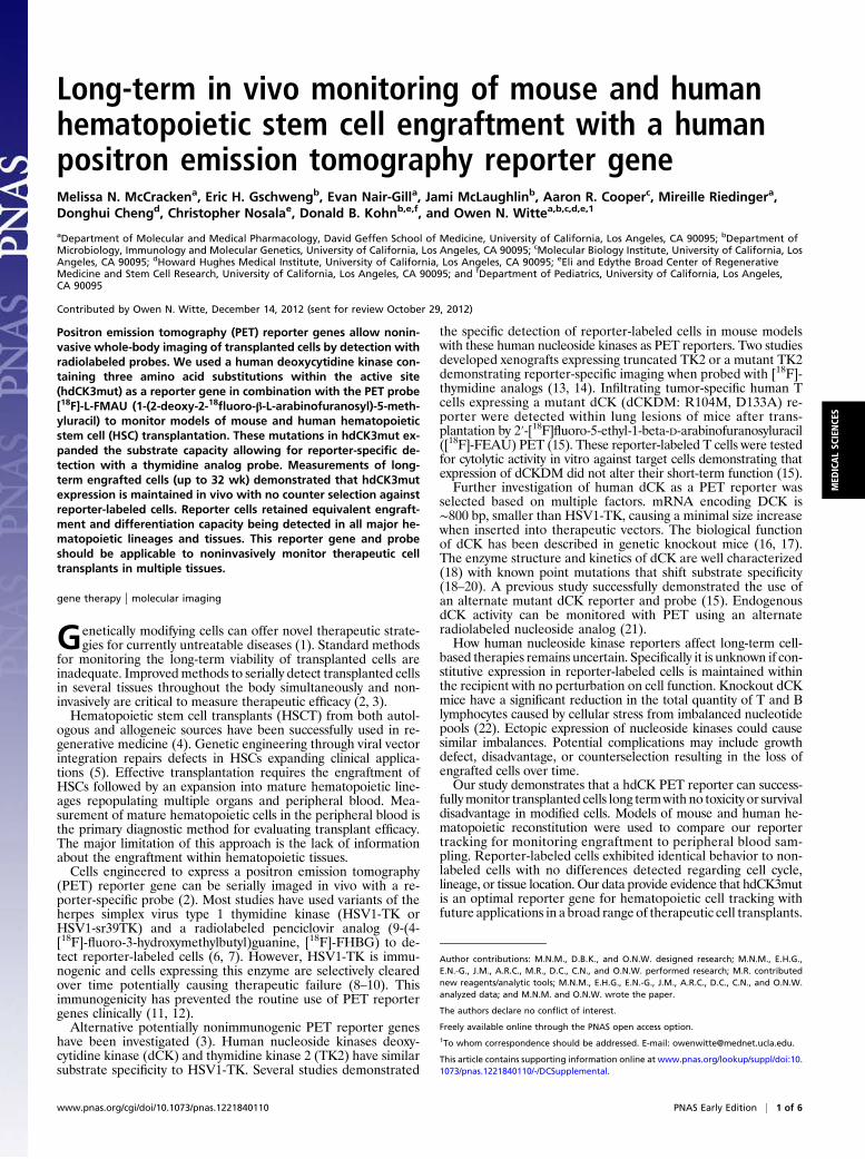
ResultsMutant Human dCK Functions as a PET Reporter When Probed withThymidine Analogs. hdCK3mut contained three point mutations(A100V, R104M, and D133A) that were chosen based on aprevious study that demonstrated a 1,100-fold increase in thy-midine activity compared with wild-type dCK in enzyme kineticassays (20). L1210-10K, a mouse leukemic cell line with no en-dogenous dCK was selected as a model cell line for in vitro studies(23). Stable expression of wild-type (WT) hdCK or hdCK3mutcoexpressed with yellow fluorescent protein (YFP) through aninternal ribosome entry site (IRES) in L1210-10K cells weregenerated to test probes for specific retention in hdCK3mutcells (Fig. 1A and vector maps in Fig. S1). Two thymidine ana-logs, 2′-fluoro-2′-deoxyarabinofuranosyl-5-ethyluracil (FEAU)and 1-(2-deoxy-2-fluoro-β-L-arabinofuranosyl)-5-methyluracil(L-FMAU), showed significant accumulation in reporter celllines compared with wild type. Retention of the probe L-FMAUwas 18-fold higher in hdCK3mut cells compared with WT hdCK(Fig. 1 B and C).Enzyme kinetic analysis further demonstrated high substrate
affinity of hdCK3mut to L-FMAU with a measured Km of ∼13μM. hdCK3mut had a fourfold lower Km for L-FMAU comparedwith the previously published dCKDM reporter (13 μM versus 56μM). A high affinity PET reporter and probe combination isoptimal because probes are administered at high specific activi-ties with low concentrations of substrate. The decreased Km ofhdCK3mut for L-FMAU demonstrates that it will achievea higher velocity at a lower substrate concentration (Table S1).Immune compromised NOD.Cg-Prkdcscid Il2rgtm1Wjl/SzJ (NSG)
mice were implanted with two s.c. grafts. The right side containedL1210-10K cells and the left side contained L1210-10k cell linesengineered to express WT hdCK or hdCK3mut. To determinetumor viability, animals were imaged by PET/CT with 2-deoxy-2-18fluoro-D-glucose ([18F]-FDG), a glucose analog used tomeasure glycolytic consumption. F-18 has a half-life of ∼110 minwith probes decayed to undetectable levels within 24 h allowingfor sequential scans with alternate probes. The following day[18F]-L-FMAU PET/CT scans detected hdCK3mut reporter
expression with signal observed within hdCK3mut grafts (Fig.1D). PET images were then quantified for total probe accumu-lation. Images were dose corrected to total radioactivity at thescan start time. Tumors were then selected in a region of interest(ROI) and the mean percent injected dose per gram (%ID/g)over the entire tumor was calculated. Tumor signal of the dCKtransduced graft is compared as the fold change in probe re-tention to the nontransduced L1210-10K tumors of each animal.Tumors expressing hdCK3mut had a 3.3-fold increase in [18F]-L-FMAU retention (P = 0.0006) compared with WT hdCK grafts(Fig. 1E and Fig. S2). These results determined that hdCK3mutand L-FMAU make a suitable PET reporter gene and probecombination for in vivo studies.
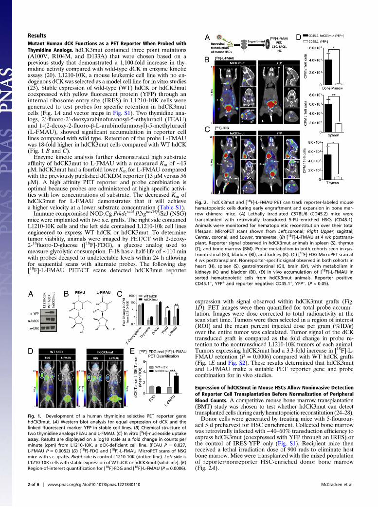
Expression of hdCK3mut in Mouse HSCs Allow Noninvasive Detectionof Reporter Cell Transplantation Before Normalization of PeripheralBlood Counts. A competitive mouse bone marrow transplantation(BMT) study was chosen to test whether hdCK3mut can detecttransplanted cells during early hematopoietic reconstitution (24–28).Donor cells were generated by treating mice with 5-flourour-
acil 5 d preharvest for HSC enrichment. Collected bone marrowwas retrovirally infected with ∼40–60% transduction efficiency toexpress hdCK3mut (coexpressed with YFP through an IRES) orthe control of IRES-YFP only (Fig. S1). Recipient mice thenreceived a lethal irradiation dose of 900 rads to eliminate hostbone marrow. Mice were transplanted with the mixed populationof reporter/nonreporter HSC-enriched donor bone marrow(Fig. 2A).
Fig. 1. Development of a human thymidine selective PET reporter genehdCK3mut. (A) Western blot analysis for equal expression of dCK and thelinked fluorescent marker YFP in stable cell lines. (B) Chemical structure oftwo thymidine analogs FEAU and L-FMAU. (C) In vitro [3H]-nucleoside uptakeassay. Results are displayed on a log10 scale as a fold change in counts perminute (cpm) from L1210-10K, a dCK-deficient cell line. (FEAU P = 0.027,L-FMAU P = 0.0052) (D) [18F]-FDG and [18F]-L-FMAU MicroPET scans of NSGmice with s.c. grafts. Right side is control L1210-10K (dotted line). Left side isL1210-10K cells with stable expression of WT dCK or hdCK3mut (solid line). (E)Region-of-interest quantification for [18F]-FDG and [18F]-L-FMAU (P = 0.0006).
Fig. 2. hdCK3mut and [18F]-L-FMAU PET can track reporter-labeled mousehematopoietic cells during early engraftment and expansion in bone mar-row chimera mice. (A) Lethally irradiated C57BL/6 (CD45.2) mice weretransplanted with retrovirally transduced 5-FU–enriched HSCs (CD45.1).Animals were monitored for hematopoietic reconstitution over their totallifespan. MicroPET scans shown from Left,coronal; Right Upper, sagittal;Center, coronal; and Lower, transverse. (B) [18F]-L-FMAU at 4 wk posttrans-plant. Reporter signal observed in hdCK3mut animals in spleen (S), thymus(T), and bone marrow (BM). Probe metabolism in both cohorts seen in gas-trointestinal (GI), bladder (Bl), and kidney (K). (C) [18F]-FDG MicroPET scan at4 wk posttransplant. Nonreporter-specific signal observed in both cohorts inheart (H), spleen (S), gastrointestinal (GI), brain (Br), with metabolism inkidneys (K) and bladder (Bl). (D) In vivo accumulation of [18F]-L-FMAU insorted hematopoietic cells from hdCK3mut animals. Reporter positive:CD45.1+, YFP+ and reporter negative: CD45.1+, YFP−. (P < 0.05).
2 of 6 | www.pnas.org/cgi/doi/10.1073/pnas.1221840110 McCracken et al.
Under standard conditions mice will display normalized en-graftment and complete blood counts (CBC) within ∼8 wk afterBMT (27). We hypothesized that early engraftment and expan-sion could be monitored by reporter imaging before normaliza-tion of peripheral blood measurements. At 4 wk post-BMT,animals received PET/CT scans with [18F]-FDG and the fol-lowing day [18F]-L-FMAU (Fig. 2 B and C).Whole-body glucose consumption was measured by [18F]-FDG
MicroPET and was indistinguishable between the two cohorts ofanimals. Weak signal was detected within the spleen and bonemarrow indicating a similar glycolytic rate across all animals (Fig.2C). The following day PET/CT with [18F]-L-FMAU detectedonly hdCK3mut cells engrafted within the spleen, thymus, andfocal areas within the bone marrow of reporter-labeled animals(Fig. 2B and Fig. S3). Animals in the control YFP cohort had nohematopoietic signal observed with [18F]-L-FMAU (Fig. 2B).Visualization of [18F]-L-FMAU in hematopoietic tissues ofhdCK3mut recipients verified that reporter imaging can monitorearly engraftment after BMT.To confirm that [18F]-L-FMAU accumulation was specific for
hdCK3mut cells, in vivo reporter and nonreporter accumulationwas measured. Donor hematopoietic cells from hdCK3mutrecipients were sorted for reporter positive or negative and thenwere counted for total radioactivity in counts per minute (cpm)normalized to cpm/1e6 cells. Cells expressing hdCK3mut hada significantly (P < 0.05) higher accumulation of [18F]-L-FMAUcompared with unlabeled cells in all hematopoietic tissues(Fig. 2D).
Reporter Labeled Mouse HSCs Retain Expression of hdCK3mut withEquivalent Engraftment and Differentiation Capacity. Overexpressionof enzymes or reporter genes can potentially cause cellular stress,developmental defects during differentiation, growth disadvantage,or transformation (29, 30). The long-term effects from forced ex-pression of hdCK3mut on mouse HSC’s engraftment and differ-entiation capacity was investigated. Reconstituted chimeric micewere evaluated to confirm that hdCK3mut expression was pre-served and that mouse HSCs expressing the reporter maintainednormal function after transplantation.
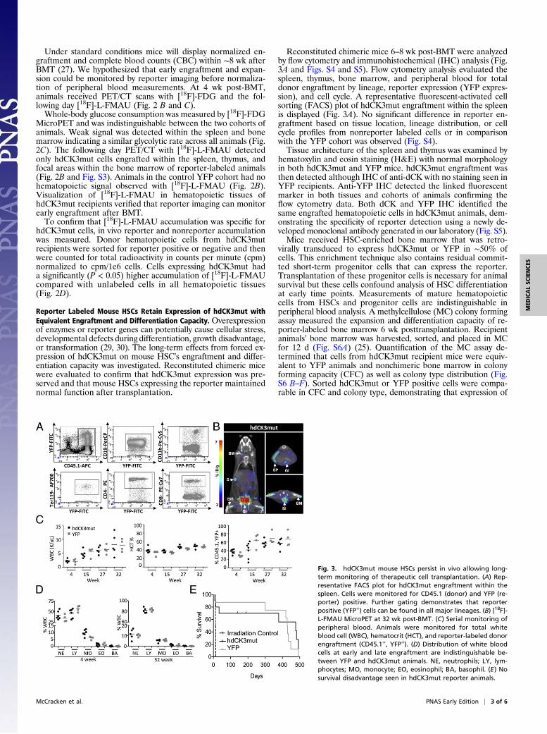
Reconstituted chimeric mice 6–8 wk post-BMT were analyzedby flow cytometry and immunohistochemical (IHC) analysis (Fig.3A and Figs. S4 and S5). Flow cytometry analysis evaluated thespleen, thymus, bone marrow, and peripheral blood for totaldonor engraftment by lineage, reporter expression (YFP expres-sion), and cell cycle. A representative fluorescent-activated cellsorting (FACS) plot of hdCK3mut engraftment within the spleenis displayed (Fig. 3A). No significant difference in reporter en-graftment based on tissue location, lineage distribution, or cellcycle profiles from nonreporter labeled cells or in comparisonwith the YFP cohort was observed (Fig. S4).Tissue architecture of the spleen and thymus was examined by
hematoxylin and eosin staining (H&E) with normal morphologyin both hdCK3mut and YFP mice. hdCK3mut engraftment wasthen detected although IHC of anti-dCK with no staining seen inYFP recipients. Anti-YFP IHC detected the linked fluorescentmarker in both tissues and cohorts of animals confirming theflow cytometry data. Both dCK and YFP IHC identified thesame engrafted hematopoietic cells in hdCK3mut animals, dem-onstrating the specificity of reporter detection using a newly de-veloped monoclonal antibody generated in our laboratory (Fig. S5).Mice received HSC-enriched bone marrow that was retro-
virally transduced to express hdCK3mut or YFP in ∼50% ofcells. This enrichment technique also contains residual commit-ted short-term progenitor cells that can express the reporter.Transplantation of these progenitor cells is necessary for animalsurvival but these cells confound analysis of HSC differentiationat early time points. Measurements of mature hematopoieticcells from HSCs and progenitor cells are indistinguishable inperipheral blood analysis. A methylcellulose (MC) colony formingassay measured the expansion and differentiation capacity of re-porter-labeled bone marrow 6 wk posttransplantation. Recipientanimals’ bone marrow was harvested, sorted, and placed in MCfor 12 d (Fig. S6A) (25). Quantification of the MC assay de-termined that cells from hdCK3mut recipient mice were equiv-alent to YFP animals and nonchimeric bone marrow in colonyforming capacity (CFC) as well as colony type distribution (Fig.S6 B–F). Sorted hdCK3mut or YFP positive cells were compa-rable in CFC and colony type, demonstrating that expression of
Fig. 3. hdCK3mut mouse HSCs persist in vivo allowing long-term monitoring of therapeutic cell transplantation. (A) Rep-resentative FACS plot for hdCK3mut engraftment within thespleen. Cells were monitored for CD45.1 (donor) and YFP (re-porter) positive. Further gating demonstrates that reporterpositive (YFP+) cells can be found in all major lineages. (B) [18F]-L-FMAU MicroPET at 32 wk post-BMT. (C) Serial monitoring ofperipheral blood. Animals were monitored for total whiteblood cell (WBC), hematocrit (HCT), and reporter-labeled donorengraftment (CD45.1+, YFP+). (D) Distribution of white bloodcells at early and late engraftment are indistinguishable be-tween YFP and hdCK3mut animals. NE, neutrophils; LY, lym-phocytes; MO, monocyte; EO, eosinophil; BA, basophil. (E) Nosurvival disadvantage seen in hdCK3mut reporter animals.
McCracken et al. PNAS Early Edition | 3 of 6
MED
ICALSC
IENCE
S
hdCK3mut does not cause a disadvantage during in vitro differ-entiation (Fig. S6E). Sorted cells from both YFP and hdCK3mutrecipients retained reporter expression during in vitro differenti-ation detected through flow cytometry, confirming the continuedexpression of hdCK3mut throughout cell development (Fig. S6F).Together these experiments demonstrate that expression of the
hdCK3mut reporter gene has no observable selective disadvan-tage on hematopoietic cell engraftment and expansion capacity ina mouse HSC transplantation model.
hdCK3mutMouse HSCs Persist in Vivo Allowing Long-TermMonitoring ofTherapeutic Cell Transplantation. Long-term effects from the expres-sion of human nucleoside reporters are poorly defined. Potentialconcerns include selective vector silencing or counterselection ofreporter-labeled cells over time.We examined whether reporter cells in BMT-recipient mice
retained the PET reporter function through serial imaging in vivo.Consecutive scans and peripheral blood analysis were obtained at4, 15, 27, and 32 wk post-BMT allowing for detection of bothshort- and long-lived reporter HSCs (27, 28).At 32 wk post-BMT hdCK3mut reporter-specific signal was
detected within the spleen and bone marrow, demonstrating long-term engraftment capability of reporter-labeled hematopoietic cells(Fig. 3B). Previous scans of the same animals demonstrated similarsignal at 15 and 27 wk (Fig. S7). Serial detection of hdCK3mutthrough PET/CT reveals that the reporter gene functions throughhematopoiesis. It is hypothesized that each sequential scan is de-tecting different hematopoietic cells that have homed to the spleen.Peripheral blood was collected at each time point and ana-
lyzed for CBC and reporter engraftment by flow cytometry. Nosignificant difference was observed in the retention of circulating
reporter positive hematopoietic cells between hdCK3mut andYFP recipients. Comparison of the total white blood cell (WBC)count and hematocrit were normal and equivalent between bothhdCK3mut and YFP after BMT (Fig. 3C). Peripheral WBCdifferential demonstrated that at both early and late engraftmentthe distribution of WBC subtypes were consistent between bothgroups (Fig. 3D).Reporter-labeled bone marrow transduced with YFP or
hdCK3mut was able to successfully rescue lethally irradiated re-cipient animals. Long-term monitoring determined there was nosurvival disadvantage for hdCK3mut recipients over YFP as in-dicated in a Kaplan–Meier survival curve (Fig. 3E). Collectivelythese experiments demonstrate that expression of hdCK3mut isan inert reporter gene capable of long-term noninvasive trackingmethod throughout the recipients’ lifespan.
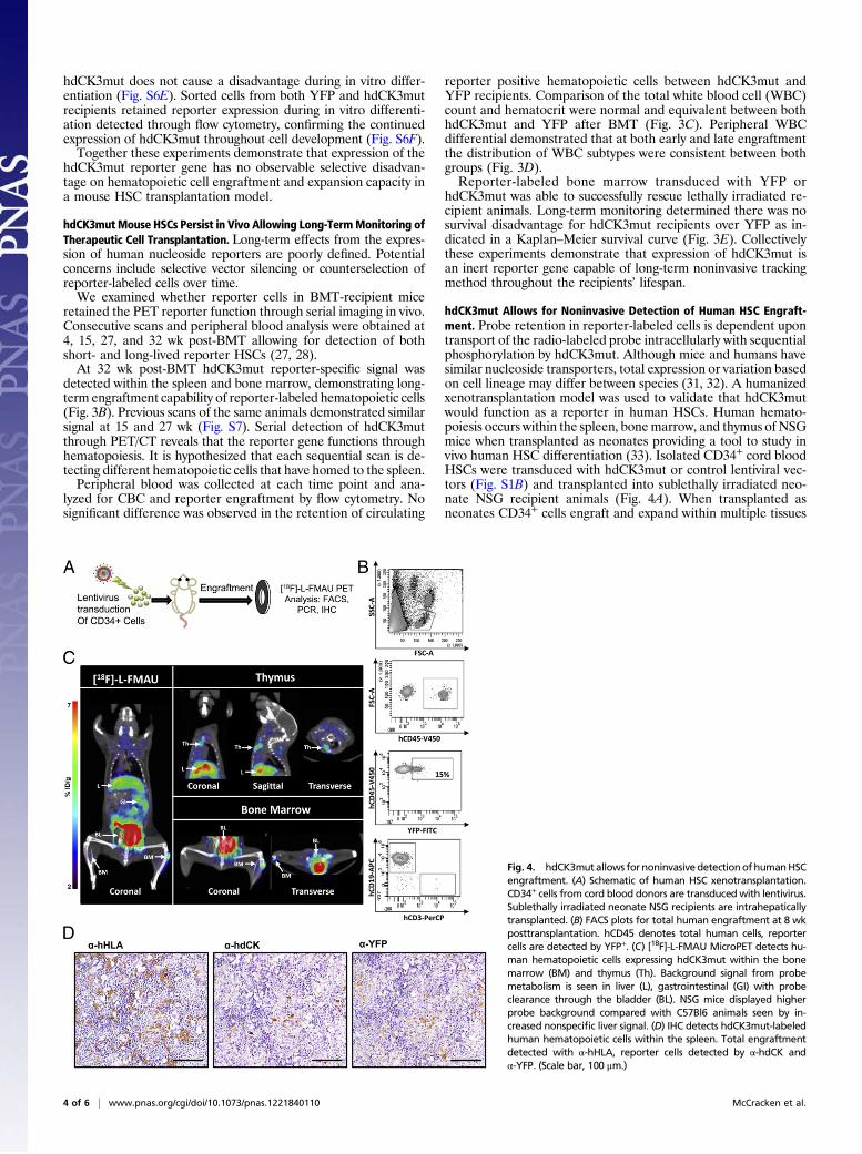
hdCK3mut Allows for Noninvasive Detection of Human HSC Engraft-ment. Probe retention in reporter-labeled cells is dependent upontransport of the radio-labeled probe intracellularly with sequentialphosphorylation by hdCK3mut. Although mice and humans havesimilar nucleoside transporters, total expression or variation basedon cell lineage may differ between species (31, 32). A humanizedxenotransplantation model was used to validate that hdCK3mutwould function as a reporter in human HSCs. Human hemato-poiesis occurs within the spleen, bonemarrow, and thymus of NSGmice when transplanted as neonates providing a tool to study invivo human HSC differentiation (33). Isolated CD34+ cord bloodHSCs were transduced with hdCK3mut or control lentiviral vec-tors (Fig. S1B) and transplanted into sublethally irradiated neo-nate NSG recipient animals (Fig. 4A). When transplanted asneonates CD34+ cells engraft and expand within multiple tissues
Fig. 4. hdCK3mutallows for noninvasivedetectionofhumanHSCengraftment. (A) Schematic of human HSC xenotransplantation.CD34+ cells from cord blood donors are transduced with lentivirus.Sublethally irradiated neonate NSG recipients are intrahepaticallytransplanted. (B) FACS plots for total human engraftment at 8 wkposttransplantation. hCD45 denotes total human cells, reportercells are detected by YFP+. (C) [18F]-L-FMAU MicroPET detects hu-man hematopoietic cells expressing hdCK3mut within the bonemarrow (BM) and thymus (Th). Background signal from probemetabolism is seen in liver (L), gastrointestinal (GI) with probeclearance through the bladder (BL). NSG mice displayed higherprobe background compared with C57Bl6 animals seen by in-creased nonspecific liver signal. (D) IHC detects hdCK3mut-labeledhuman hematopoietic cells within the spleen. Total engraftmentdetected with α-hHLA, reporter cells detected by α-hdCK andα-YFP. (Scale bar, 100 μm.)
4 of 6 | www.pnas.org/cgi/doi/10.1073/pnas.1221840110 McCracken et al.
and can home to the empty NSG thymus. These mice developpartial human hematopoietic systemswithmature humanmyeloid,T-, and B cells.At 8 wk post human HSC transplantation peripheral blood
analysis detected hdCK3mut reporter human engraftment by flowcytometry (Fig. 4B). Peripheral bloodmononuclear cells (PBMCs)were stained for human CD45 to detect total human engraftment.Additional markers were used to detect human myeloid, B-cell, T-cell, and YFP for reporter-labeled cells. MicroPET scans with[18F]-L-FMAU detected hdCK3mut cells within the thymus andbone marrow of chimeric recipient mice (Fig. 4C). This demon-strates that human cells labeled with hdCK3mut retain expressionof the reporter and are capable of engrafting after transplantation.IHC analysis of human HSC engraftment in the spleen and
thymus was performed at the experimental endpoint (Fig. 4D andFig. S8). Total human engraftment was detected with human-spe-cific HLA staining. Sequential sections verified reporter positivecells by anti-dCK and anti-YFP staining. Anti-dCK IHC in thespleen stained a fraction of the total engrafted human cells, con-sistent with the peripheral blood FACS (Fig. 4B), which revealed∼15% of human cells that were reporter positive based on YFPexpression (Fig. 4D). This supports the hypothesis that humanhematopoietic cell maturation and homing is retained in cellsexpressing hdCK3mut.
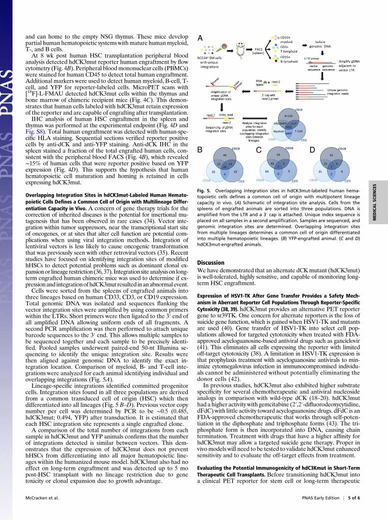
Overlapping Integration Sites in hdCK3mut-Labeled Human Hemato-poietic Cells Defines a Common Cell of Origin with Multilineage Differ-entiation Capacity in Vivo. A concern of gene therapy trials for thecorrection of inherited diseases is the potential for insertional mu-tagenesis that has been observed in rare cases (34). Vector inte-gration within tumor suppressors, near the transcriptional start siteof oncogenes, or at sites that alter cell function are potential com-plications when using viral integration methods. Integration oflentiviral vectors is less likely to cause oncogenic transformationthat was previously seen with other retroviral vectors (35). Recentstudies have focused on identifying integration sites of modifiedhHSCs to detect potential problems such as dominant clonal ex-pansionor lineage restriction (36,37). Integrationsiteanalysison long-term engrafted human chimeric mice was used to determine if ex-pressionand integrationofhdCK3mut resulted inanabnormalevent.Cells were sorted from the spleens of engrafted animals into
three lineages based on human CD33, CD3, or CD19 expression.Total genomic DNA was isolated and sequences flanking thevector integration sites were amplified by using common primerswithin the LTRs. Short primers were then ligated to the 3′ end ofall amplified DNA allowing uniform ends of all fragments. Asecond PCR amplification was then performed to attach uniquebarcode sequences to the 3′ end. This allows multiple samples tobe sequenced together and each sample to be precisely identi-fied. Pooled samples underwent paired-end 50-nt Illumina se-quencing to identify the unique integration site. Results werethen aligned against genomic DNA to identify the exact in-tegration location. Comparison of myeloid, B- and T-cell inte-grations were analyzed for each animal identifying individual andoverlapping integrations (Fig. 5A).Lineage-specific integrations identified committed progenitor
cells. Integration sites found in all three populations are derivedfrom a common transduced cell of origin (HSC) which thendifferentiated into all lineages (Fig. 5 B–D). Previous vector copynumber per cell was determined by PCR to be ∼0.5 (0.485,hdCK3mut; 0.494, YFP) after transduction. It is estimated thateach HSC integration site represents a single engrafted clone.A comparison of the total number of integrations from each
sample in hdCK3mut and YFP animals confirms that the numberof integrations detected is similar between vectors. This dem-onstrates that the expression of hdCK3mut does not preventhHSCs from differentiating into all major hematopoietic line-ages within the humanized mouse model. hdCK3mut also had noeffect on long-term engraftment and was detected up to 5 mopost-HSC transplant with no lineage restriction due to genetoxicity or clonal expansion due to growth advantage.
DiscussionWehave demonstrated that an alternate dCKmutant (hdCK3mut)is well-tolerated, highly sensitive, and capable of monitoring long-term HSC engraftment.
Expression of HSV1-TK After Gene Transfer Provides a Safety Mech-anism in Aberrant Reporter Cell Populations Through Reporter-SpecificCytoxicity (38, 39). hdCK3mut provides an alternative PET reportergene to sr39TK. One concern for alternate reporters is the loss ofsuicide gene function, which is gained when HSV1-TK and mutantsare used (40). Gene transfer of HSV1-TK into select cell pop-ulations allowed for targeted cytotoxicity when treated with FDA-approved acycloguanosine-based antiviral drugs such as ganciclovir(41). This eliminates all cells expressing the reporter with limitedoff-target cytotoxicity (38). A limitation in HSV1-TK expression isthat prophylaxis treatment with acycloguanosine antivirals to min-imize cytomegalovirus infection in immunocompromised individu-als cannot be administered without potentially eliminating thedonor cells (42).In previous studies, hdCK3mut also exhibited higher substrate
specificity for several chemotherapeutic and antiviral nucleosideanalogs in comparison with wild-type dCK (18–20). hdCK3muthad a higher activity with gemcitabine (2′,2′-difluorodeoxycytidine,dFdC)with little activity toward acycloguanosine drugs. dFdC is anFDA-approved chemotherapeutic that works through self-poten-tiation in the diphosphate and triphosphate forms (43). The tri-phosphate form is then incorporated into DNA, causing chaintermination. Treatment with drugs that have a higher affinity forhdCK3mut may allow a targeted suicide gene therapy. Proper invivomodels will need to be tested to validate hdCK3mut enhancedsensitivity and to evaluate the off-target effects from treatment.
Evaluating the Potential Immunogenicity of hdC3Kmut in Short-TermTherapeutic Cell Transplants. Before transitioning hdCK3mut intoa clinical PET reporter for stem cell or long-term therapeutic
Fig. 5. Overlapping integration sites in hdCK3mut-labeled human hema-topoietic cells defines a common cell of origin with multipotent lineagecapacity in vivo. (A) Schematic of integration site analysis. Cells from thespleens of engrafted animals are sorted into three populations. DNA isamplified from the LTR and a 3′ cap is attached. Unique index sequence isplaced on all samples in a second amplification. Samples are sequenced, andgenomic integration sites are determined. Overlapping integration sitesfrom multiple lineages determines a common cell of origin differentiatedinto multiple hematopoietic lineages. (B) YFP-engrafted animal. (C and D)hdCK3mut-engrafted animals.
McCracken et al. PNAS Early Edition | 5 of 6
MED
ICALSC
IENCE
S
transplants experimental short-lived transplanted cells need tobe investigated. The immunogenicity of HSV1-TK was foundwhen reporter-labeled lymphocytes caused a CD8 immune re-sponse selectively killing reporter cells (8–10). A similar studycould determine if expression of hdCK3mut will cause animmune response.hdCK3mut contains only three point mutations and is expec-
ted to not cause an immunogenic epitope for MHC class 1presentation. Using a predictive software for MHC class 1 pre-sentation (44), hdCK3mut has only one additional peptidefragment, which is predicted to be different fromWT hdCK. Thispeptide, which incorporates the mutated amino acid methionine atposition 104, could use the methionine as an anchoring residuewithin the MHC (45). All other amino acids that are displayed inthe MHC are natural, and therefore the peptide fragments dis-played from hdCK3mut are predicted to be recognized as “self”avoiding immune cell detection.
Summary. We demonstrate how long-term follow-up of trans-planted cells can be managed noninvasively by using reporter PETimaging. This study provides a comprehensive analysis on the inert
biological effect of hdCK3mut on hematopoietic cells. Therefore,it is anticipated that hdCK3mut monitoring will provide a safe andeffective mechanism for longitudinal monitoring of a broad rangeof transplanted cells.
Materials and MethodsDetailed information on animals, constructs and cloning, cell lines, uptakeassays, enzyme kinetic assay, grafts, mouse HSC transplant, human HSCtransplant, MicroPET imaging, peripheral blood analysis, FACS, methylcel-lulose assay, antibodies, Western blot, IHC, integration site analysis, andstatistics can be found in SI Materials and Methods.
ACKNOWLEDGMENTS. We thank Dr. Nagichettiar Satyamurthy and JefferyCollins for the radiosynthesis of [18F]-FDG and [18F]-L-FMAU and Dr.WaldemerLando, Dr. David Stout, and Darrin Williams in the Crump Institute for Molec-ular Imaging facility for their technical help with PET/CT scans. M.N.M. is sup-ported by California Institute for Regenerative Medicine Training Grant TG2-01169. E.H.G. is supported by Eli and Edythe Broad Center of RegenerativeMedicine and Stem Cell Research at UCLA. A.R.C. is supported by a PhilipWhitcome training grant. C.N. is supported by the California Institute forRegenerative Medicine Bridges Training Program. O.N.W. is supported by aCalifornia Institute for Regenerative Medicine Tools/Technology Award RT1-01126. O.N.W. is an Investigator of the Howard Hughes Medical Institute.
1. Sng J, Lufkin T (2012) Emerging stem cell therapies: Treatment, safety, and biology.Stem Cells Int 2012:521343.
2. Herschman HR (2004) PET reporter genes for noninvasive imaging of gene therapy,cell tracking and transgenic analysis. Crit Rev Oncol Hematol 51(3):191–204.
3. Serganova I, Ponomarev V, Blasberg R (2007) Human reporter genes: Potential use inclinical studies. Nucl Med Biol 34(7):791–807.
4. Gyurkocza B, Rezvani A, Storb RF (2010) Allogeneic hematopoietic cell trans-plantation: The state of the art. Expert Rev Hematol 3(3):285–299.
5. Kohn DB (2010) Update on gene therapy for immunodeficiencies. Clin Immunol135(2):247–254.
6. Gambhir SS, et al. (2000) A mutant herpes simplex virus type 1 thymidine kinase re-porter gene shows improved sensitivity for imaging reporter gene expression withpositron emission tomography. Proc Natl Acad Sci USA 97(6):2785–2790.
7. Tjuvajev JG, et al. (1998) Imaging herpes virus thymidine kinase gene transfer andexpression by positron emission tomography. Cancer Res 58(19):4333–4341.
8. Berger C, Flowers ME, Warren EH, Riddell SR (2006) Analysis of transgene-specificimmune responses that limit the in vivo persistence of adoptively transferred HSV-TK-modified donor T cells after allogeneic hematopoietic cell transplantation. Blood107(6):2294–2302.
9. Riddell SR, et al. (1996) T-cell mediated rejection of gene-modified HIV-specific cy-totoxic T lymphocytes in HIV-infected patients. Nat Med 2(2):216–223.
10. Traversari C, et al. (2007) The potential immunogenicity of the TK suicide gene doesnot prevent full clinical benefit associated with the use of TK-transduced donorlymphocytes in HSCT for hematologic malignancies. Blood 109(11):4708–4715.
11. Mercier-Letondal P, et al. (2008) Early immune response against retrovirally trans-duced herpes simplex virus thymidine kinase-expressing gene-modified T cells co-infused with a T cell-depleted marrow graft: An altered immune response? Hum GeneTher 19(9):937–950.
12. Oliveira G, Greco R, Lupo-Stanghellini MT, Vago L, Bonini C (2012) Use of TK-cells inhaploidentical hematopoietic stem cell transplantation. Curr Opin Hematol 19(6):427–433.
13. Campbell DO, et al. (2012) Structure-guided engineering of human thymidine kinase2 as a positron emission tomography reporter gene for enhanced phosphorylation ofa non-natural thymidine analog reporter probe. J Biol Chem 287(1):446–454.
14. Ponomarev V, et al. (2007) A human-derived reporter gene for noninvasive imagingin humans: Mitochondrial thymidine kinase type 2. J Nucl Med 48(5):819–826.
15. Likar Y, et al. (2010) A new pyrimidine-specific reporter gene: A mutated humandeoxycytidine kinase suitable for PET during treatment with acycloguanosine-basedcytotoxic drugs. J Nucl Med 51(9):1395–1403.
16. Choi O, et al. (2012) A deficiency in nucleoside salvage impairs murine lymphocytedevelopment, homeostasis, and survival. J Immunol 188(8):3920–3927.
17. Toy G, et al. (2010) Requirement for deoxycytidine kinase in T and B lymphocytedevelopment. Proc Natl Acad Sci USA 107(12):5551–5556.
18. Sabini E, Ort S, Monnerjahn C, KonradM, Lavie A (2003) Structure of human dCK suggestsstrategies to improve anticancer and antiviral therapy. Nat Struct Biol 10(7):513–519.
19. Hazra S, Sabini E, Ort S, Konrad M, Lavie A (2009) Extending thymidine kinase activityto the catalytic repertoire of human deoxycytidine kinase. Biochemistry 48(6):1256–1263.
20. Iyidogan P, Lutz S (2008) Systematic exploration of active site mutations on humandeoxycytidine kinase substrate specificity. Biochemistry 47(16):4711–4720.
21. Radu CG, et al. (2008) Molecular imaging of lymphoid organs and immune activationby positron emission tomography with a new [18F]-labeled 2′-deoxycytidine analog.Nat Med 14(7):783–788.
22. Austin WR, et al. (2012) Nucleoside salvage pathway kinases regulate hematopoiesisby linking nucleotide metabolism with replication stress. J Exp Med 209(12):2215–2228.
23. Jordheim LP, et al. (2004) Characterization of a gemcitabine-resistant murine leuke-mic cell line: Reversion of in vitro resistance by a mononucleotide prodrug. ClinCancer Res 10(16):5614–5621.
24. Baum C, et al. (2003) Side effects of retroviral gene transfer into hematopoietic stemcells. Blood 101(6):2099–2114.
25. Drize N, Chertkov J, Sadovnikova E, Tiessen S, Zander A (1997) Long-term mainte-nance of hematopoiesis in irradiated mice by retrovirally transduced peripheral bloodstem cells. Blood 89(5):1811–1817.
26. Stewart FM, Crittenden RB, Lowry PA, Pearson-White S, Quesenberry PJ (1993) Long-term engraftment of normal and post-5-fluorouracil murine marrow into normalnonmyeloablated mice. Blood 81(10):2566–2571.
27. Kang E, et al. (2001) In vivo persistence of retrovirally transduced murine long-termrepopulating cells is not limited by expression of foreign gene products in the fully orminimally myeloablated setting. Hum Gene Ther 12(13):1663–1672.
28. Zavidij O, et al. (2012) Stable long-term blood formation by stem cells in murinesteady-state hematopoiesis. Stem Cells 30(9):1961–1970.
29. Gambhir SS, et al. (2000) Imaging transgene expression with radionuclide imagingtechnologies. Neoplasia 2(1–2):118–138.
30. Hallek M, Wanders L, Strohmeyer S, Emmerich B (1992) Thymidine kinase: a Tumormarker with prognostic value for non-Hodgkin’s lymphoma and a broad range ofpotential clinical applications. Ann Hematol 65(1):1–5.
31. Baldwin SA, et al. (2004) The equilibrative nucleoside transporter family, SLC29.Pflugers Arch 447(5):735–743.
32. Gray JH, Owen RP, Giacomini KM (2004) The concentrative nucleoside transporterfamily, SLC28. Pflugers Arch 447(5):728–734.
33. Park CY, Majeti R, Weissman IL (2008) In vivo evaluation of human hematopoiesisthrough xenotransplantation of purified hematopoietic stem cells from umbilicalcord blood. Nat Protoc 3(12):1932–1940.
34. Corrigan-Curay J, et al. (2012) Challenges in vector and trial design using retroviralvectors for long-term gene correction in hematopoietic stem cell gene therapy. MolTher 20(6):1084–1094.
35. Biffi A, et al. (2011) Lentiviral vector common integration sites in preclinical modelsand a clinical trial reflect a benign integration bias and not oncogenic selection. Blood117(20):5332–5339.
36. Gonzalez-Murillo A, Lozano ML, Montini E, Bueren JA, Guenechea G (2008) Unalteredrepopulation properties of mouse hematopoietic stem cells transduced with lentiviralvectors. Blood 112(8):3138–3147.
37. Ronen K, et al. (2011) Distribution of lentiviral vector integration sites in mice fol-lowing therapeutic gene transfer to treat β-thalassemia. Mol Ther 19(7):1273–1286.
38. Bonini C, et al. (1997) HSV-TK gene transfer into donor lymphocytes for control ofallogeneic graft-versus-leukemia. Science 276(5319):1719–1724.
39. Ciceri F, et al. (2007) Antitumor effects of HSV-TK-engineered donor lymphocytesafter allogeneic stem-cell transplantation. Blood 109(11):4698–4707.
40. Lupo-Stanghellini MT, et al. (2010) Clinical impact of suicide gene therapy in allo-geneic hematopoietic stem cell transplantation. Hum Gene Ther 21(3):241–250.
41. Tiberghien P, et al. (1994) Ganciclovir treatment of herpes simplex thymidine kinase-transduced primary T lymphocytes: An approach for specific in vivo donor T-cell de-pletion after bone marrow transplantation? Blood 84(4):1333–1341.
42. Hébrard C, Dumontet C, Jordheim LP (2009) Development of gene therapy in associationwith clinically used cytotoxic deoxynucleoside analogues. Cancer Gene Ther 16(7):541–550.
43. Burris HA, 3rd, et al. (1997) Improvements in survival and clinical benefit with gem-citabine as first-line therapy for patients with advanced pancreas cancer: A ran-domized trial. J Clin Oncol 15(6):2403–2413.
44. Hakenberg J, et al. (2003) MAPPP: MHC class I antigenic peptide processing pre-diction. Appl Bioinformatics 2(3):155–158.
45. Reche PA, Glutting JP, Reinherz EL (2002) Prediction of MHC class I binding peptidesusing profile motifs. Hum Immunol 63(9):701–709.
6 of 6 | www.pnas.org/cgi/doi/10.1073/pnas.1221840110 McCracken et al.


![Human body - gijyuku.634tv.comgijyuku.634tv.com/pdf/English/Human-body.pdf · waste molecules and excess ions and water out of the body.[26] Human anatomy is the study of the shape](https://static.fdocuments.pl/doc/165x107/5ed289dbaf2f306b9a013d53/human-body-gijyuku634tvcomgijyuku634tvcompdfenglishhuman-bodypdf-waste.jpg)