Flow cytometry evidence about sperm competition in …rotofils/tofilski2012flow.pdf · ·...
8
Flow cytometry evidence about sperm competition in honey bee (Apis mellifera) Adam TOFILSKI 1 , Bożena CHUDA-MICKIEWICZ 2 , Krystyna CZEKOŃSKA 1 , Paweł CHORBIŃSKI 3 1 Department of Pomology and Apiculture, Agricultural University, 29 Listopada 54, 31-425 Krakow, Poland 2 Department of Apiculture, West Pomeranian University of Technology, Doktora Judyma 20, 71-466 Szczecin, Poland 3 Department of Epizootiology and Veterinary Administration with Clinic, University of Environmental and Life Sciences, pl. Grunwaldzki 45, 50-366 Wroclaw, Poland Received 9 March 2011 – Revised 21 June 2011 – Accepted 22 July 2011 Abstract – We tested whether flow cytometry can be used for assessment of viability of honey bee (Apis mellifera) sperm. The method was used to detect possible competition between the sperm of different drones. The flow cytometry analysis of semen stained with SYBR-14/propidium iodide revealed significant differences between fresh and freeze-thawed samples. The identification of populations corresponding to viable and nonviable sperm allowed us to assess the sperm viability. The comparison of single-drone semen with mixed semen of two unrelated drones showed that sperm viability was not affected by mixing, but there were differences between mixed and unmixed semen in side scatter, which correlates with shape and optical homogeneity of particles. The proportion of particles in different populations also was affected by mixing of the semen. The results suggest that there are interactions between ejaculates of different drones, possibly related to sperm competition. honey bee / Apis mellifera / spermatozoa / sperm competition 1. INTRODUCTION During a single mating flight, the honey bee (Apis mellifera) queen mates with multiple drones (Woyke 1960; Schlüns et al. 2005). Their semen is transferred to the oviducts (Woyke 1956) where it is mixed by contraction of the oviduct muscles (Page 1986). Only a small fraction of spermatozoa reaches the spermatheca and can be used later for fertiliza- tion of eggs, and the rest is expelled through the sting chamber (Laidlaw 1944). These circum- stances favor intraoviductal sperm competition (Woyciechowski and Król 1996). One mechanism of sperm competition is sperm incapacitation, by which substances present in seminal fluid kill the sperm of other males (Harshman and Prout 1994). If this mechanism operates, then mixing the semen of two or more drones should result in higher mortality of spermatozoa. Another possible mechanism of sperm competition is related to sperm polymorphism. In some species, there are two distinct types of sperm. It has been suggested that only one of those types is suitable for fertilization and that the other can block or eliminate the spermatozoa of other males (Swallow and Wilkinson 2002). At the moment, there is no data about the sperm polymorphism in honey bees. Most of the earlier attempts to confirm sperm competition in honey bees have been unsuc- Corresponding author: A. Tofilski, [email protected] Manuscript editor: Klaus Hartfelder Apidologie (2012) 43:63–70 Original article * The Author(s) 2011. This article is published with open access at Springerlink.com DOI: 10.1007/s13592-011-0089-6
Transcript of Flow cytometry evidence about sperm competition in …rotofils/tofilski2012flow.pdf · ·...

Flow cytometry evidence about sperm competitionin honey bee (Apis mellifera)
Adam TOFILSKI1, Bożena CHUDA-MICKIEWICZ
2, Krystyna CZEKOŃSKA
1,
Paweł CHORBIŃSKI3
1Department of Pomology and Apiculture, Agricultural University, 29 Listopada 54, 31-425 Krakow, Poland2Department of Apiculture, West Pomeranian University of Technology, Doktora Judyma 20, 71-466 Szczecin,
Poland3Department of Epizootiology and Veterinary Administration with Clinic, University of Environmental and Life
Sciences, pl. Grunwaldzki 45, 50-366 Wrocław, Poland
Received 9 March 2011 – Revised 21 June 2011 – Accepted 22 July 2011
Abstract – We tested whether flow cytometry can be used for assessment of viability of honey bee (Apismellifera) sperm. The method was used to detect possible competition between the sperm of different drones.The flow cytometry analysis of semen stained with SYBR-14/propidium iodide revealed significant differencesbetween fresh and freeze-thawed samples. The identification of populations corresponding to viable andnonviable sperm allowed us to assess the sperm viability. The comparison of single-drone semen with mixedsemen of two unrelated drones showed that sperm viability was not affected by mixing, but there weredifferences between mixed and unmixed semen in side scatter, which correlates with shape and opticalhomogeneity of particles. The proportion of particles in different populations also was affected by mixing of thesemen. The results suggest that there are interactions between ejaculates of different drones, possibly related tosperm competition.
honey bee / Apis mellifera / spermatozoa / sperm competition
1. INTRODUCTION
During a single mating flight, the honey bee(Apis mellifera) queen mates with multipledrones (Woyke 1960; Schlüns et al. 2005).Their semen is transferred to the oviducts(Woyke 1956) where it is mixed by contractionof the oviduct muscles (Page 1986). Only asmall fraction of spermatozoa reaches thespermatheca and can be used later for fertiliza-tion of eggs, and the rest is expelled through thesting chamber (Laidlaw 1944). These circum-stances favor intraoviductal sperm competition(Woyciechowski and Król 1996).
One mechanism of sperm competition issperm incapacitation, by which substancespresent in seminal fluid kill the sperm of othermales (Harshman and Prout 1994). If thismechanism operates, then mixing the semen oftwo or more drones should result in highermortality of spermatozoa. Another possiblemechanism of sperm competition is related tosperm polymorphism. In some species, there aretwo distinct types of sperm. It has beensuggested that only one of those types issuitable for fertilization and that the other canblock or eliminate the spermatozoa of othermales (Swallow and Wilkinson 2002). At themoment, there is no data about the spermpolymorphism in honey bees.
Most of the earlier attempts to confirm spermcompetition in honey bees have been unsuc-
Corresponding author: A. Tofilski,[email protected] editor: Klaus Hartfelder
Apidologie (2012) 43:63–70 Original article* The Author(s) 2011. This article is published with open access at Springerlink.comDOI: 10.1007/s13592-011-0089-6

cessful (Harbo 1990; Woyciechowski and Król1996; Moritz 1986; for review, see Shafir et al.2009). Analyses of patrilines in honey beecolonies showed that some drones, which matedwith the queen, were underrepresented in heroffspring (Franck et al. 1999; Haberl and Tautz1998), but it was not clear if this was the resultof sperm competition. In one experiment, theviability of sperm in the presence of seminalfluids of other males was found to be lower thanin the presence of only its own seminal fluids(den Boer et al. 2010).
Here we used flow cytometry to verifywhether the spermatozoa of two drones showhigher mortality after mixing. This methodallows quantitative analysis of semen, permitssperm viability to be assessed, and can alsodetect sperm polymorphism.
2. MATERIAL AND METHODS
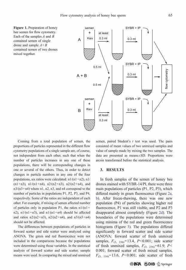
The drones used in this study came from threeunrelated colonies of honey bee (A. m. carnica). Ineach of the colonies, the queen was caged on the dronecomb for 24 h in order to obtain drones of known age.The experiment was done when the drones were at20 days of age. Semen was collected in a calibratedcapillary tube by standard technique (Laidlaw 1989;Collins and Donoghue 1999). Semen volume wasmeasured by the height of the liquid column in thecapillary. The semen was diluted 1:1,000 v:v in Kievbuffer (0.3 g glucose, 0.41 g potassium chloride,0.21 g sodium bicarbonate, 2.43 g sodium citrate per100 mL deionized water; Collins and Donoghue1999). The volume of collected semen was alwaysgreater than 0.8 μL, yielding at least 0.8 mL dilutedsperm. From each sample, 0.5 mL suspension wasmixed with the same volume of a sample collectedfrom an unrelated drone. The remaining volume (atleast 0.3 mL) of each sample was used for the analysisof unmixed semen (Figure 1).
The order in which the semen was collected andanalyzed was controlled to eliminate the effect of thetime between collection of the sperm and its analysis.For example, the order of collection was A1, B1, B2,A2, … and the order of analysis was A1, B1, A1+B1,A2+B2, B2, A2, …, where the different capital letters
indicate different unrelated colonies and thedifferent numbers indicate different drones. Thesemen of 60 drones was collected and 90 sampleswere analyzed. There were 60 samples of unmixedsemen of single drones and 30 samples of mixedsemen of two drones.
In each sample, the proportions of live and deadspermatozoa were determined by SYBR-14/propi-dium iodide (PI) fluorescent staining with the LIVE/DEAD Sperm Viability Kit (Molecular Probes L-7011). From each sample, 300 μL of diluted semenwas collected; 5 μL SYBR 14 (stock solution,deionized, sterile water 1:50) was added and thenincubated for 5 min at 36°C. Then 4 μL PI was addedand stirred. SYBR-14 stains live cells green and PIstains dead cells red. The counts were obtained byflow cytometry (Becton Dickinson FACSCalibur,USA) with a 488-nm argon laser. Green fluorescencewas measured in the LFL1 channel and red fluores-cence in the LFL3 channel, using the manufacturer’sfluorescence compensation filters. Forward scatterand side scatter were also measured. The forwardscatter tends to be more sensitive to size ofparticles and the side scatter tends to be moresensitive to shape and optical homogeneity ofparticles. Twenty thousand particles were analyzedin each sample. The groups of particles withsimilar properties are called populations. Thecytometry data were analyzed with WinMDI 2.8software. The red fluorescence, green fluorescence,forward scatter, and side scatter are reported as arbitraryunits. Measurements in those units can be comparedonly within this study.
In order to verify if flow cytometry can be used for
assessment of honey bee sperm viability, we con-
ducted freeze-thawing experiment. The experiment
allowed us to identify populations with viable and
nonviable sperm. Samples of semen from four drones
were frozen at −17°C for 2 h in order to kill the
sperm. The samples were analyzed after thawing.
Earlier work shows that honey bee sperm does not
survive freezing without cryoprotectant (Peng et al.
1992). In the freeze-thawing experiment, each of the
four samples consisted of unmixed semen. It was
assumed that live spermatozoa are in populations
which disappeared after freeze-thawing and dead
spermatozoa are in populations which appeared after
freeze-thawing.
64 A. Tofilski et al.
Coming from a total population of semen, theproportions of particles represented in the different flowcytometry populations of a single sample are, of course,not independent from each other, such that when thenumber of particles increases in any one of thesepopulations, there will be corresponding changes inone or several of the others. Thus, in order to detectchanges in particle numbers in any one of the fourpopulations, six ratios were calculated: n1/(n1+n2), n1/(n1+n3), n1/(n1+n4), n2/(n2+n3), n2/(n2+n4), andn3/(n3+n4) where n1, n2, n3, and n4 correspond to thenumber of particles in populations P1, P2, P3, and P4,respectively. Some of the ratios are independent of eachother. For example, if mixing of semen affected numberof particles only in population P1, the ratios n1/(n1+n2), n1/(n1+n3), and n1/(n1+n4) should be affectedand ratios n2/(n2+n3), n2/(n2+n4), and n3/(n3+n4)should not be affected.
The differences between populations of particles inforward scatter and side scatter were analyzed usingANOVA. The green and red fluorescence were notincluded in the comparisons because the populationswere determined using those variables. In the statisticalanalysis of forward scatter and side scatter, samplemeans were used. In comparing the mixed and unmixed
semen, paired Student’s t test was used. The pairsconsisted of mean values of two unmixed samples andvalue of sample made by mixing the two samples. Thedata are presented as means±SD. Proportions werearcsin transformed before the statistical analysis.
3. RESULTS
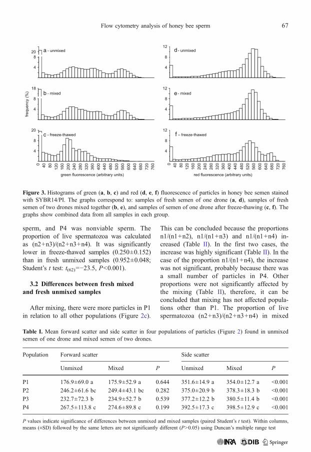
In fresh samples of the semen of honey beedrones stained with SYBR-14/PI, there were threemain populations of particles (P1, P2, P3), whichdiffered mainly in green fluorescence (Figure 2a,b). After freeze-thawing, there was one newpopulation (P4) of particles showing higher redfluorescence, P1 was still visible, and P2 and P3disappeared almost completely (Figure 2d). Theboundaries of the populations were determinedusing minima of the red and green fluorescencehistograms (Figure 3). The populations differedsignificantly in forward scatter and side scatter(ANOVA: forward scatter of fresh unmixedsamples, F(3, 236)=13.4, P<0.001; side scatterof fresh unmixed samples, F(3, 236)=61.9; P<0.001; forward scatter of fresh mixed samples,F(3, 116)=13.6, P<0.001; side scatter of fresh
Figure 1. Preparation of honeybee semen for flow cytometry.Each of the samples A and Bcontained semen of singledrone and sample A+Bcontained semen of two dronesmixed together.
Flow cytometry analysis of honey bee sperm 65

mixed samples, F(3, 116)=50.4; P<0.001). Par-ticles in P1 differed markedly from particles inall other populations, whereas particles in P2 andP3 were similar to each other (Table I).
3.1 Differences between fresh unmixedand freeze-thawed samples
Freeze-thawed samples differed significantlyfrom fresh unmixed samples in all six proportions
calculated from number of particles in the fourpopulations (Table II). After freeze-thawing, thenumber of particles in P4 increased in compar-ison to all other populations. Moreover, thenumber of particles in P2 and P3 decreased incomparison to P1. Those results (Table II)confirm the above mentioned marked differencesbetween freeze-thawed and fresh samples. Fromthe results, it can be concluded that P1 did notconsist of live cells, P2 and P3 were viable
a
c d
b
Figure 2. Green and red fluorescence of particles in honey bee semen immediately after collection (a, b, c) andafter freeze-thawing (d); a, b, d - semen of single drone; c - mixed semen of the two drones presented at a andb. Rectangles indicate four populations of particles (P1, P2, P3, P4). Each point represents one particle.
66 A. Tofilski et al.
sperm, and P4 was nonviable sperm. Theproportion of live spermatozoa was calculatedas (n2+n3)/(n2+n3+n4). It was significantlylower in freeze-thawed samples (0.250±0.152)than in fresh unmixed samples (0.952±0.048;Student’s t test: t(62)=−23.5, P<0.001).
3.2 Differences between fresh mixedand fresh unmixed samples
After mixing, there were more particles in P1in relation to all other populations (Figure 2c).
This can be concluded because the proportionsn1/(n1+n2), n1/(n1+n3) and n1/(n1+n4) in-creased (Table II). In the first two cases, theincrease was highly significant (Table II). In thecase of the proportion n1/(n1+n4), the increasewas not significant, probably because there wasa small number of particles in P4. Otherproportions were not significantly affected bythe mixing (Table II), therefore, it can beconcluded that mixing has not affected popula-tions other than P1. The proportion of livespermatozoa (n2+n3)/(n2+n3+n4) in mixed
a d
e
f – freeze-thawed
b
c
Figure 3. Histograms of green (a, b, c) and red (d, e, f) fluorescence of particles in honey bee semen stainedwith SYBR14/PI. The graphs correspond to: samples of fresh semen of one drone (a, d), samples of freshsemen of two drones mixed together (b, e), and samples of semen of one drone after freeze-thawing (c, f). Thegraphs show combined data from all samples in each group.
Table I. Mean forward scatter and side scatter in four populations of particles (Figure 2) found in unmixedsemen of one drone and mixed semen of two drones.
Population Forward scatter Side scatter
Unmixed Mixed P Unmixed Mixed P
P1 176.9±69.0 a 175.9±52.9 a 0.644 351.6±14.9 a 354.0±12.7 a <0.001
P2 246.2±61.6 bc 249.4±43.1 bc 0.282 375.0±20.9 b 378.3±18.3 b <0.001
P3 232.7±72.3 b 234.9±52.7 b 0.539 377.2±12.2 b 380.5±11.4 b <0.001
P4 267.5±113.8 c 274.6±89.8 c 0.199 392.5±17.3 c 398.5±12.9 c <0.001
P values indicate significance of differences between unmixed and mixed samples (paired Student’s t test). Within columns,means (±SD) followed by the same letters are not significantly different (P>0.05) using Duncan’s multiple range test
Flow cytometry analysis of honey bee sperm 67
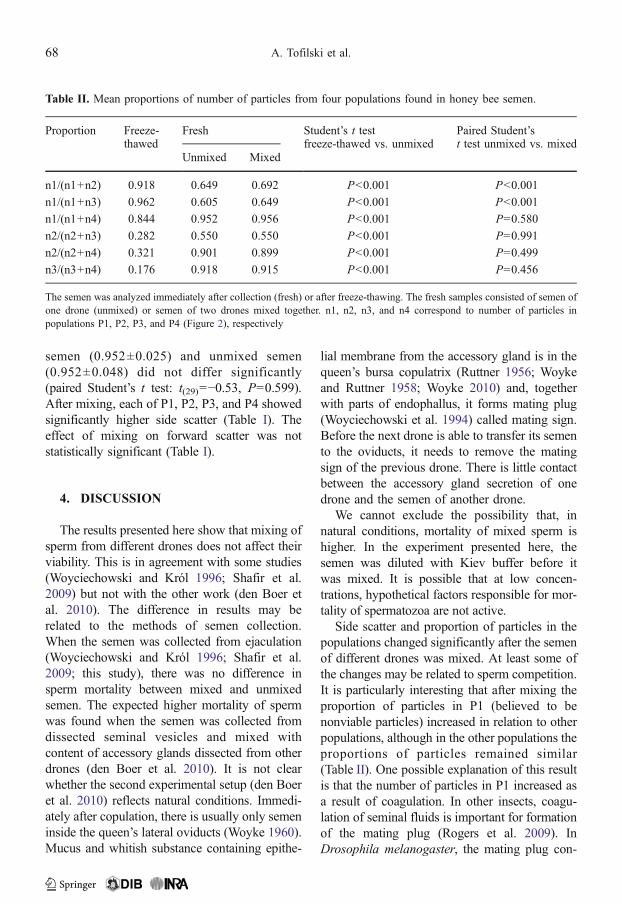
semen (0.952±0.025) and unmixed semen(0.952±0.048) did not differ significantly(paired Student’s t test: t(29)=−0.53, P=0.599).After mixing, each of P1, P2, P3, and P4 showedsignificantly higher side scatter (Table I). Theeffect of mixing on forward scatter was notstatistically significant (Table I).
4. DISCUSSION
The results presented here show that mixing ofsperm from different drones does not affect theirviability. This is in agreement with some studies(Woyciechowski and Król 1996; Shafir et al.2009) but not with the other work (den Boer etal. 2010). The difference in results may berelated to the methods of semen collection.When the semen was collected from ejaculation(Woyciechowski and Król 1996; Shafir et al.2009; this study), there was no difference insperm mortality between mixed and unmixedsemen. The expected higher mortality of spermwas found when the semen was collected fromdissected seminal vesicles and mixed withcontent of accessory glands dissected from otherdrones (den Boer et al. 2010). It is not clearwhether the second experimental setup (den Boeret al. 2010) reflects natural conditions. Immedi-ately after copulation, there is usually only semeninside the queen’s lateral oviducts (Woyke 1960).Mucus and whitish substance containing epithe-
lial membrane from the accessory gland is in thequeen’s bursa copulatrix (Ruttner 1956; Woykeand Ruttner 1958; Woyke 2010) and, togetherwith parts of endophallus, it forms mating plug(Woyciechowski et al. 1994) called mating sign.Before the next drone is able to transfer its semento the oviducts, it needs to remove the matingsign of the previous drone. There is little contactbetween the accessory gland secretion of onedrone and the semen of another drone.
We cannot exclude the possibility that, innatural conditions, mortality of mixed sperm ishigher. In the experiment presented here, thesemen was diluted with Kiev buffer before itwas mixed. It is possible that at low concen-trations, hypothetical factors responsible for mor-tality of spermatozoa are not active.
Side scatter and proportion of particles in thepopulations changed significantly after the semenof different drones was mixed. At least some ofthe changes may be related to sperm competition.It is particularly interesting that after mixing theproportion of particles in P1 (believed to benonviable particles) increased in relation to otherpopulations, although in the other populations theproportions of particles remained similar(Table II). One possible explanation of this resultis that the number of particles in P1 increased asa result of coagulation. In other insects, coagu-lation of seminal fluids is important for formationof the mating plug (Rogers et al. 2009). InDrosophila melanogaster, the mating plug con-
Table II. Mean proportions of number of particles from four populations found in honey bee semen.
Proportion Freeze-thawed
Fresh Student’s t testfreeze-thawed vs. unmixed
Paired Student’st test unmixed vs. mixed
Unmixed Mixed
n1/(n1+n2) 0.918 0.649 0.692 P<0.001 P<0.001
n1/(n1+n3) 0.962 0.605 0.649 P<0.001 P<0.001
n1/(n1+n4) 0.844 0.952 0.956 P<0.001 P=0.580
n2/(n2+n3) 0.282 0.550 0.550 P<0.001 P=0.991
n2/(n2+n4) 0.321 0.901 0.899 P<0.001 P=0.499
n3/(n3+n4) 0.176 0.918 0.915 P<0.001 P=0.456
The semen was analyzed immediately after collection (fresh) or after freeze-thawing. The fresh samples consisted of semen ofone drone (unmixed) or semen of two drones mixed together. n1, n2, n3, and n4 correspond to number of particles inpopulations P1, P2, P3, and P4 (Figure 2), respectively
68 A. Tofilski et al.
sists of PEB-me protein, which shows autofluor-escence (Lung and Wolfner 2001). In primates,there is some evidence that coagulation ofseminal fluids is related to sperm competition(Dixson and Anderson 2002). It is possible thathoney bee proteins present in semen coagulatewhen in contact with the spermatozoa of anotherdrone in order to immobilize them. The coagu-lation on the surface of spermatozoa couldexplain the differences in the side scatter betweenmixed and unmixed samples. It is not clear whatthe source of hypothetical factors causing thecoagulation is. They can be present in seminalfluid filling the space between spermatozoa andtheir origin can be seminal vesicles. Howevermore research is required to confirm the coagu-lation and identify source of the hypotheticalcoagulation factors.
Flow cytometry allowed us to detect in honeybee semen two different populations of livespermatozoa (P2 and P3). This suggests thepresence of sperm polymorphism, which hasbeen found in many systematic groups ofinvertebrates (Swallow and Wilkinson 2002).In honey bee, the morphological differencesbetween sperm populations may be small andtherefore overlooked despite a great manymicroscopic observations. Such small differ-ences can easily be detected by flow cytometry,which can measure a large number of particles.This method has been used to detect subpopu-lations of sperm in semen of boar (Pena et al.2005; Thurston et al. 2001; Abaigar et al. 1999),dog (Martinez et al. 2006), and gazelle (Abaigaret al. 1999). It is important to explain thedifferences between the populations of particlesin honey bee semen; this can be achieved bycombining flow cytometry analysis with fluo-rescence microscopy observations.
It is known from earlier studies based onmicroscopic observations that live spermatozoaare fluorescent green and the dead ones arefluorescent red when stained with SYBR-14/PI(Collins and Donoghue 1999). The same stainingmethod combined with flow cytometry giveslarge samples and makes blind counting easier(Holman 2009). Flow cytometry has been usedto study hemocytes of honey bee (de Graaf et al.
2002) and sperm of ants (Cournault and Aron2008). The freeze-thawing experiment presentedhere confirmed that flow cytometry can be usedto assess the mortality of spermatozoa. Thismethod allows quantitative analysis of semenand permits detection of sperm polymorphism.
ACKNOWLEDGEMENTS
We thank Michael Jacobs and two anonymousreviewers for helpful comments on earlier versions ofthis paper and Beata Ochał and Iwona Zbyryt fortechnical assistance. This work was supported byMinistry of Science and Higher Education grant No.N N311 292436.
Mise en évidence d’une compétition spermatiquechez l’abeille (Apis mellifera) par la méthode decytométrie en flux
Spermatozoïde / compétition spermatique / Apismellifera
Mittel Durchflusszytometrie gewonnene Erkenntnissezur Sperma-Kompetition bei der Honigbienene (Apismellifera)
Honigbiene / Spermien / Sperma-Kompetition
Open Access This article is distributed under the terms ofthe Creative Commons Attribution Noncommercial Licensewhich permits any noncommercial use, distribution, andreproduction in any medium, provided the original author(s)and source are credited.
REFERENCES
Abaigar, T., Holt, W.V., Harrison, R.A.P., del Barrio, G.(1999) Sperm subpopulations in boar (Sus scrofa)and Gazelle (Gazella dama mhorr) semen asrevealed by pattern analysis of computer-assistedmotility assessments. Biol. Reprod. 60, 32–41
Collins, A.M., Donoghue, A.M. (1999) Viability assess-ment of honey bee, Apis mellifera sperm using dualfluorescent staining. Theriogenology 51, 1513–1523
Cournault, L., Aron, S. (2008) Rapid determination ofsperm number in ant queens by flow cytometry.Insect. Soc. 55, 283–287
de Graaf, D.C., Dauwe, R., Walravens, K., Jacobs, F.J.(2002) Flow cytometric analysis of lectin-stained
Flow cytometry analysis of honey bee sperm 69

haemocytes of the honeybee (Apis mellifera). Api-dologie 33, 571–579
den Boer, S.P.A., Baer, B., Boomsma, J.J. (2010)Seminal fluid mediates ejaculate competition insocial insects. Science 327, 1506–1509
Dixson, A.F., Anderson, M.J. (2002) Sexual selection,seminal coagulation and copulatory plug formationin primates. Folia Primatol. 73, 63–69
Franck, P., Coussy, H., LeConte, Y., Solignac, R.,Garnery, L., Cornuet, J.M. (1999) Microsatelliteanalysis of sperm admixture in honeybee. InsectMol Biol. 8, 419–421
Haberl, M., Tautz, D. (1998) Sperm usage in honey bees.Behav. Ecol. Sociobiol. 42, 247–255
Harbo, J.R. (1990) Artificial mixing of spermatozoafrom honeybees and evidence for sperm competi-tion. J. Apic. Res. 29, 151–158
Harshman, L.G., Prout, T. (1994) Sperm displacementwithout sperm transfer in Drosophila melanogaster.Evolution 48, 758–766
Holman, L. (2009) Sperm viability staining in ecologyand evolution: potential pitfalls. Behav. Ecol. Socio-biol. 63, 1679–1688
Laidlaw, H.H. (1944) Artificial insemination of thequeen bee (Apis mellifera L): morphological basisand results. J. Morphol. 74, 429–465
Laidlaw Jr., H.H. (1989) Instrumental insemination ofhoney bee queens. Dadant & Sons, Hamilton
Lung, O., Wolfner, M.F. (2001) Identification andcharacterization of the major Drosophila mela-nogaster mating plug protein. Insect Biochem Mol.Biol. 31, 543–551
Martinez, I.N., Moran, J.M., Pena, F.J. (2006) Two-stepcluster procedure after principal component analysisidentifies sperm subpopulations in canine ejaculates andits relation to cryoresistance. J. Androl. 27, 596–603
Moritz, R.F.A. (1986) Intracolonial worker relationshipand sperm competition in the honeybee (Apismellifera L.). Experientia 42, 445–448
Page, R.E. (1986) Sperm utilization in social insects.Ann. Rev. Entomol. 31, 359–361
Pena, F.J., Saravia, F., Garcia-Herreros, M., Nunezmartinez,I., Tapia, J.A., Johannisson, A., Wallgren, M.,Rodriguez-Martinez, H. (2005) Identification of spermmorphometric subpopulations in two different portions
of the boar ejaculate and its relation to postthawquality. J. Androl. 26, 716–723
Peng, C.Y., Yin, C., Yin, L.R.S. (1992) Effect of rapidfreezing and thawing on cellular integrity of honeybee sperm. Physiol. Entomol. 17, 269–276
Rogers, D.W., Baldini, F., Battaglia, F., Panico, M., Dell, A.,Morris, H.R., Catteruccia, F. (2009) Transglutaminase-mediated semen coagulation controls sperm storage inthe malaria mosquito. PLoS Biol. 7, e1000272
Ruttner, F. (1956) Zur Frage der Spermaübertragung beider Bienenkönigin. Insect. Soc. 3, 351–359
Schlüns, H., Moritz, R.F.A., Neumann, P., Kryger, P.,Koeniger, G. (2005) Multiple nuptial flights, spermtransfer and the evolution of extreme polyandry inhoneybee queens. Anim. Behav. 70, 125–131
Shafir, S., Kabanoff, L., Duncan, M., Oldroyd, B.P.(2009) Honey bee (Apis mellifera) sperm competi-tion in vitro - two are no less viable than one.Apidologie 40, 556–561
Swallow, J.G., Wilkinson, G.S. (2002) The long andshort of sperm polymorphisms in insects. Biol. Rev.77, 153–182
Thurston, L.M., Watson, P.F., Mileham, A.J., Holt, W.V.(2001) Morphologically distinct sperm subpopula-tions defined by Fourier shape descriptors in freshejaculates correlate with variation in boar semenquality following cryopreservation. J. Androl. 22,382–394
Woyciechowski,M., Kabat, L., Krol, E. (1994) The functionof the mating sign in honey bees, Apis mellifera L.:new evidence. Anim. Behav. 47, 733–735
Woyciechowski, M., Król, E. (1996) On intraoviductalsperm competition in the honeybee (Apis mellifera).Folia Biol. (Krakow) 44, 1–2
Woyke, J. (1956) Anatomo-physiological changes in queen-bees returning from mating flights, and the process ofmultiple mating. Bull. Acad. Polon. Sci. 4, 81–87
Woyke, J. (1960) Natural and artificial insemination ofqueen honey bees. Pszczel. Zesz. Nauk. 4, 183–273
Woyke, J. (2010) Three substances ejected byApis melliferadrones from everted endophallus and during naturalmatings with queen bees. Apidologie 41, 613–621
Woyke, J., Ruttner, F. (1958) An anatomical study ofthe mating process in the honeybee. Bee World39, 3–18
70 A. Tofilski et al.