DNA Ligase C and Prim-PolC participate in base excision...
13
DNA Ligase C and Prim-PolC participate in base excision repair in mycobacteria Article (Published Version) http://sro.sussex.ac.uk Plocinski, Przemyslaw, Brissett, Nigel C, Bianchi, Julie, Brzostek, Anna, Korycka-Machała, Małgorzata, Dziembowski, Andrzej, Dziadek, Jaroslaw and Doherty, Aidan J (2017) DNA Ligase C and Prim-PolC participate in base excision repair in mycobacteria. Nature Communications, 1251 (2017). ISSN 2041-1723 This version is available from Sussex Research Online: http://sro.sussex.ac.uk/id/eprint/70317/ This document is made available in accordance with publisher policies and may differ from the published version or from the version of record. If you wish to cite this item you are advised to consult the publisher’s version. Please see the URL above for details on accessing the published version. Copyright and reuse: Sussex Research Online is a digital repository of the research output of the University. Copyright and all moral rights to the version of the paper presented here belong to the individual author(s) and/or other copyright owners. To the extent reasonable and practicable, the material made available in SRO has been checked for eligibility before being made available. Copies of full text items generally can be reproduced, displayed or performed and given to third parties in any format or medium for personal research or study, educational, or not-for-profit purposes without prior permission or charge, provided that the authors, title and full bibliographic details are credited, a hyperlink and/or URL is given for the original metadata page and the content is not changed in any way.
Transcript of DNA Ligase C and Prim-PolC participate in base excision...
DNA Ligase C and PrimPolC participate in base excision repair in mycobacteria
Article (Published Version)
http://sro.sussex.ac.uk
Plocinski, Przemyslaw, Brissett, Nigel C, Bianchi, Julie, Brzostek, Anna, Korycka-Machała, Małgorzata, Dziembowski, Andrzej, Dziadek, Jaroslaw and Doherty, Aidan J (2017) DNA Ligase C and Prim-PolC participate in base excision repair in mycobacteria. Nature Communications, 1251 (2017). ISSN 2041-1723
This version is available from Sussex Research Online: http://sro.sussex.ac.uk/id/eprint/70317/
This document is made available in accordance with publisher policies and may differ from the published version or from the version of record. If you wish to cite this item you are advised to consult the publisher’s version. Please see the URL above for details on accessing the published version.
Copyright and reuse: Sussex Research Online is a digital repository of the research output of the University.
Copyright and all moral rights to the version of the paper presented here belong to the individual author(s) and/or other copyright owners. To the extent reasonable and practicable, the material made available in SRO has been checked for eligibility before being made available.
Copies of full text items generally can be reproduced, displayed or performed and given to third parties in any format or medium for personal research or study, educational, or not-for-profit purposes without prior permission or charge, provided that the authors, title and full bibliographic details are credited, a hyperlink and/or URL is given for the original metadata page and the content is not changed in any way.
ARTICLE
DNA Ligase C and Prim-PolC participate in baseexcision repair in mycobacteriaPrzemysław Płociński1,2, Nigel C. Brissett1, Julie Bianchi1,4, Anna Brzostek2, Małgorzata Korycka-Machała2,Andrzej Dziembowski3, Jarosław Dziadek2 & Aidan J. Doherty1
Prokaryotic Ligase D is a conserved DNA repair apparatus processing DNA double-strand
breaks in stationary phase. An orthologous Ligase C (LigC) complex also co-exists in many
bacterial species but its function is unknown. Here we show that the LigC complex interacts
with core BER enzymes in vivo and demonstrate that together these factors constitute an
excision repair apparatus capable of repairing damaged bases and abasic sites. The poly-
merase component, which contains a conserved C-terminal structural loop, preferentially
binds to and fills-in short gapped DNA intermediates with RNA and LigC ligates the resulting
nicks to complete repair. Components of the LigC complex, like LigD, are expressed upon
entry into stationary phase and cells lacking either of these pathways exhibit increased
sensitivity to oxidising genotoxins. Together, these findings establish that the LigC complex is
directly involved in an excision repair pathway(s) that repairs DNA damage with ribonu-
cleotides during stationary phase.
DOI: 10.1038/s41467-017-01365-y OPEN
1 Genome Damage and Stability Centre, School of Life Sciences, University of Sussex, Brighton, BN1 9RQ, UK. 2 Institute of Medical Biology, Polish Academyof Sciences, Lodowa 106, 93-232 Lodz, Poland. 3 Institute of Biochemistry and Biophysics, Polish Academy of Sciences, Pawińskiego 5A, 02-106 Warsaw,Poland. 4Present address: Department of Oncology-Pathology, Cancer Center Karolinska, Karolinska Institutet, R8:04, Karolinska Universitetssjukhuset Solna,171 76 Stockholm, Sweden. Przemysław Płociński and Nigel C. Brissett contributed equally to this work. Correspondence and requests for materials should beaddressed to A.J.D. (email: [email protected])
NATURE COMMUNICATIONS |8: 1251 |DOI: 10.1038/s41467-017-01365-y |www.nature.com/naturecommunications 1
1234
5678
90
In bacteria, canonical primer synthesis during DNA replicationis carried out by enzymes from the DnaG superfamily1, 2. Incontrast, priming of replication in archaea and eukaryotes is
performed by members of the archaeo-eukaryotic primase (AEP)superfamily3, 4. However, AEPs are also widely distributed inmost bacterial species4, where they have evolved to fulfil divergentroles and have recently been reclassified as a family of poly-merases called primase-polymerases (Prim-Pols) to better reflecttheir evolutionary origins and more diverse roles in DNA meta-bolism4. The best characterised bacterial AEP is Prim-PolD(PolDom) that forms part of a multifunctional non-homologousend-joining (NHEJ) DNA break repair complex called Ligase D(LigD). In mycobacterial LigD, an AEP is fused to phosphoes-terase and ATP-dependent DNA ligase domains that, togetherwith the Ku repair factor, coordinate the sequential synapsis,processing and repair of double-strand breaks (DSBs) in sta-tionary phase5–9. However, in many other species these domainsare encoded by separate operonically associated genes6, 10.
Many bacterial species, including Actinobacteria, encode mul-tiple ATP-dependent DNA ligases and Prim-Pols similar to those
found in mycobacterial LigD. Exemplary classes of Prim-Pols andDNA ligases in selected gram-positive bacteria are shown inSupplementary Fig. 1. Mycobacterium smegmatis encodes fourdistinct primase-polymerases. Although it is known that thePrim-PolD subunit of LigD is involved in the NHEJ repaircomplex, the roles of the other stand-alone Prim-Pols remainunknown. One orthologue, MSMEG_6301 (Prim-PolC/LigC Pol/PolD) is encoded in the genomic proximity of two DNA ligasegenes (LigC1: msmeg_6302 and LigC2: msmeg_6304). AlthoughLigD’s role in NHEJ-mediated repair is firmly established7, 9, 11,the pathways in which these other ligases and Prim-Pols operatein remain unclear. Due to their similarities with the NHEJcomplex, it was proposed that DNA ligase C and operonicallyassociated Prim-PolC (LigC-Pol or PolD1) function as a “back-up” complex for the LigD pathway12, 13, 14, although this has notbeen proven. While the basic biochemical characteristics of Prim-PolC and PolD2, another closely related AEP, were partiallydescribed in a previous study12, Prim-PolC failed to act as a“back-up” of Prim-PolD during DSB repair. Another studyreported that LigC was responsible for ligation of ~20% of DSBs
PolD2
PoIA
LigC1
EndoIV
XthA
ExoIII
MPG
FPG
NTH
Protein identifierPeptNo.
M.W.[kDa]
IntensityLigC1
IntensityLigC2
IntensityPrimPolC
MS_6302 LigC1 16 39.8 3.26E+09MS_0001 DNA Pol III, beta subunit (dnaN) [2.7.7.7] 14 41.3 9.56E+08 4.98E+08 5.70E+09MS_2731 ATPase involved in DNA repair 13 48.5 3.61E+08 3.56E+08 2.10E+09MS_3839 DNA polymerase I [2.7.7.7] 31 101 2.78E+08 4.08E+08 5.06E+09MS_6896 single-stranded DNA-binding protein 4 17.4 2.48E+08 3.43E+08 2.11E+09MS_5156 base excision DNA repair protein 3 20.9 3.85E+07 1.44E+07 4.46E+08MS_1383 endonuclease IV 4 26.6 2.18E+07 6.14E+08MS_2399 uracil-DNA glycosylase (ung) [3.2.2.–] 2 26.6 1.46E+07 1.00E+08MS_3816 excinuclease ABC, B subunit (uvrB) 14 80.8 1.33E+07 8.44E+07 1.16E+09MS_5423 transcription-repair coupling factor (mfd) 9 131 3.63E+06 4.17E+06 4.00E+08MS_3883 5′–3 exonuclease 3 33.9 2.71E+07MS_5440 deoxyribonuclease 4 33.4 2.59E+07 4.07E+08MS_6304 LigC2 4 39.3 2.90E+08MS_6285 DNA Pol III gamma/tau subunit [2.7.7.7] 5 69.9 4.22E+08MS_6301 Prim-PolC 15 41.6 1.13E+10MS_2362 DNA ligase (ligA) [6.5.1.2] 16 76.3 1.69E+08 1.43E+08 2.35E+09
Nucleases
Prim-PolC
XthA ExoIII LigC1
XseB BSA PolD2
EndoIV Prim-PolC
ExoIIIA LigC1ExoIII
XseB BSA
EndoIV Prim-PolC
XthA ExoIII
XseB
EndoIV LigC1
ExoIIIA ExoIII
XseB PolD2BSA
PolD2BSA
EndoIV LigC1
MPG PolA
Ku
Ku
Ku
Ku
Nth
Nth
Nth
Nth
LigC1 FPG
MPG PolA
LigC1 FPG
MPG PolA
Prim-PolC
Prim-PolC
FPG
MPG PolA
Prim-PolC
Prim-PolC
FPG
+ Interactor + anti- Prim-PolC+ Interactor + anti- LigC1
– Interactor + anti- LigC1 – Interactor + anti- Prim-PolC
PolD2
a c
b
Primases /polymerases
DNAglycosylases
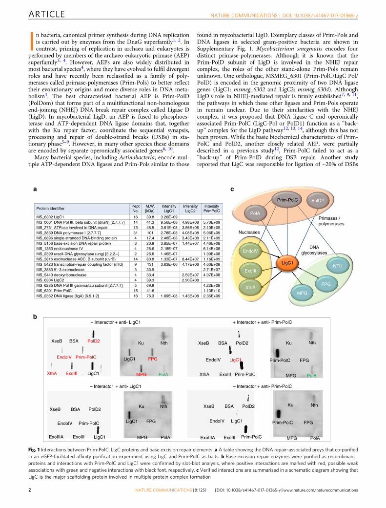
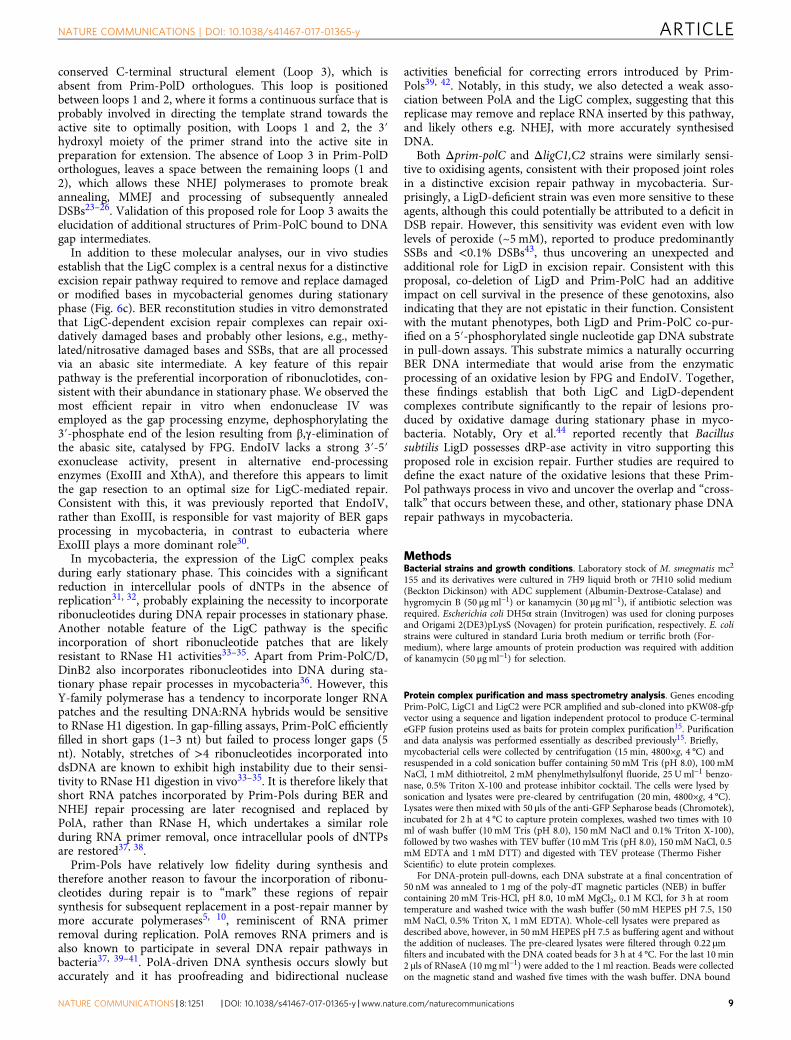
Fig. 1 Interactions between Prim-PolC, LigC proteins and base excision repair elements. a A table showing the DNA repair-associated preys that co-purifiedin an eGFP-facilitated affinity purification experiment using LigC and Prim-PolC as baits. b Base excision repair enzymes were purified as recombinantproteins and interactions with Prim-PolC and LigC1 were confirmed by slot-blot analysis, where positive interactions are marked with red, possible weakassociations with green and negative interactions with black font, respectively. c Verified interactions are summarised in a schematic diagram showing thatLigC is the major scaffolding protein involved in multiple protein complex formation
ARTICLE NATURE COMMUNICATIONS | DOI: 10.1038/s41467-017-01365-y
2 NATURE COMMUNICATIONS | 8: 1251 |DOI: 10.1038/s41467-017-01365-y |www.nature.com/naturecommunications
in a LigD mutant strain containing a mutation disrupting theligase function but possessing functional primase and exonucleasedomains that can still participate in the NHEJ14.
Here we sought to elucidate the role(s) of LigC, and its asso-ciated polymerase (Prim-PolC), in mycobacterial DNA metabo-lism. We show that components of the LigC complex act togetherto preferentially fill in short DNA gaps with ribonucleotides(rNTPs), followed by sealing of the resulting nicks. We identifythat LigC operon-encoded proteins associate with components ofthe base excision repair (BER) apparatus in vivo and, togetherwith the excision repair machinery, form repair complexes thatremove damaged DNA and abasic sites and fill in resulting gapswith rNTPs. Structural studies reveal that a conserved C-terminalelement of Prim-PolC forms an extended surface surrounding theactive site, which likely supports gap recognition and synthesis.Components of the LigC complex are expressed upon entry intostationary phase, when the intracellular pool of dNTPs is natu-rally depleted, necessitating the preferential incorporation of
rNTPs after lesion removal. Abrogation of the LigC complex,results in an increased sensitivity of mycobacterial cells to oxi-dative damaging agents. LigD-deficient cells are also sensitive tooxidative damaging agents, uncovering an unexpected duel role inboth excision and DSB repair. Together, these findings establishthat the LigC complex operates in excision repair pathways thatspecifically mends lesions and single-strand breaks in stationaryphase.
ResultsDNA Ligase C complex interacts with BER enzymes in vivo.Although LigD-associated Prim-Pols and ligases facilitate NHEJrepair of DSBs in bacteria, the functions of closely relatedorthologues remain unclear. To investigate the cellular roles ofPrim-PolC, and its operonically associated DNA ligases (LigC1and LigC2), we first sought to define their physical associationswith other proteins/complexes in mycobacterial cells. We
pss-0
PP
C
PP
D
pss-6pss-4
50 nt
1 nt Gap
14 nt
26 ntpss-6
14 ntpss-0 ATAC 3’
CCGG 3′
CCCGGG 3′
5′5′3′
5′5′3′
5′5′3′
26 nt
14 nt
26 ntpss-4
pss-0
50 nt
pss-4 pss-6
1 nt Gap/5′-
1 nt Gap/5′- 3′Ov/5′-
0.0125 0.025 0.1 0.25 0.5 1 5 10
DN
A b
ound
(%
) 1 nt Gap1 nt Gap/5′-
0.025 0.05 0.075 0.1
PPC 1 nt Gap/5′-PPD 1 nt Gap/5′-PPC 3′ Ov/5′-PPD 3′ Ov/5′-
100
80
60
40
20
0
DN
A b
ound
(%
)
100
80
60
40
20
0
Prim-PolC (μM)
Protein (μM)
P
P
P
P
P
P
PP
Prim-PolCPrim-PolC
Prim-PolDPrim-PolC Prim-PolC Prim-PolDφφ
φ PP
C
PP
D
φPP
C
PP
D
φPP
C
PP
D
φPP
C
PP
DφP
PC
PP
D
φ
a
b
c d
Fig. 2 DNA binding activity of Prim-PolC. a Prim-PolC prefers to bind to 5′ phosphorylated gaps. In EMSAs, mixtures of a single nucleotide gap containingsubstrate (30 nM), with or without phosphorylation of the 5′ end of the lesion, and 0–10 μM Prim-PolC were incubated for 20min and resolved on a nativepolyacrylamide gel. A filled triangle indicates unshifted probe, whereas the black arrow probably indicates a binary complex of Prim-PolC and DNA. Thegrey arrow indicates multiple protein monomers bound to the probe. Quantification of the EMSA data is presented. For each Prim-PolC concentration thepercentage of DNA bound (in relation to the total DNA) was calculated and compared for EMSAs containing Prim-PolC binding a 1 nucleotide gap with orwithout 5′-phosphorylation. b Prim-PolC prefers to bind to gaps rather than overhangs unlike Prim-PolD. This EMSA uses 30 nM of 5′-fluorescein labelled36-mer containing a 5′ phosphorylated single nucleotide gap or 30 nM of 5′-fluorescein labelled 36-mer containing a single-stranded 3′ overhang given byphosphorylation of the 5′-end of the gap and no primer strand. The triangles and arrow are as in a. Using the same conditions as a, this time comparingPrim-PolC to Mtu Prim-PolD (25, 50, 75 and 100 nM). Quantification of the EMSA data are presented. For each enzyme concentration the percentage ofDNA bound (in relation to the total DNA) was calculated and compared for EMSAs containing Prim-PolC or Prim-PolD binding a 5′-phosphorylated 1nucleotide gap or a 3′-overhang with a 5′-phosphorylated downstream strand. c Prim-PolC is not an NHEJ polymerase. A schematic of 5′-fluoresceinlabelled substrates used in a MMEJ activity assay. The pss-number refers to the number of bases at the end of the 3′overhang that can base pair with itself.A primer extension assay, 30 nM of the denoted substrate extended by 300 nM enzyme (PPC-Prim-PolC, PPD-Mtu Prim-PolD) in the presence of 250 µMdNTPs mix for 30min at 37 °C. The products are resolved on a 15% denaturing gel. d The same reaction products from c, after treatment with Proteinase Kand run on a 12% non-denaturing gel
NATURE COMMUNICATIONS | DOI: 10.1038/s41467-017-01365-y ARTICLE
NATURE COMMUNICATIONS |8: 1251 |DOI: 10.1038/s41467-017-01365-y |www.nature.com/naturecommunications 3
employed an eGFP-tag (enhanced green fluorescent protein)facilitated co-purification approach, in which each of the proteins(Prim-PolC, LigC1 and LigC2) were fused to eGFP to serve asbaits for the purification and identification of cellular partners.Purified complexes were cross-linked with a reversible cross-lin-ker, 3,3′-dithiobis (sulfosuccinimidyl propionate) (DSSP), washedand subjected to mass spectrometry (MS) analysis15. Complexeswere purified from cells grown to late stationary phase. Asexpected, tagged LigC and Prim-PolC were among most abun-dant “hits” in the MS samples, supporting the specificity of thepurification process. A list of polypeptides that co-purified witheach bait, known to be associated with DNA repair processes, areshown in Fig. 1a. A full list of all co-purified proteins is providedin Supplementary Data 1. Significantly, all three bait proteins co-purified with multiple repair enzymes, including the major DNAglycosylases and nucleases involved in base excision repair (BER)repair pathways. Reverse experiments using mycobacterial eGFP-tagged endonuclease IV as bait in M. bovis BCG resulted in co-purification of LigC, confirming that these two proteins form acomplex in vivo (Supplementary Data 2). Together, these in vivostudies reveal that Prim-PolC and LigC interact with BER com-ponents, suggesting that they function primarily in the repair ofdamaged bases, abasic sites and single-strand breaks (SSBs).
DNA ligase C complex interacts with BER enzymes in vitro. Tovalidate the interactions of LigC1 and Prim-PolC with compo-nents of the BER machinery identified in our pull-down studies,we expressed and purified recombinant forms of each of theseBER enzymes (Supplementary Fig. 2). Taking advantage of Prim-PolC and LigC1-specific antibodies, a slot blotting-based meth-odology was employed to authenticate the interactions betweenthe identified proteins. We also used this approach to address ifPrim-PolC or LigC1 interacted with Ku, to determine if they alsofunction in NHEJ repair. Mono-functional DNA glycosylase(MPG) was purified alongside bifunctional glycosylases (FPG andNth), that possess abasic site lyase activity, as well as several of the
key end-processing nucleases including: exodeoxyribonucleaseVIIB (ExoVIIB), endonuclease IV (EndoIV) and both exonu-clease III paralogues (ExoIII, XthA) from M. smegmatis. PolD2,the Prim-Pol with closest similarity to Prim-PolC, as well as themajor BER repair polymerase PolA, were also purified for thesein vitro interaction studies. Recombinant proteins were spottedonto nitrocellulose membrane and incubated with either Prim-PolC or LigC1, respectively, followed by extensive washing andsubsequent detection by western blotting using Prim-PolC/LigC1-specific antibodies. We observed significant signals with many ofthe BER proteins incubated with LigC1 (Fig. 1b). MPG and FPG(MutM) glycosylases exhibited the strongest interactions withLigC1. LigC1 also interacted with EndoIV, ExoIII and XthA end-processing enzymes and both Prim-PolC and PolD2. A summaryof the major interactions identified in these studies is shown inFig. 1c. In contrast, Prim-PolC only interacts with LigC1 but notwith either Ku or ExoVIIB, which therefore served as controlproteins for these experiments. Together, these data establish thatLigC appears to act as a key scaffolding protein, akin to ligase IIIin eukaryotes16, 17, required for sequential recruitment of BERrepair enzymes and therefore potentially coordinating the pro-cessing and repair of lesions and SSBs in vivo18–20. To furthervalidate the interactions with the glycosylase, we performed pull-down assays of LigC1 with the two potentially interacting gly-cosylases, FPG and MPG and observed a strong association oftag-free LigC1 with FPG coated His-trap beads and a weakerinteraction with MPG (Supplementary Fig. 3).
Previously, comparative sequence database mining analysis ofbacterial operons uncovered a genetic link between LigD and Ku,which led to the identification of the NHEJ pathway inprokaryotes21, 22. Similar in silico comparative operon analysisof many mycobacterial genomes revealed that the LigC and Prim-PolC genes are co-transcribed with the major base excision repairbifunctional glycosylase, FPG, in operons present inM. avium,M.intracellulare and Mycobacterium JS623, a close relative of M.smegmatis. Notably, these operons are also located within closegenomic proximity of other DNA excision repair enzymes (PolA
1nφ 2 3 5 ov n 1 2 3 5 n 1 2 3 5 n 1 2 3 5ov
P
+1+2
0
+3
+4
rNTPs
nφ φ
+3
+10
+2
dNTPs
1 2 3 5 ov n 1 2 3 5 n 1 2 3 5 n 1 2 3 5ov
–5′
3′3′5′
0–5 nt
5′3′
dNTPs/5′- P
dNTPs dNTPs/5′- P
PrNTPs/5′-
rNTPs PrNTPs/5′-b
a
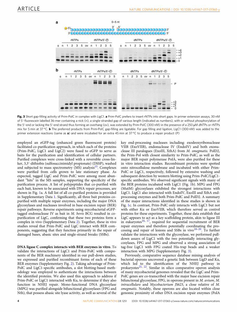
Fig. 3 Short gap-filling activity of Prim-PolC in complex with LigC1. a Prim-PolC prefers to insert rNTPs into short gaps. In primer extension assays, 30 nMof 5′-fluorescein labelled 36-mer containing a nick (n), a single-stranded gap of various length (indicated as numbers), with or without phosphorylation ofthe 5′-end or lacking the 5′-end strand thus forming an overhang (ov), was extended by Prim-PolC (300 nM) in the presence of a 250 µM dNTPs or rNTPsmix for 5 min at 37 °C. b The preferred products from Prim-PolC gap-filling are ligatable. For gap filling and ligation, LigC1 (300 nM) was added to theprimer extension reactions (same as a) and were incubated for an extra 45min at 37 °C to produce a repair product (P)
ARTICLE NATURE COMMUNICATIONS | DOI: 10.1038/s41467-017-01365-y
4 NATURE COMMUNICATIONS | 8: 1251 |DOI: 10.1038/s41467-017-01365-y |www.nature.com/naturecommunications
and UvrB; Supplementary Fig. 3). Together, these findings furtherstrengthen a functional connection between LigC-related proteinsand BER processes in mycobacteria.
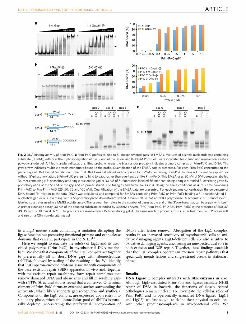
Prim-PolC preferentially binds to short gapped DNA. Follow-ing validation of the association of Prim-PolC and LigC1 withcomponents of the BER machinery, we proceeded to biochemi-cally characterize the capacity of these enzymes to process DNArepair intermediates that arise during the processing of BERsubstrates, e.g., abasic sites. We first performed electrophoreticmobility shift assays (EMSAs) to determine the optimal DNAsubstrates for binding and processing by Prim-PolC. The mostcommon BER intermediates are short DNA gaps containing aphosphate moiety on the 5′ terminus, that arise from base exci-sion and subsequent abasic site removal. We therefore testedPrim-PolC’s capacity to bind to an array of phosphorylated andnon-phosphorylated DNA substrates, containing various gaplengths. Notably, Prim-PolC’s binding affinity was significantlystimulated by the presence of a 5′-phosphate group within a gapand it bound most avidly to short gaps of 1–3 nucleotides (Fig. 2a;Supplementary Fig. 4a). In contrast with its NHEJ-specificorthologue, Prim-PolC bound much more weakly to DNA sub-strates containing overhangs (Fig. 2b).
NHEJ Prim-Pols bind strongly to 5′ phosphorylated DNAoverhangs23 and can directly induce break synapsis23–26. Todetermine if Prim-PolC also possesses end-synapsis activity, we
employed a microhomology-mediated end-joining (MMEJ) assaythat utilises substrates that mimic DSBs to measure this activityin vitro27. As expected, Prim-PolD efficiently promoted breaksynapsis and subsequent in-trans extension of MMEJ intermedi-ates (Fig. 2c, d). In contrast, Prim-PolC did not exhibit significantMMEJ activity, indicating that DSBs are not preferred substratesfor this enzyme. This identifies another significant difference insubstrate specificities between these two classes of Prim-Pols.
To cross-validate the observed DNA substrate bindingaffinities, we carried out additional DNA binding studies usingpolyA-tailed DNA substrates coated onto magnetic particles.Beads pre-coated with different DNA substrates were incubatedwith whole-cell mycobacterial lysates, washed extensively andbound preys were identified by mass spectrometry. As expected,LigD and Ku were both found to co-purify on DSB-like substrateswith free ends. However, neither Ku or LigD bound to thissubstrate when the terminal nucleotides were blocked withS-bond. Notably, Prim-PolC and LigD, but not Ku, co-purified ona substrate containing a single nucleotide gap, suggesting thatboth may contribute to the repair of such intermediates in vivo(Supplementary Fig. 4b, Supplementary Data 3).
LigC complex repairs short gapped DNA intermediates. Toestablish the functional importance of Prim-PolC’s preferentialgap binding activity, we next performed primer-template exten-sion assays using gapped substrates (1, 2, 3 or 5 nt gap) in the
XthA ExoIII EndoIV
XthA ExoIII EndoIV
··· ······ ······ ······ ···
··· ···
··· ···
··· ···
8OxoG
+UNG
dU
+FPG +Prim-PolC +LigC
Abasic site
DNA lesion
+FPG
5′5′
5′ 5′
5′ 5′5′ 5′
5′ 5′5′ 5′
LigC + PPolCLigC + PPolCLigC + PPolC FPGFPG
80xo
G
FPG
UNGUNG
UNGFPG
FPGFPG
80xo
G
80xo
G
LigC + PPolC LigC + PPolCLigC + PPolC
RNA
RNA
dU
+XthA/ExoIII/EndoIV
5′ 5′3′
3′- , 5′- -gap Fill-in product Ligated DNAP P 3′-OH, 5′- -gapP
a
b
c
P
PP
P
P P
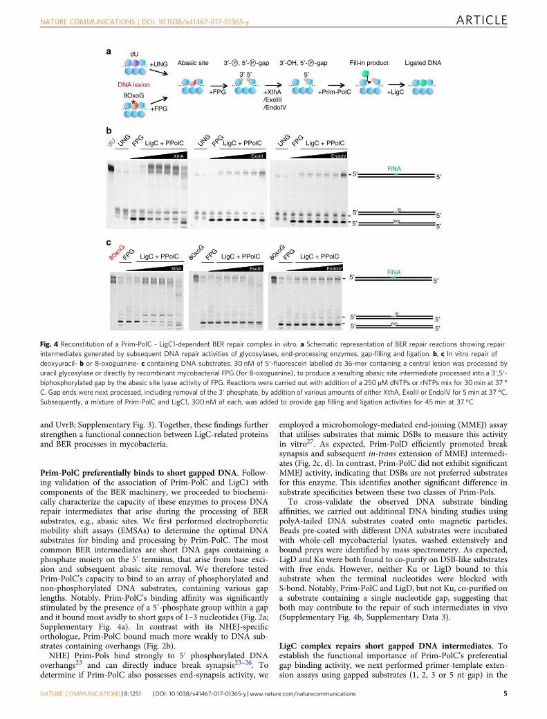
Fig. 4 Reconstitution of a Prim-PolC - LigC1-dependent BER repair complex in vitro. a Schematic representation of BER repair reactions showing repairintermediates generated by subsequent DNA repair activities of glycosylases, end-processing enzymes, gap-filling and ligation. b, c In vitro repair ofdeoxyuracil- b or 8-oxoguanine- c containing DNA substrates. 30 nM of 5′-fluorescein labelled ds 36-mer containing a central lesion was processed byuracil glycosylase or directly by recombinant mycobacterial FPG (for 8-oxoguanine), to produce a resulting abasic site intermediate processed into a 3′,5′-biphosphorylated gap by the abasic site lyase activity of FPG. Reactions were carried out with addition of a 250 µM dNTPs or rNTPs mix for 30min at 37 °C. Gap ends were next processed, including removal of the 3′ phosphate, by addition of various amounts of either XthA, ExoIII or EndoIV for 5 min at 37 °C.Subsequently, a mixture of Prim-PolC and LigC1, 300 nM of each, was added to provide gap filling and ligation activities for 45min at 37 °C
NATURE COMMUNICATIONS | DOI: 10.1038/s41467-017-01365-y ARTICLE
NATURE COMMUNICATIONS |8: 1251 |DOI: 10.1038/s41467-017-01365-y |www.nature.com/naturecommunications 5
presence of intracellular levels of divalent cations. Consistent withthe DNA binding results, Prim-PolC most efficiently processedshort DNA gaps (Fig. 3a), with its polymerisation activity sig-nificantly stimulated by the presence of a phosphate group at the5′ terminus of the gap (Fig. 3a). In common with Prim-PolD,Prim-PolC also preferentially inserted rNTPs during gap-fillingsynthesis (Fig. 3a). This intrinsic preference for NTPs over dNTPswas quantified using a nucleotide competition assay (Supple-mentary Fig. 5a)23. This assay revealed that its nucleotide pre-ference factor (F) was ~190-fold in favour of rNTP incorporation,which compares to an F-value of ~70-fold for Mt Prim-PolD(PolDom)23.
The implication of this finding is that gap-filling synthesis byPrim-PolC would preferentially place RNA, or at least a singlerNTP, on the 3′ side of the nick that must then be ligated to DNAon the opposite 5′ side. To test this prediction, we evaluated theligation specificity of LigC1 using nicked DNA substratescontaining either DNA or RNA on the 3′ side and aphosphorylated DNA strand on the 5′ side. LigC1 preferentiallyligated hybrids of RNA and DNA within the repaired gap, ratherthan more typical DNA:DNA nicks (Supplementary Fig. 5b),consistent with the previously reported ligation specificity ofLigD10, 14. As repair synthesis by Prim-PolC would result in theproduction of such hybrid RNA:DNA nicks in vitro, we nexttested whether the two enzymes co-operatively repair short DNAgaps. Together, Prim-PolC and LigC1 efficiently filled in andligated short (1−3 nt) gaps with RNA but were much less efficientat repairing longer DNA gaps (Fig. 3b), consistent with thereduced DNA binding and extension activities of Prim-PolCobserved previously on these substrates (Fig. 3a and Supplemen-tary Fig. 4a).
Reconstituting a LigC-dependent BER repair pathway in vitro.To establish whether the LigC apparatus operates as part of afunctional BER complex, we sought to reconstitute a complete
excision repair pathway in vitro using Prim-PolC, LigC1 and theinteracting BER proteins. We employed oligonucleotide sub-strates containing either deoxyuracil (dU) or 8-oxoGuanine(8-oxoG) incorporated in a central position of the labelled DNAstrand to enable us to follow the consecutive enzymatic proces-sing steps of BER (Fig. 4a). dU-containing substrates wereinitially treated with UDG glycosylase and the resulting abasic sitesubstrate was then tested with the same enzyme combinationsalso assayed on 8-oxoG containing substrates. We observed thatFPG removed the oxidatively damaged DNA base and processedthe resulting abasic site with its lyase activity (Fig. 4b, c). The3′ end of the gap was subsequently processed by either one of the3′-phosphatase and/or 3′-5′ exonuclease activities of EndoIV,ExoIII or XthA. The resulting DNA gap was subsequently filled inwith RNA by Prim-PolC and, finally, the resulting nick wasligated by LigC1. We observed that EndoIV was the most bene-ficial nuclease for ensuring proper repair in vitro as it lackedstrong 3′-5′ exonuclease activity. Together, these findings estab-lish that BER end-processing enzymes, shown to interact withLigC1 in vivo, facilitate LigC complex-mediated repair of lesionsarising from abasic site formation in vitro.
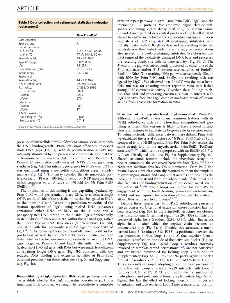
Structure of a mycobacterial LigC-associated Prim-Pol.Although Prim-PolC shares many common features with itsNHEJ orthologue, such as 5′ phosphate recognition and gap-filling synthesis, this enzyme is likely to have evolved distinctstructural features to facilitate its bespoke role in excision repair.To define molecular differences between these distinct Prim-Pols,we elucidated the crystal structure ofMs Prim-PolC (Table 1) andcompared it to a NHEJ-specific Prim-Pol. Prim-PolC retains thesame overall fold of the mycobacterial Prim-PolD (PolDom)structure23–26, which can be superposed with an RMSD of 1.712Å (over 270 aligned positions, Fig. 5a; Supplementary Fig. 6a).Shared structural features include the phosphate recognitionpocket containing the conserved basic residues (K23, K35 andN20) that facilitate this key DNA interaction. Prim-PolC alsoretains Loops 1, which is critically required to orient the template/3′ overhanging strand, and Loop 2 that accepts and positions theincoming primer strand from the adjacent side of the break andalso facilitates the binding/activation of the second metal ion inthe active site23–26. These loops are critical for Prim-PolD’sengagement with the break termini, promoting end-synapsis/MMEJ and are required for activation of the catalytic centre toallow DNA synthesis to commence25, 26.
Despite these similarities, Prim-PolC orthologues possess astrictly conserved C-terminal extension whose function has notbeen ascribed (Fig. 5b). In the Prim-PolC structure, we observethat this additional C-terminal region (aa 294–336) consists of aconserved alpha helix (residues G299−R311), which lies acrossalpha helix 4 after which the peptide chain becomes anunstructured loop (Fig. 5a, b). Notably, this structural element,termed Loop 3 (residues G313−P333), is positioned between thetwo prominent surface loops (1 and 2) that together form acontinuous surface on one side of the active site pocket (Fig. 5c,Supplementary Fig. 6b). Apical Loop 1 residues, normallyinvolved in template strand orientation23–26, are not conservedand are instead repurposed for locking Loop 3 into position(Supplementary Figs. 6b, 7). Residue P94 packs against a pocketformed of residues Y321, P322, K323 and M324 from Loop 3.This also results in Loop 1 adopting a position more proximal tothe active site. Loop 2 residue W219 interacts with Loop 3residues P316, Y317, P333 and K335 via a mixture ofhydrophobic and polar interactions (Supplementary Figs. 6b, 7).Again, this has the effect of locking Loop 3 into its currentorientation, and also reorients Loop 2 into a more distal position
Table 1 Data collection and refinement statistics (molecularreplacement)
Msm Prim-PolC
Data collectionSpace group P1Cell dimensionsa, b, c (Å) 51.55, 56.43, 64.45α, β, γ (°) 97.17, 100.2, 90.64Resolution (Å) 44.77 (1.84)a
Rsym or Rmerge 0.09 (0.60)I/σI 6.8 (1.7)Completeness (%) 96.9 (95.4)Redundancy 2.6 (2.6)RefinementResolution (Å) 44.77 (1.84)No. of reflections 59,594 (5894)Rwork/Rfree 0.1858/0.2092No. of atoms 5824Protein 5254Water 570B-factorsProtein 28.81Water 37.11R.M.S. deviationsBond lengths (Å) 0.005Bond angles (°) 0.743
aFrom 1 crystal. Values in parentheses are for highest resolution shell
ARTICLE NATURE COMMUNICATIONS | DOI: 10.1038/s41467-017-01365-y
6 NATURE COMMUNICATIONS | 8: 1251 |DOI: 10.1038/s41467-017-01365-y |www.nature.com/naturecommunications
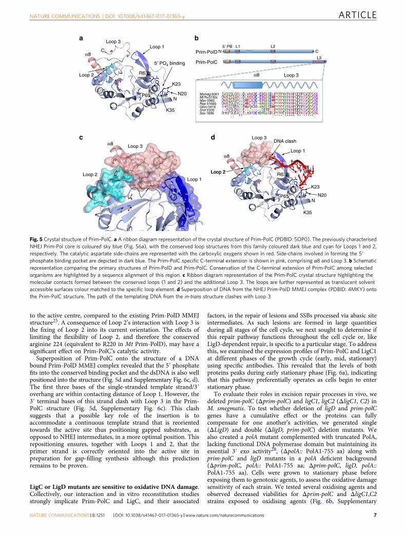
to the active centre, compared to the existing Prim-PolD MMEJstructure25. A consequence of Loop 2’s interaction with Loop 3 isthe fixing of Loop 2 into its current orientation. The effects oflimiting the flexibility of Loop 2, and therefore the conservedarginine 224 (equivalent to R220 in Mt Prim-PolD), may have asignificant effect on Prim-PolC’s catalytic activity.
Superposition of Prim-PolC onto the structure of a DNAbound Prim-PolD MMEJ complex revealed that the 5′ phosphatefits into the conserved binding pocket and the dsDNA is also wellpositioned into the structure (Fig. 5d and Supplementary Fig. 6c, d).The first three bases of the single-stranded template strand/3′overhang are within contacting distance of Loop 1. However, the3′ terminal bases of this strand clash with Loop 3 in the Prim-PolC structure (Fig. 5d, Supplementary Fig. 6c). This clashsuggests that a possible key role of the insertion is toaccommodate a continuous template strand that is reorientedtowards the active site thus positioning gapped substrates, asopposed to NHEJ intermediates, in a more optimal position. Thisrepositioning ensures, together with Loops 1 and 2, that theprimer strand is correctly oriented into the active site inpreparation for gap-filling synthesis although this predictionremains to be proven.
LigC or LigD mutants are sensitive to oxidative DNA damage.Collectively, our interaction and in vitro reconstitution studiesstrongly implicate Prim-PolC and LigC, and their associated
factors, in the repair of lesions and SSBs processed via abasic siteintermediates. As such lesions are formed in large quantitiesduring all stages of the cell cycle, we next sought to determine ifthis repair pathway functions throughout the cell cycle or, likeLigD-dependent repair, is specific to a particular stage. To addressthis, we examined the expression profiles of Prim-PolC and LigC1at different phases of the growth cycle (early, mid, stationary)using specific antibodies. This revealed that the levels of bothproteins peaks during early stationary phase (Fig. 6a), indicatingthat this pathway preferentially operates as cells begin to enterstationary phase.
To evaluate their roles in excision repair processes in vivo, wedeleted prim-polC (Δprim-polC) and ligC1, ligC2 (ΔligC1, C2) inM. smegmatis. To test whether deletion of ligD and prim-polCgenes have a cumulative effect or the proteins can fullycompensate for one another’s activities, we generated single(ΔLigD) and double (ΔligD, prim-polC) deletion mutants. Wealso created a polA mutant complemented with truncated PolA,lacking functional DNA polymerase domain but maintaining itsessential 3′ exo activity28, (ΔpolA:: PolA1-755 aa) along withprim-polC and ligD mutants in a polA deficient background(Δprim-polC, polA:: PolA1-755 aa; Δprim-polC, ligD, polA::PolA1-755 aa). Cells were grown to stationary phase beforeexposing them to genotoxic agents, to assess the oxidative damagesensitivity of each strain. We tested several oxidising agents andobserved decreased viabilities for Δprim-polC and ΔligC1,C2strains exposed to oxidising agents (Fig. 6b, Supplementary
DxDDxDDxD
Loop 2
Loop 1
Loop 3
Loop 2 Loop 1
Loop 3
Loop 2 Loop 2
Loop 1
N
K23
N20
K35
P65
R63
C
N
α8 Loop 3
α8
α8
α8
K23
N20
K35
P65
R63
Loop 3
DNA clash
- C
L3
Msmeg 6301Mt Rv3730c Mav 0362Rop 51690Gbro 0416Srot 2335Sav 1696
L15′ PB L2Prim-PolD
Prim-PolC
N -
5′ PO4 binding
a b
c d
Fig. 5 Crystal structure of Prim-PolC. a A ribbon diagram representation of the crystal structure of Prim-PolC (PDBID: 5OP0). The previously characterisedNHEJ Prim-Pol core is coloured sky blue (Fig. S6a), with the conserved loop structures from this family coloured dark blue and cyan for Loops 1 and 2,respectively. The catalytic aspartate side-chains are represented with the carboxylic oxygens shown in red. Side-chains involved in forming the 5′phosphate binding pocket are depicted in dark blue. The Prim-PolC specific C-terminal extension is shown in pink, comprising α8 and Loop 3. b Schematicrepresentation comparing the primary structures of Prim-PolD and Prim-PolC. Conservation of the C-terminal extension of Prim-PolC among selectedorganisms are highlighted by a sequence alignment of this region. c Ribbon diagram representation of the Prim-PolC crystal structure highlighting themolecular contacts formed between the conserved loops (1 and 2) and the additional Loop 3. The loops are further represented as translucent solventaccessible surfaces colour matched to the specific loop element. d Superposition of DNA from the NHEJ Prim-PolD MMEJ complex (PDBID: 4MKY) ontothe Prim-PolC structure. The path of the templating DNA from the in-trans structure clashes with Loop 3
NATURE COMMUNICATIONS | DOI: 10.1038/s41467-017-01365-y ARTICLE
NATURE COMMUNICATIONS |8: 1251 |DOI: 10.1038/s41467-017-01365-y |www.nature.com/naturecommunications 7
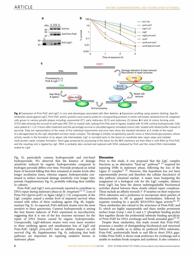
Fig. 8), particularly cumene hydroperoxide and tert-butylhydroperoxide. We observed that the kinetics of damagesensitivity induced by organic hydroperoxides compared tohydrogen peroxide differs over time. Peroxide produced an initialburst of bacterial killing that then remained at similar levels afterlonger incubation times, whereas organic hydroperoxides con-tinued to induce increased damage sensitivity over longer timeperiods (Supplementary Fig. 8), probably reflecting their stabilityin cultures.
Prim-PolC and LigC1 were previously reported to contribute toDNA repair during stationary phase inM. smegmatis12, 14. Loss ofPrim-Pol (Δprim-polC) or ligase (ΔligC1, C2) components of theLigC complex caused a similar level of impaired survival whentreated with either of these oxidising agents (Fig. 6b, Supple-mentary Fig. 8). As expected, PolA deficient strains were the mostsensitive to these genotoxins. Cumene hydroperoxide is amongthe best known inducers of PolA expression in mycobacteria,suggesting that it is one of the key enzymes necessary for therepair of DNA lesions caused by organic hydroperoxides.Unexpectedly, LigD-deficient strains were even more sensitiveto these oxidising agents and simultaneous loss of LigD andPrim-PolC (ΔligD, prim-polC) had an additive impact on cellsurvival (Fig. 6b, Supplementary Fig. 8), indicating that bothpathways are important for repairing oxidative lesions instationary phase.
DiscussionPrior to this study, it was proposed that the LigC complexfunctions as an alternative “back-up” pathway8, 14 required forrepairing DSBs in stationary phase, following the loss of theLigase D complex5, 29. However, this hypothesis has not beenexperimentally proven and therefore the cellular function(s) ofthis pathway remained unclear. A major issue hampering theassignment of a biological role for the LigC complex, distinctfrom LigD, has been the almost indistinguishable biochemicalactivities shared between these closely related repair complexes.These include an affinity towards 5′-P moieties on their respectiveDNA substrates and a preference to incorporate short patches ofribonucleotides to fill in gapped intermediates, followed byrequisite resealing by a specific RNA/DNA ligase activity24, 25.These similarities also extend to the structures of Prim-PolC andD, which are highly superposable and share similar prominentsurface loops (Loop 1 and 2) and a 5′ phosphate binding pocketthat together dictate the preferential substrate binding specificityof Prim-PolD for DNA overhangs and break-annealed gaps23–26.
Despite these similarities, this study reveals that Prim-PolCpossesses a number of distinguishing biochemical and structuralfeatures that enable us to define its preferred DNA substrates.Prim-PolC preferentially binds to and fills-in short DNA gaps.Unlike Prim-PolD, it shows weak preference for overhangs and isunable to mediate break synapsis and synthesis. It also contains a
Anti-Ku
Anti-Prim-PolC
Anti-LigC1
C
EkDa
5050
37
50
37
E/S S
0.001
0.01
0.1
1
10
100
% s
urvi
val
mc2 155
Δprim-polC
ΔligC1,C2
ΔligD
ΔligD,prim-polC
ΔpolA
ΔpolA,prim-polC
ΔpolA,ligD,prim-polC
Gap filling& ligation
Damage detection & base removalRecruitment of repair enzymes
5′
FPG
LigCExoIII/ EndoIV
5′
DNA
RNA
Ap
End processing
Prim-PolC
3′
0 1 2 3
Time of exposure (h)
Abasic siteremoval
Processing byPolA & LigA ?
ba
c Lesion
Fig. 6 Expression of Prim-PolC and LigC1 in vivo and phenotypes associated with their deletion. a Expression profiling using western blotting. Specificantibodies raised against LigC1, Prim-PolC and Ku proteins were used to probe for corresponding proteins in whole-cell lysates obtained fromM. smegmatiscells grown to various growth phases including, exponential (E”), early stationary (E/S) and stationary (S) phase. b A plot of colony forming units(CFU) data showing the survival of wild-type (MC 155) or mutant cells, lacking Prim-Pols and/or ligases, treated with 10mM cumene hydroperoxide. Cellswere plated at 1, 2 or 3 hours after treatment and the percentage survival is calculated against untreated control cells, treated with diluted buffer instead ofperoxide. Data are representative of the mean of five individual experiments and error bars show the standard deviation. c A model of the repairof a damaged base by the LigC-dependent excision repair complex. The damage is initially recognised by specific mono or bifunctional glycosylases, whoseactivity results in the formation of an abasic site intermediate. LigC is recruited early to the lesion to coordinate later repair steps and mediatemulti-protein repair complex formation. Short gaps produced by processing of the lesion by the BER machinery are then filled in with RNA by Prim-PolCand the resulting nick is ligated by LigC. RNA is probably later excised and replaced with DNA catalysed by PolA and the nicked DNA intermediatesealed by LigA
ARTICLE NATURE COMMUNICATIONS | DOI: 10.1038/s41467-017-01365-y
8 NATURE COMMUNICATIONS | 8: 1251 |DOI: 10.1038/s41467-017-01365-y |www.nature.com/naturecommunications
conserved C-terminal structural element (Loop 3), which isabsent from Prim-PolD orthologues. This loop is positionedbetween loops 1 and 2, where it forms a continuous surface that isprobably involved in directing the template strand towards theactive site to optimally position, with Loops 1 and 2, the 3′hydroxyl moiety of the primer strand into the active site inpreparation for extension. The absence of Loop 3 in Prim-PolDorthologues, leaves a space between the remaining loops (1 and2), which allows these NHEJ polymerases to promote breakannealing, MMEJ and processing of subsequently annealedDSBs23–26. Validation of this proposed role for Loop 3 awaits theelucidation of additional structures of Prim-PolC bound to DNAgap intermediates.
In addition to these molecular analyses, our in vivo studiesestablish that the LigC complex is a central nexus for a distinctiveexcision repair pathway required to remove and replace damagedor modified bases in mycobacterial genomes during stationaryphase (Fig. 6c). BER reconstitution studies in vitro demonstratedthat LigC-dependent excision repair complexes can repair oxi-datively damaged bases and probably other lesions, e.g., methy-lated/nitrosative damaged bases and SSBs, that are all processedvia an abasic site intermediate. A key feature of this repairpathway is the preferential incorporation of ribonuclotides, con-sistent with their abundance in stationary phase. We observed themost efficient repair in vitro when endonuclease IV wasemployed as the gap processing enzyme, dephosphorylating the3′-phosphate end of the lesion resulting from β,γ-elimination ofthe abasic site, catalysed by FPG. EndoIV lacks a strong 3′-5′exonuclease activity, present in alternative end-processingenzymes (ExoIII and XthA), and therefore this appears to limitthe gap resection to an optimal size for LigC-mediated repair.Consistent with this, it was previously reported that EndoIV,rather than ExoIII, is responsible for vast majority of BER gapsprocessing in mycobacteria, in contrast to eubacteria whereExoIII plays a more dominant role30.
In mycobacteria, the expression of the LigC complex peaksduring early stationary phase. This coincides with a significantreduction in intercellular pools of dNTPs in the absence ofreplication31, 32, probably explaining the necessity to incorporateribonucleotides during DNA repair processes in stationary phase.Another notable feature of the LigC pathway is the specificincorporation of short ribonucleotide patches that are likelyresistant to RNase H1 activities33–35. Apart from Prim-PolC/D,DinB2 also incorporates ribonucleotides into DNA during sta-tionary phase repair processes in mycobacteria36. However, thisY-family polymerase has a tendency to incorporate longer RNApatches and the resulting DNA:RNA hybrids would be sensitiveto RNase H1 digestion. In gap-filling assays, Prim-PolC efficientlyfilled in short gaps (1–3 nt) but failed to process longer gaps (5nt). Notably, stretches of >4 ribonucleotides incorporated intodsDNA are known to exhibit high instability due to their sensi-tivity to RNase H1 digestion in vivo33–35. It is therefore likely thatshort RNA patches incorporated by Prim-Pols during BER andNHEJ repair processing are later recognised and replaced byPolA, rather than RNase H, which undertakes a similar roleduring RNA primer removal, once intracellular pools of dNTPsare restored37, 38.
Prim-Pols have relatively low fidelity during synthesis andtherefore another reason to favour the incorporation of ribonu-cleotides during repair is to “mark” these regions of repairsynthesis for subsequent replacement in a post-repair manner bymore accurate polymerases5, 10, reminiscent of RNA primerremoval during replication. PolA removes RNA primers and isalso known to participate in several DNA repair pathways inbacteria37, 39–41. PolA-driven DNA synthesis occurs slowly butaccurately and it has proofreading and bidirectional nuclease
activities beneficial for correcting errors introduced by Prim-Pols39, 42. Notably, in this study, we also detected a weak asso-ciation between PolA and the LigC complex, suggesting that thisreplicase may remove and replace RNA inserted by this pathway,and likely others e.g. NHEJ, with more accurately synthesisedDNA.
Both Δprim-polC and ΔligC1,C2 strains were similarly sensi-tive to oxidising agents, consistent with their proposed joint rolesin a distinctive excision repair pathway in mycobacteria. Sur-prisingly, a LigD-deficient strain was even more sensitive to theseagents, although this could potentially be attributed to a deficit inDSB repair. However, this sensitivity was evident even with lowlevels of peroxide (~5 mM), reported to produce predominantlySSBs and <0.1% DSBs43, thus uncovering an unexpected andadditional role for LigD in excision repair. Consistent with thisproposal, co-deletion of LigD and Prim-PolC had an additiveimpact on cell survival in the presence of these genotoxins, alsoindicating that they are not epistatic in their function. Consistentwith the mutant phenotypes, both LigD and Prim-PolC co-pur-ified on a 5′-phosphorylated single nucleotide gap DNA substratein pull-down assays. This substrate mimics a naturally occurringBER DNA intermediate that would arise from the enzymaticprocessing of an oxidative lesion by FPG and EndoIV. Together,these findings establish that both LigC and LigD-dependentcomplexes contribute significantly to the repair of lesions pro-duced by oxidative damage during stationary phase in myco-bacteria. Notably, Ory et al.44 reported recently that Bacillussubtilis LigD possesses dRP-ase activity in vitro supporting thisproposed role in excision repair. Further studies are required todefine the exact nature of the oxidative lesions that these Prim-Pol pathways process in vivo and uncover the overlap and “cross-talk” that occurs between these, and other, stationary phase DNArepair pathways in mycobacteria.
MethodsBacterial strains and growth conditions. Laboratory stock of M. smegmatis mc2
155 and its derivatives were cultured in 7H9 liquid broth or 7H10 solid medium(Beckton Dickinson) with ADC supplement (Albumin-Dextrose-Catalase) andhygromycin B (50 μg ml−1) or kanamycin (30 μg ml−1), if antibiotic selection wasrequired. Escherichia coli DH5α strain (Invitrogen) was used for cloning purposesand Origami 2(DE3)pLysS (Novagen) for protein purification, respectively. E. colistrains were cultured in standard Luria broth medium or terrific broth (For-medium), where large amounts of protein production was required with additionof kanamycin (50 μg ml−1) for selection.
Protein complex purification and mass spectrometry analysis. Genes encodingPrim-PolC, LigC1 and LigC2 were PCR amplified and sub-cloned into pKW08-gfpvector using a sequence and ligation independent protocol to produce C-terminaleGFP fusion proteins used as baits for protein complex purification15. Purificationand data analysis was performed essentially as described previously15. Briefly,mycobacterial cells were collected by centrifugation (15 min, 4800×g, 4 °C) andresuspended in a cold sonication buffer containing 50 mM Tris (pH 8.0), 100 mMNaCl, 1 mM dithiotreitol, 2 mM phenylmethylsulfonyl fluoride, 25 Uml−1 benzo-nase, 0.5% Triton X-100 and protease inhibitor cocktail. The cells were lysed bysonication and lysates were pre-cleared by centrifugation (20 min, 4800×g, 4 °C).Lysates were then mixed with 50 μls of the anti-GFP Sepharose beads (Chromotek),incubated for 2 h at 4 °C to capture protein complexes, washed two times with 10ml of wash buffer (10 mM Tris (pH 8.0), 150 mM NaCl and 0.1% Triton X-100),followed by two washes with TEV buffer (10 mM Tris (pH 8.0), 150 mM NaCl, 0.5mM EDTA and 1 mM DTT) and digested with TEV protease (Thermo FisherScientific) to elute protein complexes.
For DNA-protein pull-downs, each DNA substrate at a final concentration of50 nM was annealed to 1 mg of the poly-dT magnetic particles (NEB) in buffercontaining 20 mM Tris-HCl, pH 8.0, 10 mM MgCl2, 0.1 M KCl, for 3 h at roomtemperature and washed twice with the wash buffer (50 mM HEPES pH 7.5, 150mM NaCl, 0.5% Triton X, 1 mM EDTA). Whole-cell lysates were prepared asdescribed above, however, in 50 mM HEPES pH 7.5 as buffering agent and withoutthe addition of nucleases. The pre-cleared lysates were filtered through 0.22 µmfilters and incubated with the DNA coated beads for 3 h at 4 °C. For the last 10 min2 µls of RNaseA (10 mgml−1) were added to the 1 ml reaction. Beads were collectedon the magnetic stand and washed five times with the wash buffer. DNA bound
NATURE COMMUNICATIONS | DOI: 10.1038/s41467-017-01365-y ARTICLE
NATURE COMMUNICATIONS |8: 1251 |DOI: 10.1038/s41467-017-01365-y |www.nature.com/naturecommunications 9
protein complexes were then eluted by DNaseI digestion (NEB) for 1 h at 37 °C indedicated buffer.
The resultant protein mixture was precipitated with pyrogallol red–molybdate(PRM; 0.05 mM pyrogallol red, 0.16 mM sodium molybdate, 1 mM sodium oxalate,50 mM succinic acid, pH 2.5), collected by centrifugation and subjected to astandard trypsin digestion. The resulting peptide mixtures were applied to a nano-HPLC RP-18 column (Waters) using an acetonitryle gradient in the presence oftrifluoroacetic acid. The column was directly linked to the ion source and theOrbitrap (Thermo Scientific) was operated in a data-dependent mode.
The MaxQuant (v1.3.0.5) computational proteomics platform was used toprocess raw MS files. Integrated Andromeda search engine and apropriate M.smegmatis or M. bovis BCG protein databases were used to search against thefragmentation spectra. Carbamidomethylation of cysteines, N-terminal acetylationand oxidations were set as possible peptide modifications. One per cent falsediscovery rate was applied to all protein and peptide identifications. Humanderived contaminants and random protein identifications were excluded from theresults files. Proteins considered as valid identifications were identified by at leasttwo peptides.
Purification of multiple DNA repair proteins. Genes encoding Prim-PolC,PolD2, LigC1, LigC2, MPG, FPG, NTH, EndoIV, ExoIII, XthA, XseVIIB and PolAwere PCR amplified using primers flanked with restriction digestion sites requiredfor in frame cloning into pET28 vector (Supplementary Table 1). All the proteinswere designed to contain a N-terminal His-tag, except for FPG, which was ren-dered inactive unless tagged at its C terminus. Proteins were purified according toroutine laboratory procedures using ÄKTA purifier and compatible columnspurchased from GE healthcare. Briefly, pre-cleared cell lysates obtained afteroverexpression of recombinant proteins in E. coli Origami B pLysS strain wereloaded onto Ni Sepharose (Qiagen) column in Tris buffers (50 mM Tris pH 8.0),washed extensively and eluted in a gradient of imidazole. Proteins were next loadedonto an ion exchange column (SP or Q (GE Healthcare), depending on the iso-electric point (pI)) eluted in a gradient of NaCl and further purified on preparativegel filtration columns (S200 or S75 (GE Healthcare), depending on the molecularmass of purified protein). Quality of proteins after each and every purification stepwas checked using sodium dodecyl sulfate (SDS) polyacrylamide gel electrophoresis(PAGE).
EMSA. Assays were carried out essentially as described previously10, 25. Briefly,EMSAs were employed to determine optimal substrates for Prim-PolC activity andwere set up in 50 mM Tris-HCl (pH 7.5), 0.1 mg ml−1 of BSA (NEB Biolabs), 1 mMDTT, 5% glycerol, with indicated 30 nM 6-carboxyfluorescein (6-FAM) labelledDNA substrates and different concentrations of Prim-PolC in a 15 μl reactionvolume. Incubation mixtures were kept on ice for 30 min and were resolved bynative gel electrophoresis on a 5% polyacrylamide gel (80:1 (w/w) acrylamide/bisacrylamide).
Polymerisation and DNA repair assays. DNA extension reaction mixture con-tained 50 mM Tris-HCl (pH 7.5), 5 mM MgCl2, 100 μM MnCl2, 30 nM 6-FAMlabelled DNA substrate, 250 μM NTPs and indicated DNA repair proteins, in atotal volume of 20 μl. Reactions were further supplemented with 1 mM ATP, 1 mMDTT and 0.1 mgml−1 bovine serum albumin (BSA) for DNA repair reconstitutionreactions. After a set incubation time at 37 °C, reactions were terminated by addingstop buffer solution (95% (v/v) formamide, 0.09% (w/v) bromophenol blue, 20 mMEDTA and 600 nM unlabelled oligonucleotide, identical to labelled strand).Resulting DNA extension or repair products were resolved for 2 h at constantwattage of 20W, on TBE-buffered 15% polyacrylamide gels containing 8M urea.Detection of fluorescently labelled oligonucleotide products was carried out usingFujifilm FLA-5100 fluorescent image scanner and EMSA data were quantifiedusing Quantity One (Bio-Rad).
Antibodies and western blotting. Antibodies were generated against M. smeg-matis proteins (Ku, LigC1 or Prim-PolC) using a 28 day rabbit immunisationprogram (Eurogentec), using ~1 mg full-length recombinant protein (two appli-cations), with serum collected at different time points. Recombinant proteins (5 µgper lane) were resolved by 10% SDS-PAGE and proteins transfer onto poly-vinylidene fluoride (PVDF) membrane, which was subsequently blocked with Tris-buffered saline and Tween 20 (TBST) and 5% (w/v) non-fat dried milk. Membranebands containing the recombinant protein were excised, washed twice in TBST andincubated overnight at 4 °C with 500 µl of serum. Serum was next removed andmembranes washed (x4) with 500 µl of TBST. Purified immunoglobulins bound tothe membranes were eluted with 150 µl of glycine buffer (500 mM NaCl, 50 mMglycine, 0.5% Tween 20, 0.1% BSA, pH 2–3) and the blot vortex pulsed beforeadding 25 µl of 1 M Tris pH 8.8.
For western blotting analysis, the primary antibodies (anti- Ku, Prim-PolC andLigC1) were used at a dilution of 1/1000 and the secondary antibody (HRP-conjugated Anti-Rabbit IgG; Abcam ab6721) at a dilution of 1:5000 dilution.Uncropped versions of all western blots and gels can be found in SupplementaryFig. 9.
Far-western analysis. This method was used to test for protein−protein interac-tions in vitro. Recombinant proteins were first slot-blotted using a slot blotmanifold (GE Healthcare) onto a methanol-wetted PVDF membrane according tothe manufacturer’s instructions. Typically, a fixed concentration of recombinantprotein (50 ng for the probed protein and 3 μg for the possible interactors) wasblotted onto the membrane. The membrane was first blocked with Tris-bufferedsaline (TBS: 280 mM NaCl, 20 mM Tris) containing 0.05% (v/v) Tween 20 and 5%(w/v) non-fat dried milk. For far-western analysis, the membrane was then incu-bated with blocking buffer containing 5 μg ml−1 of the candidate interactingrecombinant protein. The membrane was then washed, probed with primary (1/1000 dilution) and secondary (1/5000 dilution) antibodies, and subjected to che-miluminescent detection. The primary antibody used was specific for the candidateinteracting recombinant protein, and chemiluminescence detected if interactionoccurred between the two proteins.
Pull-down assays. Pull-down assays were used to confirm chosen protein−proteininteractions. Equimolar amounts of all proteins were used at 5 nM per pull-down.Recombinant FPG-6xHis or 6xHis-MPG bait proteins were first bound to nickel-nitrilotriacetic acid (NTA) coupled resin in a Tris-based buffer (50 mM, pH 8.0)containing 150 mM NaCl, for 1 h at 4 °C. The protein-coated beads were washedand interacted with tag-free LigC (6xHis-LigC subjected to a standard thrombindigestion) or BSA, for further 2 h. The beads were next washed extensively andprotein complexes were eluted with buffer containing 300 nM imidazole. Theprotein complexes were visualised by SDS-PAGE followed by Coomassie staining.
Crystallisation and X-ray structure determination. Crystals of the apo Prim-PolC were grown at 285 K by vapour diffusion as sitting drops. The protein wasscreened at a concentration of 8 mgml−1 and 0.5 μL of protein solution was mixedwith 0.5 μL of crystallisation buffer (0.05 M Tris-HCl (pH 8.0), 0.2 M ammoniumchloride, 0.01 M calcium chloride, 30% (w/v) PEG 4000). Prior to data collection,crystals were soaked in mother liquor containing 20% ethylene glycol. 0.9795 ÅX-ray diffraction data were collected at 100 K using a synchrotron source at stationI02 Diamond Light Source, Didcot, UK. The diffraction data were processed withxia245 with additional processing by programs from the CCP4 suite (CollaborativeComputational Project, Number 4, 1994). The statistics for data processing aresummarised in Table 1. Initial phases were obtained by molecular replacement withPHASER46 using Prim-PolD (2IRY) as a search model23. Iterative cycles of modelbuilding and refinement were performed using Coot47 and Phenix48. A finalrefined model at 1.84 Å resolution, with an Rfactor of 18.58% and Rfree of 20.92%was obtained. 99.1% of residues are in preferred regions with the remainder (0.9%)in allowed regions according to Ramachandran statistics. Structural images wereprepared with CCP 4mg49. The structure of apo Prim-PolC is deposited in theProtein Data Bank under accession code 5OP0.
Construction of mutant strains lacking DNA repair genes. Targeted genereplacement strategy was employed to generate unmarked Δprim-polC, ΔligC1,ΔligC2, ΔligD and ΔpolA:: PolA1-755aa mutants and strains with simultaneousremoval of multiple of these genes as previously published50, 51. Briefly, shortfragments corresponding to 3′ and 5′ end of each genes, flanked with surroundinggenomic region of about 1 kb at each side (primers listed in SupplementaryTable 1), were ligated together out of frame and sub-cloned into a suicidal p2NILdelivery vector, merged with crossing over event selection cassette from pGOAL17.Multistep screening led to double crossing over strains selection and resulted ingene inactivation, which was confirmed by Southern blotting and PCR, for eachrecombination event.
Phenotypic analysis of M. smegmatis strains. M. smegmatis MC2 155 wild-type(ATCC 700084) and mutant strains were grown to late stationary phase in 7H9liquid media, with supplements, typically 4 days after reaching optical density (OD600 nm) of 3.0. The cells were collected and suspended in PBS containing Tween 80(0.05% (v/v)), to prevent clumping, at OD 600 of 0.1. They were next treated with10 mM cumene hydroperoxide (from 1M stock in 50% ethanol), 50 mM tert-butylhydroperoxide (from 5M stock in water), or 5 mM hydrogen peroxide (from 0.5Mstock in water), respectively, for various amount of time with incubation at 37 °C.Untreated cells suspension was used to control overall viability of each strain. Atindicated time points 100 μls of each cell suspension (diluted ten fold) wastransferred onto 7H10 solid agar and plates were incubated for 72 h before read outof cell survival was recorded.
Data availability. All data are provided in full in the results section and theSupplementary Information accompanying this paper, or from the authors uponrequest. The crystal structure has been deposited in the Protein Data Bank and canbe accessed using accession code 5OP0.
Received: 24 October 2016 Accepted: 13 September 2017
ARTICLE NATURE COMMUNICATIONS | DOI: 10.1038/s41467-017-01365-y
10 NATURE COMMUNICATIONS | 8: 1251 |DOI: 10.1038/s41467-017-01365-y |www.nature.com/naturecommunications
References1. Bouche, J. P., Zechel, K. & Kornberg, A. dnaG gene product, a rifampicin-resistant
RNA polymerase, initiates the conversion of a single-stranded coliphage DNA toits duplex replicative form. J. Biol. Chem. 250, 5995–6001 (1975).
2. Keck, J. L., Roche, D. D., Lynch, A. S. & Berger, J. M. Structure of the RNApolymerase domain of E. coli primase. Science 287, 2482–2486 (2000).
3. Frick, D. N. & Richardson, C. C. DNA primases. Annu. Rev. Biochem. 70, 39–80(2001).
4. Guilliam, T. A., Keen, B. A., Brissett, N. C. & Doherty, A. J. Primase-polymerases are a functionally diverse superfamily of replication and repairenzymes. Nucleic Acids Res. 43, 6651–6664 (2015).
5. Pitcher, R. S. et al. NHEJ protects mycobacteria in stationary phase against theharmful effects of desiccation. DNA. Repair 6, 1271–1276 (2007).
6. Weller, G. R. et al. Identification of a DNA nonhomologous end-joiningcomplex in bacteria. Science 297, 1686–1689 (2002).
7. Della, M. et al. Mycobacterial Ku and ligase proteins constitute a two-component NHEJ repair machine. Science 306, 683–685 (2004).
8. Gong, C. et al. Mechanism of nonhomologous end-joining in mycobacteria: alow-fidelity repair system driven by Ku, ligase D and ligase C. Nat. Struct. Mol.Biol. 12, 304–312 (2005).
9. Pitcher, R. S., Brissett, N. C. & Doherty, A. J. Nonhomologous end-joining inbacteria: a microbial perspective. Annu. Rev. Microbiol. 61, 259–282 (2007).
10. Bartlett, E. J., Brissett, N. C. & Doherty, A. J. Ribonucleolytic resection isrequired for repair of strand displaced nonhomologous end-joiningintermediates. Proc. Natl Acad. Sci. USA 110, E1984–E1991 (2013).
11. Bowater, R. & Doherty, A. J. Making ends meet: repairing breaks in bacterialDNA by non-homologous end-joining. PLoS Genet. 2, e8 (2006).
12. Zhu, H., Bhattarai, H., Yan, H. G., Shuman, S. & Glickman, M. S.Characterization of Mycobacterium smegmatis PolD2 and PolD1 as RNA/DNAPolymerases Homologous to the POL Domain of Bacterial DNA Ligase D.Biochemistry 51, 10147–10158 (2012).
13. Matthews, L. A. & Simmons, L. A. Bacterial nonhomologous end joiningrequires teamwork. J. Bacteriol. 196, 3363–3365 (2014).
14. Bhattarai, H., Gupta, R. & Glickman, M. S. DNA ligase C1 mediates the LigD-independent nonhomologous end-joining pathway of Mycobacteriumsmegmatis. J. Bacteriol. 196, 3366–3376 (2014).
15. Plocinski, P. et al. Identification of protein partners in mycobacteria using asingle-step affinity purification method. PLoS ONE 9, e91380 (2014).
16. Caldecott, K. W., Tucker, J. D., Stanker, L. H. & Thompson, L. H.Characterization of the XRCC1-DNA ligase III complex in vitro and its absencefrom mutant hamster cells. Nucleic Acids Res. 23, 4836–4843 (1995).
17. Leppard, J. B., Dong, Z., Mackey, Z. B. & Tomkinson, A. E. Physical andfunctional interaction between DNA ligase IIIalpha and poly(ADP-Ribose)polymerase 1 in DNA single-strand break repair. Mol. Cell Biol. 23, 5919–5927(2003).
18. De, A. & Campbell, C. A novel interaction between DNA ligase III and DNApolymerase gamma plays an essential role in mitochondrial DNA stability.Biochem. J. 402, 175–186 (2007).
19. Tomkinson, A. E. & Sallmyr, A. Structure and function of the DNA ligasesencoded by the mammalian LIG3 gene. Gene 531, 150–157 (2013).
20. Fan, J. & Wilson, D. M. 3rd Protein-protein interactions and posttranslationalmodifications in mammalian base excision repair. Free Radical Biol. Med. 38,1121–1138 (2005).
21. Doherty, A. J., Jackson, S. P. & Weller, G. R. Identification of bacterialhomologues of the Ku DNA repair proteins. FEBS Lett. 500, 186–188 (2001).
22. Aravind, L. & Koonin, E. V. Prokaryotic homologs of the eukaryotic DNA-end-binding protein Ku, novel domains in the Ku protein and prediction of aprokaryotic double-strand break repair system. Genome Res. 11, 1365–1374(2001).
23. Pitcher, R. S. et al. Structure and function of a mycobacterial NHEJ DNA repairpolymerase. J. Mol. Biol. 366, 391–405 (2007).
24. Brissett, N. C. et al. Structure of a preternary complex involving a prokaryoticNHEJ DNA polymerase. Mol. Cell 41, 221–231 (2011).
25. Brissett, N. C. et al. Molecular basis for DNA double-strand break annealingand primer extension by an NHEJ DNA polymerase. Cell Rep. 5, 1108–1120(2013).
26. Brissett, N. C. et al. Structure of a NHEJ polymerase-mediated DNA synapticcomplex. Science 318, 456–459 (2007).
27. Kent, T., Chandramouly, G., Mcdevitt, S. M., Ozdemir, A. Y. & Pomerantz, R. T.Mechanism of microhomology-mediated end-joining promoted by humanDNA polymerase θ. Nat. Struct. Mol. Biol. 22, 230–237 (2015).
28. Gordhan, B. G., Andersen, S. J., De Meyer, A. R. & Mizrahi, V. Construction byhomologous recombination and phenotypic characterization of a DNApolymerase domain polA mutant of Mycobacterium smegmatis. Gene 178,125–130 (1996).
29. Wilson, T. E., Topper, L. M. & Palmbos, P. L. Non-homologous end-joining:bacteria join the chromosome breakdance. Trends Biochem. Sci. 28, 62–66(2003).
30. Puri, R. V., Singh, N., Gupta, R. K. & Tyagi, A. K. Endonuclease IV Is the majorapurinic/apyrimidinic endonuclease in Mycobacterium tuberculosis and isimportant for protection against oxidative damage. PLoS ONE 8, e71535(2013).
31. Buckstein, M. H., He, J. & Rubin, H. Characterization of nucleotide pools as afunction of physiological state in Escherichia coli. J. Bacteriol. 190, 718–726(2008).
32. Jacewicz, A. & Shuman, S. Biochemical characterization of mycobacteriumsmegmatis RnhC (MSMEG_4305), a bifunctional enzyme composed ofautonomous N-terminal type I RNase H and C-terminal acid phosphatasedomains. J. Bacteriol. 197, 2489–2498 (2015).
33. Minias, A. E. et al. RNase HI Is essential for survival of mycobacteriumsmegmatis. PLoS ONE 10, e0126260 (2015).
34. Nowotny, M., Gaidamakov, S. A., Crouch, R. J. & Yang, W. Crystal structures ofRNase H bound to an RNA/DNA hybrid: substrate specificity and metal-dependent catalysis. Cell 121, 1005–1016 (2005).
35. Jacewicz, A. & Shuman, S. Biochemical characterization of mycobacteriumsmegmatis RnhC (MSMEG_4305), a bifunctional enzyme composed ofautonomous N-terminal type I RNase H and C-terminal acid phosphatasedomains. J. Bacteriol. 197, 2489–2498 (2015).
36. Ordonez, H., Uson, M. L. & Shuman, S. Characterization of three mycobacterialDinB (DNA polymerase IV) paralogs highlights DinB2 as naturallyadept at ribonucleotide incorporation. Nucleic Acids Res. 42, 11056–11070(2014).
37. Ogawa, T. & Okazaki, T. Function of RNase H in DNA replication revealed byRNase H defective mutants of Escherichia coli. Mol. Genet. Genom. 193,231–237 (1984).
38. Minias, A. E., Brzostek, A. M., Minias, P. & Dziadek, J. The deletion of rnhB inMycobacterium smegmatis does not affect the level of RNase HII substrates orinfluence genome stability. PLoS ONE 10, e0115521 (2015).
39. Heyneker, H. L. & Klenow, H. Involvement of Escherichia coli DNApolymerase-I-associated 5' in equilibrium 3' exonuclease in excision-repair ofUV-damaged DNA. Basic Life Sci. 5A, 219–223 (1975).
40. Friedberg, E. C. The eureka enzyme: the discovery of DNA polymerase. Nat.Rev. Mol. Cell Biol. 7, 143–147 (2006).
41. Tait, R. C., Harris, A. L. & Smith, D. W. DNA repair in Escherichia colimutants deficient in DNA polymerases I, II and-or 3. Proc. Natl Acad. Sci. USA71, 675–679 (1974).
42. Bailly, V. & Verly, W. G. The excision of AP sites by the 3'-5' exonucleaseactivity of the Klenow fragment of Escherichia coli DNA polymerase I. FEBSLett. 178, 223–227 (1984).
43. Ismail, I. H., Nyström, S., Nygren, J. & Hammarsten, O. Activation of ataxiatelangiectasia mutated by DNA strand break-inducing agents correlates closelywith the number of DNA double strand breaks. J. Biol. Chem. 280, 4649–4655(2005).
44. de Ory, A. et al. Identification of a conserved 5'-dRP lyase activity in bacterialDNA repair ligase D and its potential role in base excision repair. Nucleic AcidsRes. 44, 1833–1844 (2016).
45. Winter, G. xia2: an expert system for macromolecular crystallography datareduction. J. Appl. Crystallogr. 43, 186–190 (2010).
46. Mccoy, A. J. et al. Phaser crystallographic software. J. Appl. Crystallogr. 40,658–674 (2007).
47. Emsley, P., Lohkamp, B., Scott, W. G. & Cowtan, K. Features anddevelopment of Coot. Acta Crystallogr. Sec. D: Biol. Crystallogr. 66, 486–501(2010).
48. Adams, P. D. et al. PHENIX: a comprehensive Python-based system formacromolecular structure solution. Acta Crystallogr. Sec. D: Biol. Crystallogr.66, (213–221 (2010).
49. McNicholas, S., Potterton, E., Wilson, K. S. & Noble, M. E. Presenting yourstructures: the CCP4mg molecular-graphics software. Acta Crystallogr. Sec. D:Biol. Crystallogr. 67, 386–394 (2011).
50. Parish, T. & Stoker, N. G. Use of a flexible cassette method to generate a doubleunmarked Mycobacterium tuberculosis tlyA plcABC mutant by genereplacement. Microbiology 146(Pt 8), 1969–1975 (2000).
51. Plocinski, P. et al. Mycobacterium tuberculosis CwsA interacts withCrgA and Wag31, and the CrgA-CwsA complex is involved in peptidoglycansynthesis and cell shape determination. J. Bacteriol. 194, 6398–6409(2012).
AcknowledgementsWe thank Dr M. Roe for assistance with X-ray data collection. A.J.D.’s laboratorywas supported by grants from Biotechnology and Biological Sciences Research Council(BB/F013795/1, BB/J018643/1 and BB/M004236/1). P.P. was supported by a mobilitygrant from the Polish Ministry of Science and Higher Education (1073/MOB/2013/0:Mobilnosc Plus). Funding for open access charge: Research Councils UK(RCUK).
NATURE COMMUNICATIONS | DOI: 10.1038/s41467-017-01365-y ARTICLE
NATURE COMMUNICATIONS |8: 1251 |DOI: 10.1038/s41467-017-01365-y |www.nature.com/naturecommunications 11
Author contributionsA.J.D. designed the project, directed the experimental work, performed database analysisand wrote the manuscript. P.P. and N.C.B. contributed to the project design, performedand designed experiments, performed computational analyses and co-wrote the manu-script. P.P. performed mass spectrometry analysis. N.C.B. performed X-ray data proces-sing, model building and analysis. J.B. generated LigC and Prim-PolC expressionplasmids, purified proteins, measured protein expression levels in cells and performed theinitial biochemical and phenotype studies. A.B. and M.K.-M. constructed mutant strainsinM. smegmatis and assisted P.P. with CFU experiments. A.D. and J.D. helped with studydesign and directed parts of the experimental work (mass spectrometry analysis of proteincomplexes and generation of mutant strains and phenotypic analysis, respectively).
Additional informationSupplementary Information accompanies this paper at doi:10.1038/s41467-017-01365-y.
Competing interests: The authors declare no competing financial interests.
Reprints and permission information is available online at http://npg.nature.com/reprintsandpermissions/
Publisher's note: Springer Nature remains neutral with regard to jurisdictional claims inpublished maps and institutional affiliations.
Open Access This article is licensed under a Creative CommonsAttribution 4.0 International License, which permits use, sharing,
adaptation, distribution and reproduction in any medium or format, as long as you giveappropriate credit to the original author(s) and the source, provide a link to the CreativeCommons license, and indicate if changes were made. The images or other third partymaterial in this article are included in the article’s Creative Commons license, unlessindicated otherwise in a credit line to the material. If material is not included in thearticle’s Creative Commons license and your intended use is not permitted by statutoryregulation or exceeds the permitted use, you will need to obtain permission directly fromthe copyright holder. To view a copy of this license, visit http://creativecommons.org/licenses/by/4.0/.
© The Author(s) 2017
ARTICLE NATURE COMMUNICATIONS | DOI: 10.1038/s41467-017-01365-y
12 NATURE COMMUNICATIONS | 8: 1251 |DOI: 10.1038/s41467-017-01365-y |www.nature.com/naturecommunications