
Contemporary aspects of prostate cancer grading · stopnia złośliwości raka definiuje się...
5
Post N Med 2016; XXIX(11): 841-845 841 ©Borgis Conflict of interest Konflikt interesów None Brak konfliktu interesów *Łukasz Nyk 1 , Michał A. Skrzypczyk 2 , Stanisław Szempliński 2 , Mieszko Kozikowski 1 , Wojciech Michalak 1 , Sebastian Piotrowicz 1 , Szymon Kawecki 1 , Tomasz Dzik 3 , Maciej Wysocki 4 , Andrzej Borówka 1 , Jakub Dobruch 1, 2 Contemporary aspects of prostate cancer grading Współczesne kryteria oceny złośliwości raka stercza 1 1 st Unit of Didactics, Department of Urology, Centre of Postgraduate Medical Education, European Health Centre Otwock Head of Department: Jakub Dobruch, MD, PhD 2 2 nd Unit of Didactics, Department of Urology, Centre of Postgraduate Medical Education, Professor W. Orłowski Independent Public Teaching Hospital, Warsaw Head of Department: Jakub Dobruch, MD, PhD 3 Pathomorphology Division, Międzylesie Specialist Hospital, Warsaw Head of Division: Tomasz Dzik, MD, PhD 4 Pathomorphology Division, Bielany Hospital, Warsaw Head of Division: Jan Faryna, MD, PhD Summary Prostate cancer (PCa) incidence in Poland and many countries all over the world is second only to lung cancer incidence. Analysis of epidemiology data indicates a gradual increase in PCa incidence and mortality in recent decades, with the growth rate of mor- tality being smaller than the growth rate of incidence. Carcinogenesis is a complicated biological process and usually starts with mutation of normal cells to precancerous chang- es (atypical small acinar proliferation and prostatic intraepithelial neoplasia) and after that to carcinoma in situ and invasive cancer. Malignancy is defined according to rules devised by Donald Gleason. The “Gleason score” is a system that grades malignancy according to 5 Gleason patterns, with 1 being the least, and 5 being the most malignant. PCas are differentiated mainly by architecture and, to a lesser degree, neoplastic cell characteristics. Recently, a growing interest in magnetic resonance imaging application for predicting PCa malignancy has been observed. Streszczenie Rak gruczołu krokowego (ang. prostate cancer – PCa) zarówno pod względem roz- poznawalności, jak i śmiertelności jest jednym z najczęstszych nowotworów u mężczyzn w Polsce oraz w większości państw na świecie. Karcynogeneza w gruczole krokowym jest zjawiskiem złożonym biologicznie. Najczęściej rozpoczyna się od mutacji w komór- kach nabłonka gruczołu krokowego i powstania zmian przednowotworowych: nowotwo- rzenia śródnabłonkowego (PIN) oraz atypowego rozrostu drobnozrazikowego (ASAP). Doprowadza to do powstania zmian dysplastycznych o charakterze raka przedinwazyjne- go (łac. carcinoma in situ – CIS), a następnie do powstanie raka inwazyjnego. Określenie stopnia złośliwości raka definiuje się według skali Gleasona, której istotą jest podział na 5 kategorii różniących się między sobą głównie architektoniką i – w mniejszym stopniu – wyglądem komórek nowotworowych. Obecnie coraz częściej w piśmiennictwie wspo- mina się o zastosowaniu badań obrazowych, w tym głównie rezonansu magnetyczne- go (MRI) w prognozowaniu złośliwości raka gruczołu krokowego. Prostate carcinoma (PCa) is one of the most com- mon cancers in men both in terms of incidence and mortality in Poland and worldwide (1). Prostate cancer predominantly develops (70%) in the peripheral zone of the prostate gland. Approx. 10-15% of PCas are found in the transitional zone, and 15-20% in the central zone (2). Carcinogenesis within the prostate gland is a bio- logically complex phenomenon. It typically begins with precancerous changes in the epithelium and progress- es to invasive cancer. The processes commonly asso- ciated with precancerous changes or co-existing with cancer include prostatic intraepithelial neoplasia (PIN) and typical small acinar proliferation (ASAP). Original- ly, three PIN forms were recognized, differing by the degree of cellular abnormality and the percentage of abnormal epithelial cells: PIN 1 (benign), PIN 2 (mod- erate) and PIN 3 (severe) (3). Currently, just 2 PIN types Adres/address: *Łukasz Nyk Klinika Urologii CMKP Europejskie Centrum Zdrowia Otwock ul. Borowa 14/18, 05-400 Otwock [email protected] Keywords prostate carcinoma (PCa), grade, Gleason score (Gl.s.), International Society of Urological Pathology (ISUP), prostatic intraepithelial neoplasia (PIN), atypical small acinar proliferation (ASAP) Słowa kluczowe rak gruczołu krokowego, złośliwość raka, skala Gleasona, Międzynarodowe Towarzystwo Patologii Urologicznej, nowotworzenie śródnabłonkowe w sterczu, atypowy rozrost drobnozrazikowy
Transcript of Contemporary aspects of prostate cancer grading · stopnia złośliwości raka definiuje się...
Post N Med 2016; XXIX(11): 841-845
841
©Borgis
Conflict of interestKonflikt interesów
NoneBrak konfliktu interesów
*Łukasz Nyk1, Michał A. Skrzypczyk2, Stanisław Szempliński2, Mieszko Kozikowski1, Wojciech Michalak1, Sebastian Piotrowicz1, Szymon Kawecki1, Tomasz Dzik3, Maciej Wysocki4, Andrzej Borówka1, Jakub Dobruch1, 2
Contemporary aspects of prostate cancer grading
Współczesne kryteria oceny złośliwości raka stercza
11st Unit of Didactics, Department of Urology, Centre of Postgraduate Medical Education, European Health Centre Otwock Head of Department: Jakub Dobruch, MD, PhD22nd Unit of Didactics, Department of Urology, Centre of Postgraduate Medical Education, Professor W. Orłowski Independent Public Teaching Hospital, Warsaw Head of Department: Jakub Dobruch, MD, PhD3Pathomorphology Division, Międzylesie Specialist Hospital, Warsaw Head of Division: Tomasz Dzik, MD, PhD4Pathomorphology Division, Bielany Hospital, Warsaw Head of Division: Jan Faryna, MD, PhD
S u m m a r y
Prostate cancer (PCa) incidence in Poland and many countries all over the world is second only to lung cancer incidence. Analysis of epidemiology data indicates a gradual increase in PCa incidence and mortality in recent decades, with the growth rate of mor-tality being smaller than the growth rate of incidence. Carcinogenesis is a complicated biological process and usually starts with mutation of normal cells to precancerous chang-es (atypical small acinar proliferation and prostatic intraepithelial neoplasia) and after that to carcinoma in situ and invasive cancer. Malignancy is defined according to rules devised by Donald Gleason. The “Gleason score” is a system that grades malignancy according to 5 Gleason patterns, with 1 being the least, and 5 being the most malignant. PCas are differentiated mainly by architecture and, to a lesser degree, neoplastic cell characteristics. Recently, a growing interest in magnetic resonance imaging application for predicting PCa malignancy has been observed.
S t r e s z c z e n i e
Rak gruczołu krokowego (ang. prostate cancer – PCa) zarówno pod względem roz-poznawalności, jak i śmiertelności jest jednym z najczęstszych nowotworów u mężczyzn w Polsce oraz w większości państw na świecie. Karcynogeneza w gruczole krokowym jest zjawiskiem złożonym biologicznie. Najczęściej rozpoczyna się od mutacji w komór-kach nabłonka gruczołu krokowego i powstania zmian przednowotworowych: nowotwo-rzenia śródnabłonkowego (PIN) oraz atypowego rozrostu drobnozrazikowego (ASAP). Doprowadza to do powstania zmian dysplastycznych o charakterze raka przedinwazyjne-go (łac. carcinoma in situ – CIS), a następnie do powstanie raka inwazyjnego. Określenie stopnia złośliwości raka definiuje się według skali Gleasona, której istotą jest podział na 5 kategorii różniących się między sobą głównie architektoniką i – w mniejszym stopniu – wyglądem komórek nowotworowych. Obecnie coraz częściej w piśmiennictwie wspo-mina się o zastosowaniu badań obrazowych, w tym głównie rezonansu magnetyczne-go (MRI) w prognozowaniu złośliwości raka gruczołu krokowego.
Prostate carcinoma (PCa) is one of the most com-mon cancers in men both in terms of incidence and mortality in Poland and worldwide (1).
Prostate cancer predominantly develops (70%) in the peripheral zone of the prostate gland. Approx. 10-15% of PCas are found in the transitional zone, and 15-20% in the central zone (2).
Carcinogenesis within the prostate gland is a bio-logically complex phenomenon. It typically begins with
precancerous changes in the epithelium and progress-es to invasive cancer. The processes commonly asso-ciated with precancerous changes or co-existing with cancer include prostatic intraepithelial neoplasia (PIN) and typical small acinar proliferation (ASAP). Original-ly, three PIN forms were recognized, differing by the degree of cellular abnormality and the percentage of abnormal epithelial cells: PIN 1 (benign), PIN 2 (mod-erate) and PIN 3 (severe) (3). Currently, just 2 PIN types
Adres/address:
*Łukasz NykKlinika Urologii CMKPEuropejskie Centrum Zdrowia Otwockul. Borowa 14/18, 05-400 [email protected]
Keywords
prostate carcinoma (PCa), grade, Gleason score (Gl.s.), International Society of Urological Pathology (ISUP), prostatic intraepithelial neoplasia (PIN), atypical small acinar proliferation (ASAP)
Słowa kluczowe
rak gruczołu krokowego, złośliwość raka, skala Gleasona, Międzynarodowe Towarzystwo Patologii Urologicznej, nowotworzenie śródnabłonkowe w sterczu, atypowy rozrost drobnozrazikowy

842
Łukasz Nyk et al.
are differentiated, namely low grade and high grade (4). Low grade prostatic intraepithelial neoplasia (LGPIN) has been determined to differ in nature from high grade prostatic intraepithelial neoplasia (HGPIN), as it is as-sociated with cancer only in isolated cases (5, 6), and does not constitute a separate pathomorphological en-tity (7). PIN is diagnosed based on multiple clearly de-fined architectural and cytological criteria (8). Several features make PIN a precancerous change. It occurs in the prostate in the fourth and fifth decade of life, and its prevalence grows with age. PIN precedes PCa by a min-imum of 5-10 years (9). It is found in approx. 60-90% of PCas and is frequently situated near (< 2 mm) in-vasive cancer site (5, 9-12). As opposed to PCa, PIN retains an intact or fragmented basal cell layer, hence its presence is not associated with elevated PSA lev-el in blood serum. The percentage of HGPIN found in needle prostate biopsy without coexisting PCa ranges from 0.15-16% (5, 13-20). Prevalence of PCa identified in repeat biopsy of a gland where previously HGPIN was found ranges from 22-100% (5, 6, 18-23).
Atypical small acinar proliferation (ASAP) is a patho-logical change of the prostatic epithelium that may be indicative of PCa. ASAP consists in the presence of foci of small, atypical glands suspicious for cancer, yet not referred to as cancerous, since their basal membrane is preserved (24). The percentage of ASAP diagnosed in needle biopsy of the prostate gland, without coex-istence of cancer, ranges from 1.5-6.3% (5, 13-20), whereas the prevalence of PCa identified in a follow-up biopsy of a prostate gland where ASAP was previously detected ranges from 22-100% (5, 6, 18-23).
Owing to the different morphology of the changes listed above, it should be remembered that when a fol-low-up prostatic biopsy is conducted, the location of cancerous tissue may differ from the location of pre-viously identified HGPIN, whereas every repeat pros-tatic biopsy of a gland where ASAP was previously identified should heavily focus on the areas where it was found (21, 23, 25, 26). The process of carcinogen-esis (fig. 1) involves mutation of cells in the afore men-tioned precancerous changes, which, in turn, leads
to dysplastic changes resulting with carcinoma in situ (CIS), and ultimately with invasive carcinoma. The ma-jority of prostatic cancers are multifocal. The multiple foci typically arise in various prostatic zones and are characterized by varying histological grades (27-29).
A crucial element of tissue core examination is iden-tifying their malignancy score. Malignancy is defined according to the rules developed by Donald Glea-son (1920-2008) that he originally described in 1966 in the journal “Cancer Chemotherapy Report”, in his paper that had previously been rejected by two major urological journals (30-32). PCa grading according to Gleason scale is at present a universally used method of PCa evaluation.
As a young pathologist, Donald Gleason (fig. 2) worked on the histological interpretation of prostatic cancer at the Minneapolis Veterans Administration Medical Center from 1962. He based his study on the results of 280 prostatic biopsies performed between 1960-1964 (33). The scale he proposed has been in common use since 1978, when it was adopted by the American Cancer Society and ap-proved by the WHO (33, 34).
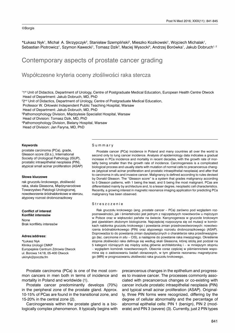
Gleason scale recognizes 5 PCA malignancy pat-terns, ranging from 1 (least malignant) to 5 (most ma-lignant) differing predominantly by the architectural features, and, to a lesser degree, by the appearance of the cancerous cells (fig. 3) (35).
Fig. 1. Carcinogenesis of the prostate gland
Fig. 2. Donald Gleason (34)
Contemporary aspects of prostate cancer grading
843
There are also other grading systems that are much less frequently used to assess the histological advancement of PCa, including systems by Mosto-fi, Böcking and Anderson (MDAH) (tab. 1). Mostofi scale identifies 3 degrees of glandular differentiation and nuclear anaplasia, as does Böcking scale. MDAH system comprises four grades and is based the per-centage of tumor that forms the gland (35). See table below for the comparison of different systems of PCA grading.
Tab. 1. PCa grading systems
Grade 1 Grade 2 Grade 3
Gleason score 2, 3, 4, 5 6, 7 8, 9, 10
Mostofi 1 2 3
Böcking 1 2 3
MDAH 1 2, 3 4
While assessing tumour grade in Gleason scale, the pathologist identifies the most common and the next-
most common pattern in a given tumour, assigning each of them with a numerical value (1-5). The sum of those two numbers comprises Gleason score (Gl.s.) reflecting the global malignancy of the prostate cancer found in the gland, e.g. Gl.s. 5 = 3+ 2 (Gl.s. 7 = 4 + 3) means that the most common histological pattern iden-tified by the pathologist in the tumour is 3, and the next-most common Gleason pattern is 2. If only one histological pattern is found, two identical numbers are added, or the number is multiplied by two (e.g. Gl.s. 6 = 3 + 3 or 3 x 2).
For PCa malignancy patterns see figures 4-10.
Fig. 3. 5 patterns of prostate cancer grading (35)
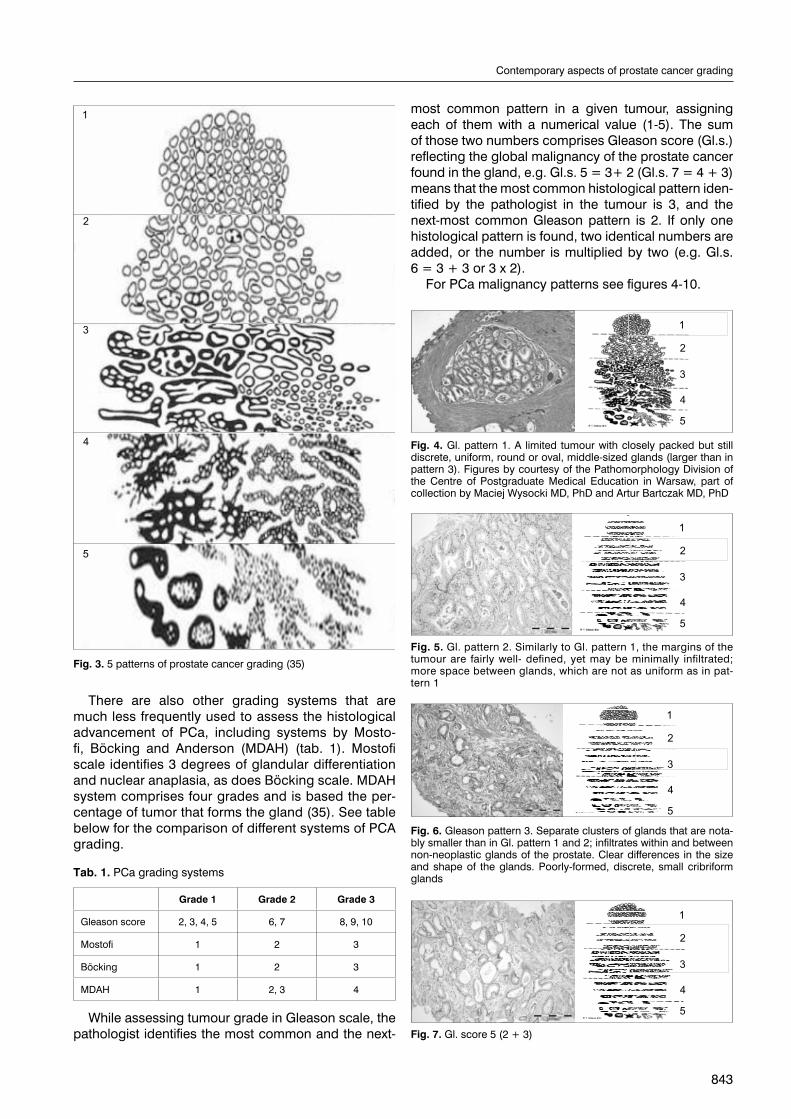
Fig. 4. Gl. pattern 1. A limited tumour with closely packed but still discrete, uniform, round or oval, middle-sized glands (larger than in pattern 3). Figures by courtesy of the Pathomorphology Division of the Centre of Postgraduate Medical Education in Warsaw, part of collection by Maciej Wysocki MD, PhD and Artur Bartczak MD, PhD
Fig. 5. Gl. pattern 2. Similarly to Gl. pattern 1, the margins of the tumour are fairly well- defined, yet may be minimally infiltrated; more space between glands, which are not as uniform as in pat-tern 1
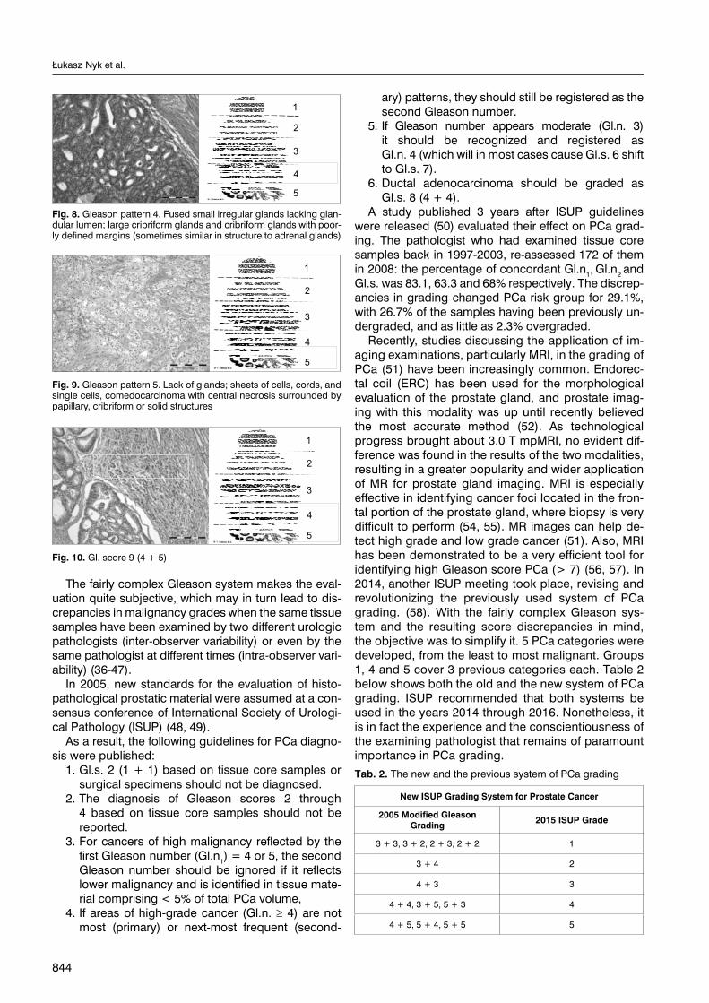
Fig. 6. Gleason pattern 3. Separate clusters of glands that are nota-bly smaller than in Gl. pattern 1 and 2; infiltrates within and between non-neoplastic glands of the prostate. Clear differences in the size and shape of the glands. Poorly-formed, discrete, small cribriform glands
Fig. 7. Gl. score 5 (2 + 3)
844
Łukasz Nyk et al.
The fairly complex Gleason system makes the eval-uation quite subjective, which may in turn lead to dis-crepancies in malignancy grades when the same tissue samples have been examined by two different urologic pathologists (inter-observer variability) or even by the same pathologist at different times (intra-observer vari-ability) (36-47).
In 2005, new standards for the evaluation of histo-pathological prostatic material were assumed at a con-sensus conference of International Society of Urologi-cal Pathology (ISUP) (48, 49).
As a result, the following guidelines for PCa diagno-sis were published:
1. Gl.s. 2 (1 + 1) based on tissue core samples or surgical specimens should not be diagnosed.
2. The diagnosis of Gleason scores 2 through 4 based on tissue core samples should not be reported.
3. For cancers of high malignancy reflected by the first Gleason number (Gl.n1) = 4 or 5, the second Gleason number should be ignored if it reflects lower malignancy and is identified in tissue mate-rial comprising < 5% of total PCa volume,
4. If areas of high-grade cancer (Gl.n. ≥ 4) are not most (primary) or next-most frequent (second-
ary) patterns, they should still be registered as the second Gleason number.
5. If Gleason number appears moderate (Gl.n. 3) it should be recognized and registered as Gl.n. 4 (which will in most cases cause Gl.s. 6 shift to Gl.s. 7).
6. Ductal adenocarcinoma should be graded as Gl.s. 8 (4 + 4).
A study published 3 years after ISUP guidelines were released (50) evaluated their effect on PCa grad-ing. The pathologist who had examined tissue core samples back in 1997-2003, re-assessed 172 of them in 2008: the percentage of concordant Gl.n1, Gl.n2 and Gl.s. was 83.1, 63.3 and 68% respectively. The discrep-ancies in grading changed PCa risk group for 29.1%, with 26.7% of the samples having been previously un-dergraded, and as little as 2.3% overgraded.
Recently, studies discussing the application of im-aging examinations, particularly MRI, in the grading of PCa (51) have been increasingly common. Endorec-tal coil (ERC) has been used for the morphological evaluation of the prostate gland, and prostate imag-ing with this modality was up until recently believed the most accurate method (52). As technological progress brought about 3.0 T mpMRI, no evident dif-ference was found in the results of the two modalities, resulting in a greater popularity and wider application of MR for prostate gland imaging. MRI is especially effective in identifying cancer foci located in the fron-tal portion of the prostate gland, where biopsy is very difficult to perform (54, 55). MR images can help de-tect high grade and low grade cancer (51). Also, MRI has been demonstrated to be a very efficient tool for identifying high Gleason score PCa (> 7) (56, 57). In 2014, another ISUP meeting took place, revising and revolutionizing the previously used system of PCa grading. (58). With the fairly complex Gleason sys-tem and the resulting score discrepancies in mind, the objective was to simplify it. 5 PCa categories were developed, from the least to most malignant. Groups 1, 4 and 5 cover 3 previous categories each. Table 2 below shows both the old and the new system of PCa grading. ISUP recommended that both systems be used in the years 2014 through 2016. Nonetheless, it is in fact the experience and the conscientiousness of the examining pathologist that remains of paramount importance in PCa grading.
Tab. 2. The new and the previous system of PCa grading
New ISUP Grading System for Prostate Cancer
2005 Modified Gleason Grading 2015 ISUP Grade
3 + 3, 3 + 2, 2 + 3, 2 + 2 1
3 + 4 2
4 + 3 3
4 + 4, 3 + 5, 5 + 3 4
4 + 5, 5 + 4, 5 + 5 5
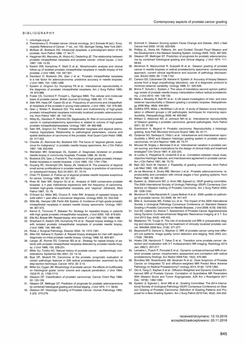
Fig. 8. Gleason pattern 4. Fused small irregular glands lacking glan-dular lumen; large cribriform glands and cribriform glands with poor-ly defined margins (sometimes similar in structure to adrenal glands)
Fig. 9. Gleason pattern 5. Lack of glands; sheets of cells, cords, and single cells, comedocarcinoma with central necrosis surrounded by papillary, cribriform or solid structures
Fig. 10. Gl. score 9 (4 + 5)
Contemporary aspects of prostate cancer grading
845
B I B L I O G R A P H Y
1. onkologia.org.pl.2. Theodorescu D: Prostate cancer, clinical oncology. [In:] Schwab M (ed.): Ency-
clopedic Reference of Cancer. 1st ed., vol. 720. Springer Verlag, New York 2001.3. McNeal JE, Bostwick DG: Intraductal dysplasia: a premalignant lesion of the
prostate. Hum Pathol 1986; 17: 64-71.4. Haggman MJ, Macoska JA, Wojno KJ, Oesterling JE: The relationship between
prostatic intraepithelial neoplasia and prostate cancer: critical issues. J Urol 1997; 158: 12-22.
5. Keetch DW, Humphrey P, Stahl D et al.: Morphometric analysis and clinical follow-up of isolated prostatic intraepithelial neoplasia in needle biopsy of the prostate. J Urol 1995; 154: 347-351.
6. Davidson D, Bostwick DG, Qian J et al.: Prostatic intraepithelial neoplasia is a risk factor for adenocarcinoma: predictive accuracy in needle biopsies. J Urol 1995; 154: 1295-1299.
7. Epstein JI, Grignon DJ, Humphrey PA et al.: Interobserver reproducibility in the diagnosis of prostatic intraepithelial neoplasia. Am J Surg Pathol 1995; 19: 873-886.
8. Foster CS, Cornford P, Forsyth L, Djamgoz MBA: The cellular and molecular basis of prostate cancer. British Journal of Urology 1999; 83: 171-194.
9. Sakr WA, Haas GP, Cassin BJ et al.: Frequency of carcinoma and intraepitheli-al neoplasia of the prostate in young male patients. J Urol 1993; 150: 379-385.
10. Qian J, Wollan P, Bostwick DG: The extent and multicentricity of high-grade prostatic intraepithelial neoplasia inclinically localized prostatic adenocarcino-ma. Hum Pathol 1997; 28: 143-148.
11. Wiley EL, Davidson P, McIntire DD, Sagalowsky AI: Risk of concurrent prostate cancer in cystoprostatectomy specimens is related to volume of high-grade prostatic intraepithelialneoplasia. Urology 1997; 49: 692-696.
12. Sakr WA, Grignon DJ: Prostatic intraepithelial neoplasia and atypical adeno-matous hyperplasia. Relationship to pathological parameters, volume and spatial distribution of carcinoma of the prostate. Anal Quant Cytol Histol 1998; 20: 417-423.
13. Cheville JC, Reznicek MJ, Bostwick DG: The focus of “atypical glands, suspi-cious for malignancy” in prostatic needle biopsy specimens. Am J Clin Pathol 1997; 108: 633-640.
14. Weinstein MH, Greenspan DL, Epstein JI: Diagnoses rendered on prostate needle biopsy in community hospitals. Prostate 1998; 35: 50-55.
15. Bostwick DG, Qian J, Frankel K: The incidence of high grade prostatic intraepi-thelial neoplasia in needle biopsies. J Urol 1995; 154: 1791-1794.
16. Ouyang RC, Kenwright DN, Nacey JN, Delahunt B: The presence of atypical small acinar proliferations in prostate needle biopsy is predictive of carcinoma on subsequent biopsy. BJU Int 2001; 87: 70-74.
17. Chan TY, Epstein JI: Follow-up of atypical prostate needle biopsies suspicious for cancer. Urology 1999; 53: 351-355.
18. Khan W, Sakr WA, Grignon DJ et al.: Analysis of 3300 consecutive prostate biopsies: a 4 year institutional experience with the frequency of carcinoma, isolated high-grade intraepithelial neoplasia, and “atypical” (Abstract). Mod Pathol 1997; 10: 79.
19. O’Dowd GJ, Miller MG, Orozco R, Veltri RW: Analysis of repeated biopsy re-sults within 1 year after a noncancer diagnosis. Urology 2000; 55: 553-559.
20. Wills ML, Hamper UM, Partin AW, Epstein JI: Incidence of high-grade prostatic intraepithelial neoplasia in sextant needle biopsy specimens. Urology 1997; 49: 367-373.
21. Kamoi K, Troncoso P, Babaian RJ: Strategy for repeated biopsy in patients with high grade prostatic intraepithelial neoplasia. J Urol 2000; 163: 819-823.
22. Ellis WJ, Brawer MK: Repeat biopsy: who needs it? J Urol 1995; 153: 1496-1498.23. Shephard D, Keetch DW, Humphrey PA et al.: Repeat biopsy strategy in men
with isolated prostatic intraepithelial neoplasia of prostate needle biopsy. J Urol 1996; 156: 460-463.
24. Rosai J: Surgical Pathology. Elsevier 2004; 18: 1372-1381.25. Allen EA, Kahane H, Epstein JI: Repeat biopsy strategies for men with atypical
diagnoses on initial prostate needle biopsy. Urology 1998; 52: 803-807.26. Langer JE, Rovner ES, Coleman BG et al.: Strategy for repeat biopsy of pa-
tients with prostatic intraepithelial neoplasia detected by prostate needle biop-sy. J Urol 1996; 155: 228-231.
27. Miller GJ, Torkko KC: Natural history of prostate cancer – epidemiologic con-siderations. Epidemiol Rev 2001; 23: 14-18.
28. Byar DP, Mostofi FK: Carcinoma of the prostate: prognostic evaluation of certain pathologic features in 208 radical prostatectomies: examined by the step-section technique. Cancer 1972; 30: 5-13.
29. Miller GJ, Cygan JM: Morphology of prostate cancer: the effects of multifocality on histological grade, tumor volume and capsule penetration. J Urol 1994; 152(5 Pt. 2): 1709-1713.
30. Gleason DF: Classification of prostatic carcinomas. Cancer Chem Rep 1966; 50: 125-128.
31. Gleason DF, Mellinger GT: Prediction of prognosis for prostatic adenocarcinoma by combined histological grading and clinical staging. J Urol 1974; 111: 58-64.
32. Gleason DF: Histologic Grading of Prostate Cancer. Human Pathology 1992; 3 (23): 273-279.
33. Schmidt C: Gleason Scoring system Faces Change and Debate. JNCI J Natl Cancer Inst 2009; 101(9): 622-629.
34. Phillips JL, Sinha AA: Patterns, Art, and Context: Donald Floyd Gleason and the Development o the Gleason Grading System. Urology 2009; 74(3): 497-503.
35. Gleason DF, Mellinger GT. Prediction of prognosis for prostatic adenocarcino-ma by combined histological grading and clinical staging. J Urol 1974; 111: 58-64.
36. Montironi R, Mazzuccheli R, Scarpelli M et al.: Gleason grading of prostate cancer in needle biopsies or radical prostatectomy specimens: contemporary approach, current clinical significance and sources of pathology discrepan-cies. BJUInt 2005; 95: 1146-1152.
37. Carlson GD, Calvanese CB, Kahane H, Epstein JI: Accuracy of biopsy Gleason scores from a large uropathology laboratory: use of a diagnostic protocol to minimize observer variability. Urology 1998; 51: 525-529.
38. Brimo F, Schultz L, Epstain J: The value of mandatory second opinion pathol-ogy review of prostate needle biopsy interpretation before radical prostatecto-my. J Urol 2010; 184: 126-130.
39. Melia J, Moseley R, Ball RY et al.: A UK based investigation of inter- and intra- observer reproducibility o Gleason grading o prostatic biopsies. Histopatholo-gy 2006 May; 48(6): 644-654.
40. Griffiths DFR, Melia J, McWilliam LJU et al.: A study of Gleason score interpre-tation in different groups o UK pathologists; techniques for improving repro-ducibility. Histopathology 2006; 48: 655-662.
41. William C, Allsbrook WC Jr, Johnson MH et al.: Interobserver reproducibility of Gleason grading o prostatic carcinoma: general pathologists. Hum Pathol 2001; 32: 81-88.
42. Svanholom H, Mygind H: Prostatic carcinoma. Reproducibility o histologic grading. Acta Path Microbiol Immunol Scand 1985; 93: 67-71.
43. Ozdamar SO, Sarikaya S, Yildiz L et al.: Intraobserver and interobserver repro-ducibility o WHO and Gleason histologic grading system in prostatic adeno-carcinomas. Int Urol Nephrol 1996; 28: 73-77.
44. McLean M, Srigley J, Banerjee D et al.: Interobserver variation in prostate can-cer scoring: are there implications for the design of clinical trials and treatment strategies? Clin Oncol 1997; 9: 222-225.
45. di Loreto C, Fitzpatrick B, Underhill S et al.: Correlation between visual clues, objective histologic features, and interobservere agreement in prostate cancer. Am J Clin Pathol 1991; 96: 70-75.
46. Bain GO, Koch M, Hanson J: Feasibility of grading carcinomas. Arch Pathol Lab Med 1982; 106: 265-267.
47. de las Morenas A, Siroky MB, Merriam J et al.: Prostatic adenocarcinoma: re-producibility and correlation with clinical stages o four grading systems. Hum Pathol 1988; 19: 595-597.
48. Epstein JL, Allsbroock WC Jr, Amin MB, Egevad LL: ISUP grading committee. The 2005 International Society of Urologic Pathology (ISUP) Consensus Con-ference on Gleason trading of Prostatic Carcinoma. Am J Surg Pathol 2005; 29: 1228-1242.
49. Montironi R, Vela-Navarrete R, Lopez-Beltran A et al.: 2005 update on patholo-gy of prostate cancer. EU 2006; 49: 441-447.
50. Billis A, Guimaraes MS, Freitas LLL et al.: The Impact of the 2005 International Society o Urological Pathology Concensus Conference on Standard Gleason Grading o Prostatic Carcinoma on Needle Biopsies. J Urol 2008; 4(18): 548-553.
51. Vos EK, Litjens GJ, Kobus T: Assessment of Prostate Cancer Aggressiveness Using Dynamic Contrast-enhanced Magnetic Resonance Imaging at 3 T. Eur Urol 2013 Sep; 64(3): 448-455.
52. Masterson TA, Touijer K: The role of endorectal coil MRI in preoperative stag-ing and decision-making for the treatment of clinically localized prostate can-cer. MAGMA 2008 Nov; 21(6): 371-377.
53. Beyersdorff D, Darsow U, Stephan C: MRI of prostate cancer using tree differ-ent coil systems: image quality, tumor detection and staging. Rofo 2003 Jun; 175(6): 799-805.
54. Hoeks CM, Hambrock T, Yakar D et al.: Transition zone prostate cancer: de-tection and localization with 3-T multiparametric MR imaging. Radiology 2013 Jan; 266(1): 207-217.
55. Lemaitre L, Puech P, Poncelet E et al.: Dynamic contrast-enhanced MRI of an-terior prostate cancer: morphometric assessment and correlation with radical prostatectomy findings. Eur Radiol 2009 Feb; 19(2): 470-480.
56. Borofsky MS, Rosenkrantz AB, Abraham N et al.: Does Suspicion of Prostate Cancer on Integrated T2 and diffusion-weighted MRI Predict More Adverse Pathology on Radical Prostatectomy? Urology 2013; 81(6): 1279-1283.
57. Oto A, Yang C, Kayhan A et al.: Diffusion-Weighted and Dynamic Contrast-En-hanced MRI of Prostate Cancer: Correlation of Quantitative MR Parameters With Gleason Score and Tumor Angiogenesis. AJR Am J Roentgenol 2011 Dec; 197(6): 1382-1390.
58. Epstein JI, Egevad L, Amin MB et al.; Grading Committee: The 2014 Interna-tional Society of Urological Pathology (ISUP) Consensus Conference on Glea-son Grading of Prostatic Carcinoma: Definition of Grading Patterns and Pro-posal for a New Grading System. Am J Surg Pathol 2016 Feb; 40(2): 244-252.
received/otrzymano: 12.10.2016accepted/zaakceptowano: 03.11.2016


![Nr 10 (709–XXX) PAŹDZIERNIK 2017 Tom LXII POLIMERYyadda.icm.edu.pl/yadda/element/bwmeta1.element.baztech-1df7bd5… · ści wymiarów różniących nanosfery od mikrosfer [26].](https://static.fdocuments.pl/doc/165x107/6092eeb7b9474645d73fc6cb/nr-10-709axxx-padziernik-2017-tom-lxii-ci-wymiarw-rnicych-nanosfery.jpg)