ACTA BIOLOGIAE EXPERIMENTALIS - rcin.org.pl filepolish academy of sciences the nencki institute of...
45
POLISH ACADEMY OF SCIENCES THE NENCKI INSTITUTE OF EXPERIMENTAL BIOLOGY ACTA BIOLOGIAE EXPERIMENTALIS (Acta Biol Exper., Warsaw) Vol. XXII. No. 4 Founded by KAZIMIERZ BIAŁASZEWICZ E d i t o r - i n - Chief JERZY KONORSKI (Warszawa) Managing Editor STEFAN BRUTKOWSKI (Warszawa) Editorial Board JAN DEMBOWSKI (Warszawa), STANISŁAW DRYL (Warszawa), JANINA HURYNOWICZ (Toruń), JERZY KREINER (Kraków), LILIANA LUBIŃSKA (Warszawa), LUCJAN STĘPIEŃ (Warszawa), WANDA WYRWICKA (Łódź) PAŃSTWOWE WYDAWNICTWO NAUKOWE WARSZAWA 1962 http://rcin.org.pl
Transcript of ACTA BIOLOGIAE EXPERIMENTALIS - rcin.org.pl filepolish academy of sciences the nencki institute of...

P O L I S H A C A D E M Y O F S C I E N C E S THE NENCKI INSTITUTE OF EXPERIMENTAL BIOLOGY
ACTA BIOLOGIAE EXPERIMENTALIS
(Acta Biol Exper., Warsaw)
V o l . X X I I . N o . 4
Founded by
KAZIMIERZ BIAŁASZEWICZ
E d i t o r - i n - C h i e f
JERZY KONORSKI (Warszawa)
M a n a g i n g E d i t o r
STEFAN BRUTKOWSKI (Warszawa)
E d i t o r i a l B o a r d
JAN DEMBOWSKI (Warszawa), STANISŁAW DRYL (Warszawa), JANINA HURYNOWICZ (Toruń), JERZY KREINER (Kraków), LILIANA LUBIŃSKA (Warszawa), LUCJAN STĘPIEŃ (Warszawa),
WANDA WYRWICKA (Łódź)
P A Ń S T W O W E W Y D A W N I C T W O N A U K O W E W A R S Z A W A 1 9 6 2
http://rcin.org.pl

ACTA BIOLOGIAE EXPERIMENTALIS publish original papers on physiology and anatomy of the central nervous system with special reference to the physiology of the higher nervous activity. In addition, experimental investigations on animil behaviour and related disciplines are included. The journal appears quarterly in issues of about 80 pages.
Editorial and other correspondence should be addressed to the Managing Editor, Docent Dr Stefan Brutkowski, Dept. of Neurophysiology, The Nencki Institute of Experimental Bidogy, Pasteura 3, Warsaw 22, Poland.
Subscriptions orders should be sent to: Export and Import Entreprise "Ruch", War-szava, Wilcza 46, Poland. Cables: Exprimruch, Warszawa. Payments to the account of: Na-odowy Bank Polski No. 1534-6-71. Orders for back volumes and sets should be addressed to "Ruch" as well.
NOTICE TO AUTHORS
Manuscripts should be typewritten in triple spacing on one side of the paper and must be fully corrected. Authors will be responsible for the payment of any sum charged for the correction of the printer's proof. Original and one carbon copy should be sent. The English lanjuage is preferred and the authors are urged to submit their papers in the English lan-guage. However, manuscripts in French or German will also be accepted.
All illustrations must be properly lettered or labelled. Figures should be numbered consecutively with Arabic numerals and furnished with author's name, journal's volume and abbreviated tittle of the article on their back side. The legend describing an illustration mist be typed on a separate sheet and should not be appended to the drawing. Authors should indicate the size and the approximate position of text figures.
The Editors reserve the privilege of returning to the author for revision approved ma-nuscript and illustrations which are not in a finished form ready for the printer. Pages that aie heavilly corrected should be retyped.
References to literature should be typed in alphabetical order on a separate sheet. Nimes and initials of the authors with full titles and the first page of the paper should be given. Abbrevations for titles of journals are according to the World List of Scientific Periodicals.
Twenty five gratis reprints are given to each author. Additional copies up to 150 are ob ained according to rates which will be sent to the author as soon as the manuscript has been acccpted.
http://rcin.org.pl
' O L 1 S H A C A D E M Y O F S C 1 E N C E 8 T H E N E N C K I I N S T I T U T E O F E X P E R I M E N T A L B I O L O G Y
ACTA BIOLOGIAE EXPERIMENTAL^
(Acta Biol E x p e r . , W a r s a w )
Vol. XXII. No. 4
Founded by
KAZIMIERZ BIAŁASZEWIOZ
E d i t o r - i n - C h i e f
JERZY KONORSKI (Warszawa)
M a n a g i n g E d i t o r
STEFAN BRUTKOWSKI (Warszawa)
E d i t o r i a l B o a r d
JAN DEMBOWSKI (Warszawa), STANISŁAW DRYL (Warszawa), JANINA HURYNOWICZ (Toruń), JERZY KREINER (Kraków). LILIANA LUBIŃSKA (Warszawa), LUCJAN STĘPIEŃ (Warssawa).
WANDA WYRWICKA (Łódź)
P A Ń S T W O W E W Y D A W N I C T W O N A U K O W E
W A R S Z A W A 1 9 6 2
http://rcin.org.pl
Copyright 1962 by Państwowe Wydawnictwo Naukowe
Warszawa
Printed in Poland to the order
by Warszawska Drukarnia Naukowa Warszawa Śniadeckich 8
Nakład 700 + 100 egz. Ark. wyd. 3,25. Ark. druk. 2,5 Papier druk. sat. III kl. 80 g, 70 X 100. Oddano do składu 14.IX.1962 r. Podp. do druku i druk ukończono w grudniu 1962 r. Zam 383 H-90.. Cena zł 14—
http://rcin.org.pl

T H E BRAIN VENTRICLES IN T H E DOG
M. HORODYSKA and J. KREINER
Department of Comparative Neuroanatomy, Jagellonian University, Cracow and
Laboratory of Neuroanatomy, The Nencki Institute of Experimental Biology, Warsaw, Poland
(Received March 18, 1962)
The aim of this investigation was to study the real shape of the ventricle system in the dog's brain. Similar work was done for the human brain ( R e t z i u s 1900, Brück 1933), and also for the brain of the horse ( D e x l e r 1903). However, the brain ventricles in the dog have not been described so far.
MATERIAL AND METHODS
The shape of the brain ventricles can be studied with two methods. Either a cast can be made by filling them with Wood's metal or any suitable injection mass, and then removing the lissue, or by making a model from a series of sections. According to the literature (F rank-Marcus 1933) the methods of making casts do not seem reliable. The soft walls of the ventricles resist neither the heavy metal nor the pressure of the injected mass, and thus the casts are much enlarged, if not disformed.
Therefore, we preferred to make a model. Our model was made of cardboard, 2 mm thick. We based on a series of horizontal sections of a brain fixed in formalin and stained according to the method of Weigert-Wolters. Every fourth section was drawn in a 10 times magnification on the cardboard, cut out and carefully sticked together. The chorioid plexuses protruding into the ventricles were considered to be interior to the ventricle resp., and were not drawn. Some of the series of sagittal and frontal sections of the dog's brain were used for the control.
THE THIRD VENTRICLE (Figs. 2, 3 and 4)
The ventricular system in the dog's brain shows the same parts as those which are known in the human brain: the central canal in the spinal cord, the fourth ventricle, the aqueduct, and two lateral ventricles inside the hemispheres. A detailed description we will begin with the third ventricle situated in the centre of the ventricular system.
http://rcin.org.pl

As it is usual in mammals, the third ventricle of the dog is a vertical cleft within diencephalon. Its side walls are formed of the two thalami. In the middle, the thalami fuse forming the interthalamic adhesion (commissura mollis, massa intermedia). Thus, the general outline of real shape of the third ventricle becomes similar to a flattened ring (Figs. 2 and 3). Orally, the third ventricle is closed by the lamina terminalis, caudally, by the mass of mesencephalon, and ventrally, by the basis hypothalami. Dorsally, the third ventricle is covered by the ependymal lamina, forming the chorioid plexus.
One must not imagine that the ring-like shape of the third ventricle is very regular. Its external contour is disturbed by many diverticula. A large diverticulum is seen in its ventral part: the ring widens here, and the ventricle protrudes between the hypothalamic nuclear complexes of the left and right sides reaching the optic chiasm, infundibulum and mammillary bodies. Some recessus appear here in the vicinity of these bodies, the most pronounced being the preoptic and infundibular recessus. The ventricle widens here over the optic chiasm, while in the middle of the hypo-thalamus its walls nearly stick together. In the oral part, the ventricle is limited by the lamina terminalis. Its outline is disturbed here by the mass of the anterior commissure protruding a little into the ventricle, and thus forming two shallow diverticula under and over the commissure.
The dorsal portion (Figs. 3 and 4) is the most interesting part of the third ven-tricle. The rounded masses of the two thalami diverge here, and thus the ventricle widens here considerably, its sections becoming similar to the letter T. The roof
Fig. 1. Lateral view of the whole model seen from the right side.
http://rcin.org.pl
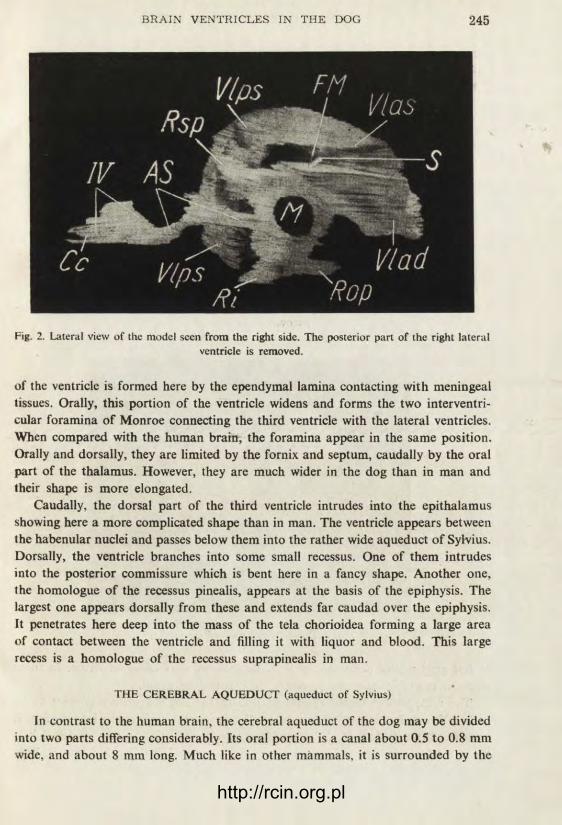
Fig. 2. Lateral view of the model seen from the right side. The posterior part of the right lateral ventricle is removed.
of the ventricle is formed here by the ependymal lamina contacting with meningeal tissues. Orally, this portion of the ventricle widens and forms the two interventri-cular foramina of Monroe connecting the third ventricle with the lateral ventricles. When compared with the human brain, the foramina appear in the same position. Orally and dorsally, they are limited by the fornix and septum, caudally by the oral part of the thalamus. However, they are much wider in the dog than in man and their shape is more elongated.
Caudally, the dorsal part of the third ventricle intrudes into the epithalamus showing here a more complicated shape than in man. The ventricle appears between the habenular nuclei and passes below them into the rather wide aqueduct of Sylvius. Dorsally, the ventricle branches into some small recessus. One of them intrudes into the posterior commissure which is bent here in a fancy shape. Another one, the homologue of the recessus pinealis, appears at the basis of the epiphysis. The largest one appears dorsally from these and extends far caudad over the epiphysis, it penetrates here deep into the mass of the tela chorioidea forming a large area of contact between the ventricle and filling it with liquor and blood. This large recess is a homologue of the recessus suprapinealis in man.
THE CEREBRAL AQUEDUCT (aqueduct of Sylvius)
In contrast to the human brain, the cerebral aqueduct of the dog may be divided into two parts differing considerably. Its oral portion is a canal about 0.5 to 0.8 mm wide, and about 8 mm long. Much like in other mammals, it is surrounded by the
http://rcin.org.pl
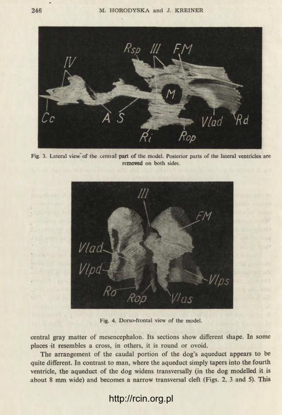
Fig. 3. Lateral view'of the cential part of the model. Posterior parts of the lateral ventricles are removed on both sides.
Fig. 4. Dorso-frontal view of the model.
central gray matter of mesencephalon. Its sections show different shape. In some places -it resembles a cross, in others, it is round or ovoid.
The arrangement of the caudal portion of the dog's aqueduct appears to be quite different. In contrast to man, where the aqueduct simply tapers into the fourth ventricle, the aqueduct of the dog widens transversally (in the dog modelled it is about 8 mm wide) and becomes a narrow transversal cleft (Figs. 2, 3 and 5). This
http://rcin.org.pl

Fig. 5. Dorsal view of the model of the fourth ventricle and the cerebral aqueduct.
is caused by the peculiar arrangement of the quadrigeminal plate which bends here dorsad and lifts the attached oral portion of the anterior medullary velum of the cerebellum. Thus, the aqueduct widens largely caudad and dorsad forming an accessory ventricle limited latero-dorsally by the diverging brachia coniunctiva. This space secondarily is narrowed from the caudal side by the pressure of the lobulus centralis cerebelli bulging into this hollow space and pushing the lamina of the velum medulläre. Thus, in its oral part, the velum assumes a position in the frontal plane, while its middle and caudal portions form a kind of garland approach-ing the cerebellum.
Considering the position ventrally from the quadrigeminal lamina, and orally from the velum medulläre, and situation of the lobulus centralis cerebelli behind it, we regard this space as belonging to the aqueduct. In favour of this conception there speaks the fact that in man the limit between the aqueduct and the IV-th ventricle is situated just at the lips of a fissure between the lobulus centralis and the lingula cerebelli. In the dog's brain this point appears in ventral part of the widening of the aqueduct just described.
THE FOURTH VENTRICLE (Figs. 1, 2, 3 and 5)
The space caudally from the fissure between the lobulus centralis and the lingula is considered the fourth ventricle proper. The ventricular space widens here more and more, and may be divided into two portions: the anterior, extending in approxi-mately dorsal direction, and the posterior, situated horizontally. Pars anterior is found between the lingula orally and the nodulus cerebelli caudally. This is the
http://rcin.org.pl

same situation as that of the recessus superior or fastigiura ventriculi in man, but in the dog's brain this part of the fourth ventricle appears much more expanded laterad where it passes into the wide recessus laterales without any marked boundary. The horizontal pars posterior is again a narrow cleft stretched transversally between the fossa rhomboidea and the uvula cerebelli. Caudad it passes into the central canal of the spinal cord.
THE LATERAL VENTRICLES (Figs. J, 2, 3, 4, 6 and 7)
The lateral ventricles in the dog, much like in other mammals are hidden within the hemispheres. They communicate with the third ventricle through the oblongue. in the dog relatively large, foramen interventriculare of Monroe.
The appearance of the lateral ventricles in the dog is rather difficult to describe. They are clefts of fancy shape influenced largely by the neighbouring structures, especially by the Amnion's horn and the fimbria, and by the striatal nuclei, the caudate and the amygdala.
In the human anatomy, it is generally accepted to divide the lateral ventricle nto the central part, and the anterior, inferior and posterior horns. This can be
Fig. 6. Latero-ventral view of the posterior part of the right lateral vcntricle.
hardly applied to the dog's brain. Therefore, we divide it only into two parts: the anterior, being a homologue of the anterior horn in man, and the posterior com-prising the ventricle caudally from the foramen of Monroe.
The shape of the anterior part is similar to the corresponding region in man. The ventricular space is found here between the septum pellucidum medially, and the bulky caudate nucleus laterally. Dorsally, the ventricle slopes laterad over the
http://rcin.org.pl

caudate. Its roof is here formed of the fibers of the callosal commissure. Ventrally. the ventricle continues in a small recess being a vestige of the ventriculus bulbi olfactorii which in some dogs is obliterated, and in others it remains in a form of a narrow canal penetrating to the olfactory bulb.
The shape of the posterior part of the lateral ventricle is related largely to the formation of the Ammon's horn. The ventricle accompanies this formation along
Fig. 7. Oro-ventral view of the posterior part of the right lateral ventricle.
its whole length in its caudal and dorsal aspect. Therefore, in the model a large grove (Figs. 6 and 7) is seen along the whole ventricle which is the impression of the Ammon's horn accompanied by a smaller furrow caused by the fimbria. The external surface of the ventricle, formed of the fibers of the semioval centre is convex and smooth (Fig. 1) with only one shallow concavity in its lower portion. This is the impression of the amygdaloid nuclear complex.
CONCLUSIONS AND SUMMARY
The authors describe the ventricular system of the dog's brain basing on a card-board model in 10x magnification. The third ventricle in the dog's brain appears in the form of a flat ring with some recessus homologue to those found in man. The recessus suprapinealis is found to be much larger in the dog than in man. The arrangement of the cerebral aqueduct appears different from that in man. Its caudal portion in the dog's brain widens considerably due to the different position of the quadrigeminal lamina, and it forms an accessory ventricle. The fourth
http://rcin.org.pl
ventricle extends more transversally than in man, and shows no limit between the central part and the lateral recessus. The whole fourth ventricle may be divided into pars anterior, extending dorsad, and pars posterior extending horizontally. The lateral ventricles are divided into pars anterior, homologue and similar to the anterior horn in man, and pars posterior caudally from the inter-ventricular foramen. This part accompanies the formation of the Ammon's horn which largely influences the shape of the ventricle. The interventricular foramen of Monroe is relatively larger than in man and more oblongue.
ABBREVIATIONS
Am — impression of the amygdala AS — cerebral aqueduct (aqueduct of Sylvius) CA — impression of the Ammon's horn Cc — central canal of the spinal cord Fi — impression of the fimbria FM — foramen of Monroe M — massa intermedia thalami Ri — recessus infundibularis RL — recessus lateralis of the fourth ventricle Ro — recessus olfactorius Rop — recessus opticus Rsp — recessus suprapinealis S — the plain of section Vlad — anterior part of the right lateral
ventricle Vlas — anterior part of the left lateral ven-
tricle Vlpd — posterior part of the right lateral ventricle Vlps — posterior part of the left lateral
ventricle III — the third ventricle IV — the fourth ventricle
REFERENCES
BRÜCK Ch. 1933 — Das Ventrikelsystem des menschlichen Gehirns. J. Psych. Neur. 45, 545. DEXLER H. 1903— Beiträge zur Kenntnis des feineren Baues des Zentralnervensystems der
Ungulaten. Morph. Jahrb. 32, 288. FRANK J. and MARCUS G. 1933 — Verfahren zur Herstellung von Metallausgüssen des Hirn-
ventrikelsystems. Anat. Anz. 76, 1. RETZIUS A. 1900 —Die Gestalt der Hirnventrikel des Menschen. Biol. Untersuch. IX.
http://rcin.org.pl
T R A N S F E R OF T H E ' C O N D I T I C N E D AVOIDANCE REACTION TO THE U N C O N D I T I O N E D NOXIOUS STIMULI
E. FONBERG
Department of Neurophysiology, The Nencki Institute of Experimental Biology, Warsaw, Poland
(Received April 6, 1962) *
The fact that the avoidance reaction is sustained for months or even years without being reinforced in the classical, Pavlovian sense of the word evokes much interest and discussion.
It is well known that a noxious reinforcement is usually applied only in the first period of the avoidance training. The avoidance reaction once established to a given conditioned stimulus (CS) remains stable and occurs regularly to this particular stimulus, without being reinforced. According to the most popular theories ( M i l l e r and D o l l a r d 1941, M i l l e r 1948, M o w r e r 1950, S o l o m o n and W y n n e 1953, 1954, and others), the CS by having been associated with the U S during the initial training, has acquired properties similar to those of an un-conditioned stimulus (US). Therefore, the termination of the CS acts in the same way as the termination of the US, serving as reinforcement for the reaction being performed at that time. In this case, the mechanism of avoidance should be the same as that of escape, the performance of the avoidance movement being an escape from the "aver^ive" CS, identical with escape from the US.
Our method of differentiation and extinction of avoidance reaction was based on such an assumption ( F o n b e r g 1952). In this method the CS outlasted the performance of avoidance reaction by several seconds, such extension of the CS served as a lack of reinforcement and led to extinction of the reaction ( B r e g a d z e 1953, F o n b e r g 1958 a, S o l t y s i k e t al. 1960). The role of termination of the CS as a reinforcing factor for avoidance reactions has also been proved by other ex-periments ( K a m i n 1956, 1957, K a m i n el al. 1959, M o w r e r and L a m o r e a u x 1942, S o l o m o n , K a m i n and W y n n e 1953, S i d m a n and B o r e n 1957, and
http://rcin.org.pl

others). However, the question arises, does the CS really evoke a state similar to that evoked by the US? It is possible that the defensive excitement produced by the CS has a rather general, "anonymous" character, independent of the quality of noxious reinforcement. After months of avoidance training without the use of this reinforcement, the CS may no longer signalize a particular US, and may evoke only an indefinite defensive or fear state not connected with any particular noxious stimulus. This last point of view seems to be supported by the experiments of K o n o r s k i and Mi l l er (1936), who have shown that an animal may perform an instrumental avoidance reaction not only to the CSi signalizing the same noxious agent as that used in the original training, but also to those CSi which signalize different noxious agents. Furthermore, the experiments by F o n b e r g (1956, 1958 c, 1959) have demonstrated that a previously trained avoidance reaction appears spontaneously in neurotic states in which neither conditioned nor unconditioned defensive stimuli were used.
This last group of experiments seems to support the supposition that the defen-sive excitement produced by the CS is unspecific, that is, it is not related to the type of reinforcement.
More detailed studies on the transfer of avoidance reaction ( F o n b e r g 1957, 1958 a, b, 1961) show, however, that only when the animal manifests a strong and continuous defensive reaction to the whole experimental situation (consisting of a general motor restlessness, attempts to escape from the stand and numerous inter-trial reactions), does he perform the trained avoidance movement to heterogenous CSi. But if the dog is calm, it performs the avoidance reaction only to those stimuli to which it was trained, and if to other CSi, only to those signalizing the original noxious reinforcement. These results would suggest that the defensive state evoked by the CS is specific and somehow related to the kind of reinforcement.
The question of whether the CS during the initial training acquires properties similar to those of the US associated with it remained still unanswered. If, in fact, these two states are similar, and, in consequence, related to the excitation of the same cerebral structures, the reaction performed in both states should be similar.
The method most widely used to establish the avoidance reaction is to train it on the basis of the escape reaction. This does not permit us to distinguish the responses to CS and US, since in both cases the motor reaction is the same. Due to the method of K o n o r s k i and Mi l l er (1933, see below in procedure) we are able to separate these two reactions.
The aim of the present paper is to study, by the aid of this method, the relations between the states evoked by the CS and the US. If these states are identical, the avoidance reactions evoked regularly by the CS should appear also to the US with which the particular CS have been associated. If, however, the sensations produced by the CS and US are qualitatively different, the transfer of the avoidance reaction should not occur to the US.
http://rcin.org.pl

PROCEDURE
Experiments were performed on 5 dogs in a regular soundproof reflex-chamber. P re l iminary t r a in ing . At first, the dogs were trained to perform an avoidance reaction
in response to the CS. The avoidance training was carried out by the method of K o n o r s k i and Miller (1933). The animals were taught to perform a particular movement in response to a given
stimulus in order to avoid a noxious reinforcement which otherwise accompanied this stimulus. Different movements were used as the avoidance reaction, viz. lifting the right fore leg, right hind leg, left foxe leg, and barking reaction. Each dog was trained to perform only one of these reactions. Acoustic and visual stimuli were applied as CSi, and air-puff into the ear, or electric shock to the skin, or injection of acid into the mouth served as USi (see Table I). During the initial training the CS was in random order either followed by the negative reinforcement, or by a passive movement produced by the experimenter. In the case of barking the reaction was provoked by the imitation of the sound of barking. Immediate cessation of the CS followed the passive movement and noxious reinforcement was not applied. With time, the animal responded to CS actively, avoiding the noxious reinforcement. The avoidance reaction was established to a 100% criterion of performance and thereafter trained for at least three more months.
Table I
No. of Dog
Conditioned Avoidance Stimulus Reaction
1
Original Other
Unconditioned . . . _ . , noxious Stimuli Stimulas
1 Buzzer Lifting of the left fore leg Air-puff Electric shock, Acid
2 Splash Lifting of the right hind leg Air-puff Electric shock Acid
3 Splash Barking Air-puff Electric shock Acid
4 Buzzer Lifting of the right hind leg Air-puff Electric shock Acid
!
! 5 !
Light Lifting of the right fore leg Electric shock Air-puff Acid
Test experiments .
1) Tests were made to see whether the avoidance reaction would appear to the same US as used in the avoidance training when it was applied alone, without being preceded by the CS.
2) The reaction to other noxious stimuli was also tested. For example, if during the avoidance training an air-puff into the ear was used as reinforcement, then, in the test experiments, the effect of electric shock and acid injection was observed.
The procedure of test experiments was as follows: Every day a normal avoidance session was carried out in which 6 to 15 avoidance trials were given. Test sessions were conducted about once a week and consisted of the regular avoidance trials with test trials irregularly interspersed. In the test trials the noxious stimuli were not preceded by CSi. The duration of the US was usually 10 to 20 seconds. In addition, the effect of prolongation of the US was tested. In 3 dogs, both the original and new USi were applied for 40, or 60 seconds. Total number of test trials was 10 to 20 for each kind of US.
http://rcin.org.pl

RESULTS
As seen in Fig. 1, the (instrumental) avoidance reaction trained to the presen-tation of CS to avoid a certain U S and established in 100%, does not appear as a rule to the US itself. This applies to the U S which had been used in the avoidance training, as well as to other kinds of noxious stimuli. Only dog No . 4 performed the avoidance reaction in 85% to the original US, and in 25% to the other USi . In this dog, however, the test experiments evoked a general defensive excitement due to the unexpected use of noxious stimuli among the normal avoidance trials. This state was manifested both by the general restlessness and in the numerous intertrial reactions (Fig. 2c). Because of their great frequency, it may be just accidental that the performance of avoidance reaction coincided with the tested US. In other dogs, even in a state of general defensive excitement the action of U S produced either a decrease or, even more often, the complete disappearance of the intertrial avoidance reactions (Figs. 2d and e). Usually, the dogs reacted to the unexpected U S by general motor excitement. Sometimes, however, they remained frozen in a motionless position. When such USi as an air-puff into the ear, or an injection of the acid were applied, it happened that the dogs tried to tear off the tubes fastened to the ear or to the cheek.
The prolongation of the US did not, as a rule, evoke the avoidance movement (Fig. 2e). Usually, if the avoidance reaction did not occur within the first few second of the action of the noxious stimulus, it did not appear at all in spite of its prolon-gation.
DISCUSSION
The results of our experiments clearly show that the animal does not try to escape from the U S by performing the same reaction as was trained to avoid it, if the trained movement is not identical with the unconditioned reaction to the US.
It is possible that this fact is mainly due to the Konorski and Miller method of the avoidance training. If the avoidance movement is trained in such a way that it is transferred from the escape reaction, it preserves some of the qualities of the unconditioned response. So the avoidance movement is a transformed natural escape reaction from the noxious stimulus, or at least a reaction synergic with it. In the method of Konorski and Miller the movement used as avoidance reaction is d e l i b e r a t e l y d i f f e r e n t from the unconditioned response to the noxious agent. The incompatibility of these two reactions may be the cause of the antagonism in their occurrence and may explain the fact that the avoidance reaction is inhibited during the action of the US. This does not explain, however, why the avoidance movement fails to appear even when the animal is not performing any visible un-conditioned reaction (Fig. 2b). Also the conditioned barking is not antagonistic to the natural defensive response to the US. Even in such cases where the reaction to the U S is well manifested and quite different from the artificially established
http://rcin.org.pl

avoidance movement, the animal is able to perform them both together, or, at
least, in succession. Therefore, the discrepancy in the occurrence of these reactions
is not due to a physical inability to perform them both. It may be rather due to the
fact that different systems are mediating these two reactions, or that the pattern
of the excitation of various brain areas is different, and this leads to different per-
formance.
If the states evoked by the CS and the U S were similar, the reactions mediated by these states should occur in conformity. As this is not the case, it seems that
% 100 - — — — -
^ 80
<o
SO
I 1 40 <S3
§¿7
£ 'A a
1 i 1
/ 2 3 4 5
Fig. 1. Transfer of avoidance reaction to the noxious USi White blocks represent the percent of avoidance reaction to the CS to which this reaction was trained. Striped blocks, the avoidance reaction to the original US used in the avoidance training; black blocks, the reaction to other noxious stimuli. The avoidance reaction does appear to the original US only in dog No. 4 (85%). It is nil in dogs Nos. 3 and 5, and 10% of performance in dogs Nos. 1 and 2. To the other noxious stimuli the avoidance reaction does not appear at all in three
dogs, and appears only in 25% in dog No. 4.
the noxious US itself produces quite a different state from the state of its expectancy
evoked by the CS. As shown in our previous experiments ( F o n b e r g 1958 a, b,
1959), if we introduce a new CS reinforced by the US (even different from the one
used in the avoidance training), the degree of transfer of avoidance reaction to such
a CS is much greater than that to the US in our present experiment. This transfer
occurs especially, when the animal is in a state of general defensive excitement.
In such a state, if two different reactions are trained to two different CSi, the animal
may perform both of them to each CS even if the movements are incompatible
( F o n b e r g 1961). It was expected that the action of the U S evoking a high level
of general defensive excitement would provide good conditions for the appearance
of the avoidance reaction. But as we have seen, the results of our experiments show
that, in spite of a high degree of defensive state, the avoidance reaction did not
appear to the US. On the contrary, even if the intertrial reactions were present,
they were inhibited during the action of the US (Fig. 2d).
http://rcin.org.pl

a
10 D-1
r ^
J 1 1 i i i . , CS — us
C
kxxJ U j l j l J U j l ^
CS us CS us
ULLl, -A
_» m4—*—-yK-
US us
Fig. 2. Partial kymographic records of the test experiments Line I — Avoidance reaction. Line II — Control leg. Line III — CSi and USi. Line IV — Time in seconds.
a. Experiments carried out on dogs Nos. 1 and 5. The CS evokes a regular avoidance reaction, and the US (the same one as used in the avoidance training i.e. air-puff into the ear in dog No. 1 and an electric shock on dog No. 5) does evoke only the unconditioned reaction of tearing off the tube fastened to the ear (dog No. 1), or withdrawal of the lep to which
the electrodes were connected (dog No. 5). The avoidance reaction does not appear to the US. b. Experiment carried out on dog No. 2. The CS evokes the regular avoidance reaction and the noxious stimulus — an
electric shock on the back — does neither evoke the avoidance movement nor any other motor reaction. c. Dog No. 4 performs the avoidance reaction to the US. He does, however, perform it during intertrial intervals too.
d. Intertrial reactions are abolished during the action of the tested US. e. Prolongation of the US does not provoke the avoidance reaction to appear.
http://rcin.org.pl

So, we may conclude that the states evoked by any CSi reinforced by different USi have more in common with each other than those evoked by the CS and the US, even if the latter have been associated in the avoidance training. Therefore, we cannot say that during the initial training the CS had acquired by classical condi-tioning properties similar to those of the US, and, in consequence, we cannot consider the CS as a substitute for the US. The independence of the avoidance reaction from the state evoked by the US indicates that the mechanism of avoidance must be different from that of escape and of classical conditioning. Even if the escape from the CS is an important factor in training and stabilization of the avoidance reaction, it must have a quite different mechanism from the escape from the US.
The data provided by S o l t y s i k and Zie l i r i sk i (1961), S o l o m o n and W y n n e (1954) give more evidence indicating that the escape and avoidance reactions have different features, and therefore may be mediated by different or even antagonistic mechanisms. As shown by the present experiments this may be due to the fact that the states evoked by the CS and the US are different. The unconditioned "fear" evoked by the U S must have quite a different nature from the conditioned fear, the expectancy of impending noxious stimulation. It is also possible that there may exist various kinds of fear both conditioned and unconditioned (if we can call by the same name the different kinds of aversive and unpleasant sensations). In our previous work ( F o n b e r g 1958 b) an assumption was made that the defensive center is not uniform and that there may exist various defensive and fear centers more or less connected with each other. The experiments by W y r w i c k a et al. (1961) on brain stimulation in goats and our experiments on the stimulation of hypothalamus which are now in progress, seem to support this point of view.
SUMMARY
The aim of this paper was to study the relation between the conditioned stimulus (CS) and unconditioned stimulus (US) in avoidance training. Experiments were performed on 5 dogs. The results show that the avoidance reaction which appears in 100% to the CS does not appear as a rule to the same US which had been associated with the CS in the original training. It neither appears to the other kinds of noxious stimuli. The sources of this discrepancy are disscussed. It was concluded that the state produced by the CS seemed to be different from the one produced by the US, and that CS is not simply a substitute of the US.
The author wishes to thank Professor J. Konorski for his valuable criticism and helpful advice.
REFERENCES
BREGADZE A. N. 1953 —- On the problem of the elaboration of the conditioned defensive reflex in dogs (in Russian) Trudy Inst. Fiziol. im. I. S. Beritashvili, 9, 53.
FONBERG E. 1952 — Unpublished data. Presented at the Meeting of the Department of Neuro-physiology, The Nencki Institute of Experimental Biology, Lodz.
http://rcin.org.pl
FONBERG E. 1956 — On the manifestation of conditioned defensive reactions in stiess. Bull. Soc. Sci. Lettr. Lodz. Cl. Ill, 7, 1.
FONBERG E. 1957 — Concerning the mechanism of conditioned defensive reactions occurring in experimental neuroses (in Polish). Acta Physiol. Polon., 8, 321.
FONBERG E. 1958a — On the defensive conditioned reflexes type II (in Russian). Proceedings of the International Symposium on the Central and Peripheral Mechanisms of the Motor Activity of the Animals, Osieczna, Poland 1958. Published by the Academy of Sciences of the USRR, Moscow 1960, 124.
FONBERG E. 1958b — Transfer of instrumental avoidance reactions in dogs. Bull. Acad. Polon. Sci., 6, 353.
FONBERG E. 1958c — The manifestation of the defensive reactions in neurotic states. Acta Biol. Exper., 18, 89.
FONBERG E. 1959 — The manifestation of the defensive reactions in neurotic states (in Polish). Doctor's thesis (in press).
FONBERG E. 1961—On the transfer of two different defensive conditioned reflexes type II. Bull. Acad. Polon. Sci., 9, 47.
KAMIN L. J. 1956 — The effect of termination of the CS and avoidance of the US on avoidance learning. J. comp. physiol. Psychol., 49, 420.
KAMIN L.J. 1957 — The gradient of delay of secondary reeward in avoidance learning tested on avoidance trials only. J. comp. physiol. Psychol., 50, 450.
KAMIN L. J., CAMPBELL D., JUDD R., RYAN T. and WALKER J. 1959 — Two determinants of the emergence of anticipatory avoidance. J. comp. physiol. Psychol., 52, 202.
MILLER N. E. and DOLLARD J. 1941—Social learning and imitation. New Haven, Yale Univ. Press.
MILLER N. E. 1948 — Studies of fear as an acquirable drive. J. Exper. Psychol. 38, 89. MOWRER O. H. 1950 — Learning theory and personality dynamics. Ronald Comp. Press, New
York. MOWRER O. H. and LAMOREAUX R. R. 1946 — Fear as an intervening variable in avoidance
conditioning. J. comp. physiol. psychol. 39, 29. KONORSKI J. and MILLER S. 1933 —Les principles fondamentaux de la théorie physiologique
des mouvements acquis. Varsovie. (In Polish with French Summary). KONORSKI J. and MILLER S. 1936 —Conditioned reflexes of the motor analyzer. Trudy Fiziol.
Lab. im. I .P. Pavlova (In Russian), 6, 119. KONORSKI J. 1948—Conditioned reflexes and neuron organization. Cambridge. SIDMAN M. and BOREN J.J . 1957 — The relative aversiveness of warning signal and shock
in an avoidance situation. J. abnorm. soc., Psychol., 55, 339. SOLOMON R. L. and WYNNE L. C. 1953—Traumatic avoidance learning: Acquisition in
normal dogs. Psychol. Monogr., 67, 354. SOLOMON R. L. and WYNNE L. C. 1954— Traumatic avoidance learning: The principles of
anxiety conservation and partial irreversibility. Psych. Rev., 61, 353. SOLOMON R. L., KAMIN L.J. and WYNNE L. C. 1953—Traumatic avoidance learning:
The outcomes of several extinction procedures with dogs. J. abnorm. soc. Psychol., 48, 291. SOLTYSIK S. I960 — Studies on the avoidance conditioning. Acta Biol. Exper., 20, 171. SOLTYSIK S. and ZIELINSKI K. 1961 — The role of afferent feed-back in conditioned avoidance
reflex. International Symposium in Liblice, Czechoslovakia (in press). * WYRWICKA W. and DOBRZECKA C. 1961 — On the transfer of defensive conditioned reactions
established to the electrical stimulation of diencephalon in goats. Bull. Acad. Polon. Sci., 9, 51.
http://rcin.org.pl
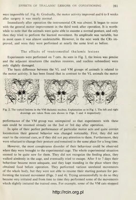
THE EFFECT OF LESIONS IN THE VENTROMEDIAL
A N D / O R VENTROLATERAL THALAMIC NUCLEI O N I N S T R U M E N T A L
C O N D I T I O N E D REFLEXES IN CATS
R. ÏARNECKI
Department of Neurophysiology, The Nencki Institute of Expe»imental Biology,
Warsaw, Poland
(Received April 6, 1962)
One of the major problems concerning the mechanisms of instrumental con-
ditioned reflexes (CRs) is that of the role of sensory feedback generated by the
performance of the trained movement in the formation and preservation of these
reflexes. According to the results obtained by J a n k o w s k a (1959), and G ô r s k a
and J a n k o w s k a (1959), deafferentation of the limb involved in the performance
of the instrumental CR does not lead to its abolition, although the movement
itself becomes more or less defective. However, it could be supposed that, instead
of the peripheral feedback, a shortened central feedback might be responsible for
the preservation of the CR. Such a feedback may be represented either by the py-
ramidal-leminiscal loop, or by the cortico-cerebellar loop.
In previous experiments of this series ( T a r n e c k i 1962) the medial-leminiscal
pathways were bilaterally damaged in cats. It has been found that this lesion docs
not abolish the instrumental CR. The present paper is concerned with the problem
of the effects of lesions located in the ventrolateral (VL) and ventromedial (VM)
thalamic nuclei. The connections of the VL nucleus are relatively well known,
but the VM nucleus has been insufficiently explored so far. It has been, however,
anticipated that this nucleus might belong to the same functional system as the
VL nucleus.
MATERIAL AND EXPERIMENTAL PROCEDURE
Experiments were performed on 24 cats in the age of 1.5 to 2 years. Details of the procedure were described previously (Tarnecki 1952). Ia brief, after the animils had been adjusted to the experimintal conditions, the instrununtal CR. consisting of scratching-like movements of the right hindleg was established. This was nude by reinforcing with food the natural scratching movements
http://rcin.org.pl

induced by placing a piece of cotton-wool into the right ear. 30 reinforced trials were given in each experimental session, and the duration of the session was taken as the measure of the rate of the animal's performance.
The instrumental response established in this way had the following character. The cat assumed the same posture which he held in normal scratch-reflex, i.e. he was sitting with the body slightly inclined to the left, so as to have the right hindleg free for the performance of the movement. Then, he protracted the leg towards his ear, and eventually made a few rhythmic movements imitating scratching. The skin of the body or head was, however, never being touched.
When the instrumental CR was fixed, and the animal performed the scratch-like movement of the hindleg at the maximal rate, pausing only while eating food presented after each response, symmetrical bilateral thalamic lesions were placed stereotaxically in one stage under the nembutal anesthesia (35 mg./kg.). Lesions were made by passing unipolar DC of 2.5 mA within 2 min.
Three types of lesions were performed: a) within the VL nucleus, b) within the VM nucleus, and c) in both nuclei.
The CR experiments usually were resumed 2 to 5 days after surgery, unless the condition of the animal was unsatisfactory. In those cases in which the instrumental CR was absent the session was discontinued and the nexpone performed after a few days. If the CR did not recover within a period of 6 weeks, a retraining was carried out by placing the cotton-wool into the animal's ear.
After the completion of the postoperative testing the cat was sacrificed, his brain perfused with formalin and removed. The location of the lesion was determined by sectioning the brains and staining the sections with the Nissl technique.
RESULTS
T h e e f f e c t s o f v e n t r o - l a t e r a l t h a l a m i c l e s i o n s
In 7 cats the lesion was located in the lateral part of the VL nucleus. However, as seen in Fig. 1, the ventral part of the central lateral and paracentral nucleus, and the dorsal part of the ventral postero-lateral nucleus was also damaged.
During the first 7 to 10 days after operation the motor activity of the cats was very strongly impaired. They walked with great difficulty and the movements of
Fig. 1. The typical lesions in the VL thalamic nucleus in three different cats. The total destruction of the tissue is shown in black, the partial destruction is denoted by stippling.
their limbs were clearly discoordinated. Due to an increase of the extensor tonus the animals walked as on stilts. The movements were often hypermetric and showed intentional tremor. Sometimes, the animals fell down since two opposite legs were lifted in the same time. The skilled movements, even simple (e.g. grasping food),
http://rcin.org.pl
were impossible (cf. Fig. 6). Gradually, the motor activity improved and 6 to 8 weeks after surgery it was nearly normal.
Immediately after operation the instrumental CR was absent. It began to occur with the general motor improvement in the third week after operation. It is worth-while to note that the animals were quite able to assume a normal posture, and only then they tried to perform the learned movement. Its amplitude was variable, but in most cases it was almost undetectable. However, in the next week the CRs im-proved, and soon they were performed at nearly the same level as before.
T h e e f f e c t s o f v e n t r o m e d i a l t h a l a m i c l e s i o n s
Experiments were performed on 7 cats. As seen in Fig. 2, the lesion was precise and the adjacent structures (the nucleus reuniens, and nucleus submedius) were only slightly damaged.
The basic difference between the VL and VM groups of animals is related to the motor activity. It has been found that in contrast to the VL animals the motor
Fig. 2. The typical lesions in the VM thalamic nucleus. Explanation as in Fig. 1. The left and right drawings are taken from cats shown in Figs. 3 and 4 respectively.
performance of the VM group was unimpaired so that experiments with these cats could be resumed already on the 2nd or 3rd day after operation.
In spite of their perfect performance of particular motor acts and quite correct locomotion their general behavior was changed noticeably. First, they did not react to the external cues, as if they did not pay attention to them. Furthermore, they were reluctant to change their posture and remained in the same place for a long time.
However, the most conspicuous disorder of their behaviour could be observed when they were brought to the experimental cage. The whole experimental situation seemed to be quite new for them. They did not recognize the feeding place, they walked aimlessly in the cage, and eventually tried to escape. After 5 to 7 days their behaviour became more adequate, and they kept standing in the place where they obtained food before operation. They performed various unrelated movements of the whole body, but they were not able to resume their starting posture for per-forming the trained movement (Figs. 3 and 4). Trying unsuccessfully to do so they became very irritated and from time to time they performed some of the movements which slightly imitated the trained ones. For example, some of the VM cats stopped
http://rcin.org.pl
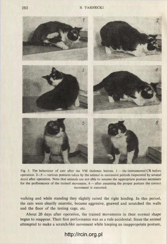
Fig. 3. The behaviour of cats after the VM thalamic lesions. 1 — the instrumental CR before operation. 2—5 —various postures taken by the animal in successive periods (separated by several days) after operation. Note that animals are not able to assume the appropriate posture necessary for the performance of the trained movement. 6 — after assuming the proper posture the correct
movement is executed.
walking and while standing they slightly raised the right hindleg. In this period, the cats were clearily neurotic, became aggresive, gnawed and scratched the walls and the floor of the testing cage, etc.
About 20 days after operation, the trained movements in their normal shape began to reappear. Their first performance was as a rule accidental. Since the animal attempted to make a scratch-like movement while keeping an inappropriate posture,
http://rcin.org.pl

Fig. 4. Explanations as in Fig. 3
he often lost his balance and fell on the opposite side. In such a position the per-formance of the trained movement was quite feasible and the animal made use of it. If the posture was not changed, the animal was able to perform a long series of regular movements in quick succession. However, any change of the posture, either spontaneous or induced, disturbed the performance of the trained movements until the animal accidentally resumed the proper position. After a few experimental sessions the animal learned to assume the proper posture more and more promptly. Although this posture often was different from, and not as convenient as, that used before operation (Figs. 3 and 4), the movements were performed quite skilfully and rapidly.
http://rcin.org.pl
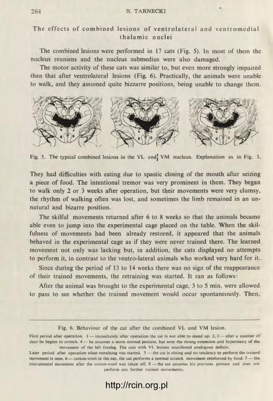
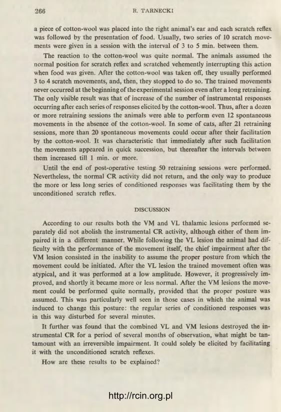
The e f f e c t s o f c o m b i n e d l e s i o n s o f v e n t r o l a t e r a l a n d v e n t r o m e d i a l t h a l a m i c n u c l e i
The combined lesions were performed in 17 cats (Fig. 5). In most of them the nucleus reuniens and the nucleus submedius were also damaged.
The motor activity of these cats was similar to, but even more strongly impaired than that after ventrolateral lesions (Fig. 6). Practically, the animals were unable to walk, and they assumed quite bizzarre positions, being unable to change them.
Fig. 5. The typical combined lesions in the VL and] VM nucleus. Explanation as in Fig. 1.
They had difficulties with eating due to spastic closing of the mouth after seizing a piece of food. The intentional tremor was very prominent in them. They began to walk only 2 or 3 weeks after operation, but their movements were very clumsy, the rhythm of walking often was lost, and sometimes the limb remained in an un-natural and bizarre position.
The skilful movements returned after 6 to 8 weeks so that the animals became able even to jump into the experimental cage placed on the table. When the skil-fulness of movements had been already restored, it appeared that the animals behaved in the experimental cage as if they were never trained there. The learned movement not only was lacking but, in addition, the cats displayed no attempts to perform it, in contrast to the ventro-lateral animals who worked very hard for it.
Since during the period of 13 to 14 weeks there was no sign of the reappearance of their trained movements, the retraining was started. It ran as follows:
After the animal was brought to the experimental cage, 3 to 5 min. were allowed to pass to see whether the trained movement would occur spontaneously. Then,
Fig. 6. Behaviour of the cat after the combined VL and VM lesion. First period after operation. 1 — immediately after operation the cat is not able to stand up. 2, 3 — after a number of days he begins to crouch. 4 — he assumes a more normal posture, but note the strong extension and hypermetry of the
movement of the left foreleg. The cats with VL lesions manifested analogous defects. Later period after operation when retraining was started. 5 — the cat is sitting and no tendency to perform the trained movement is seen. 6 — cotton-wool in the ear, the cat performs a normal scratch movement reinforced by food. 7 — the instrumental movement after the cotton-wool was taken off. 8 — the cat assumes his previous posture and does no!
perform any further trained movements.
http://rcin.org.pl

http://rcin.org.pl
a piece of cotton-wool was placed into the right animal's ear and each scratch reflex was followed by the presentation of food. Usually, two series of 10 scratch move-ments were given in a session with the interval of 3 to 5 min. between them.
The reaction to the cotton-wool was quite normal. The animals assumed the normal position for scratch reflex and scratched vehemently interrupting this action when food was given. After the cotton-wool was taken off, they usually performed 3 to 4 scratch movements, and, then, they stopped to do so. The trained movements never occurred at the beginning of the experimental session even after a long retraining. The only visible result was that of increase of the number of instrumental responses occurring after each series of responses elicited by the cotton-wool. Thus, after a dozen or more retraining sessions the animals were able to perform even 12 spontaneous movements in the absence of the cotton-wool. In some of cats, after 21 retraining sessions, more than 20 spontaneous movements could occur after their facilitation by the cotton-wool. It was characteristic that immediately after such facilitation the movements appeared in quick succession, but thereafter the intervals between them increased till 1 min. or more.
Until the end of post-operative testing 50 retraining sessions were performed. Nevertheless, the normal CR activity did not return, and the only way to produce the more or less long series of conditioned responses was facilitating them by the unconditioned scratch reflex.
DISCUSSION
According to our results both the VM and VL thalamic lesions performed se-parately did not abolish the instrumental CR activity, although either of them im-paired it in a different manner. While following the VL lesion the animal had dif-ficulty with the performance of the movement itself, the chief impairment after the VM lesion consisted in the inability to assume the proper posture from which the movement could be initiated. After the VL lesion the trained movement often was atypical, and it was performed at a low amplitude. However, it progressively im-proved, and shortly it became more or less normal. After the VM lesions the move-ment could be performed quite normally, provided that the proper posture was assumed. This was particularly well seen in those cases in which the animal was induced to change this posture: the regular series of conditioned responses was in this way disturbed for several minutes.
It further was found that the combined VL and VM lesions destroyed the in-strumental CR for a period of several months of observation, what might be tan-tamount with an irreversible impairment. It could solely be elicited by facilitating it with the unconditioned scratch reflexes.
How are these results to be explained?
http://rcin.org.pl
According to our observations, it appears that the performance of the trained instrumental movements depends on two factors. First, in order to perform the given movement the animal must assume a proper posture. In our case this posture consisted of sitting down, and loosening the right hindleg. After assuming this posture the animal must be able to perform the trained movement. Our results indicate that the lateral part of the VL nucleus is concerned with the latter capacity, while the VM nucleus is rather concerned with enabling the animal to assume parti-cular postures. When one of these structures is destroyed the trained movement is abolished only temporarily. It seems likely that in the case of the VL lesion other structures may provide the animal with the appropriate feedback necessary for the performance of the movement, while after VM lesion the required posture is taken by accident. However, if both the centre controlling posture and that con-trolling the movement are impaired, the prerequisites for its performance are lost.
In this respect, our results are similar to those obtained on dogs with ablations of the sensori-motor cortex. ( S t ę p i e ń , S t ę p i e ń and K o n o r s k i 1960 a, b). While the impairment following the VM nucleus lesion resembles that described after ablation of the premotor cortex, the lesion of the VL nucleus produces similar deficits to those which are seen after ablation of the motor cortex. The connections between the motor cortex and the VL nucleus are well known. Whether or not direct pathways are present between the VM nucleus and the premotor area remains to be elucidated*.
However, it must be taken into account that according to the opinion of most authors (cf. J a s p e r 1961) the VM nucleus belongs to the group of the midline nuclei and represents the rostral part of the thalamic reticular system. There is some evidence indicating that lesions in this system located caudally to the VM nucleus produce a great disorder of instrumental CRs. C a r d o (1961) found that destruction of the para-fascicular nucleus in rats permanently affected the instrumental avoidance reflexes, although after administration of amphetamine the animals were able to recover their correct performance. Therefore, it may be supposed that the mechanism of the impairment of CRs in our experiments after the VM lesions might be considered as due to a decrease of facilitatory influence of the thalamic reticular system on those structures which are responsible for maintenance of these reflexes.
The comparison between the cats subjected to VL and VM lesions, or VL lesion alone, and those subjected to leminiscal lesions ( T a r n e c k i 1962) should be com-mented upon. After extensive leminiscal lesions the motor activity of the animals seems to be much more incapacitated than after VL lesions. The animals are hardly able to walk, but the trained movement is well preserved. After VL lesions the motor activity is preserved from the very beginning, but its coordination is much impaired.
* According to the data reported by McLardy (1950), the VM nucleus in man projects to the ventral part of area 6 of the cortex.
http://rcin.org.pl

SUMMARY
1) The effects of bilateral lesions in the ventrolateral, and ventro-medial thalamic nuclei on the performance of alimentary instrumental CRs were studied in cats. The CRs were established by placing a piece of cotton-wool into the animal's right ear and reinforcing by food every unconditioned scratch response performed by the right hindleg.
2) It was found that after damage of either the lateral part of the VL nucleus, or the whole VM nucleus the instrumental reflex was temporarily abolished but reappeared after several weeks without any additional training. However, when the VL and VM nuclei were destroyed jointly the instrumental CR was abolished and did not reappear. The training consisting in evoking unconditioned scratch movements and reinforcing them by food had only partial effect: after a series of unconditioned scratch movements, the instrumental response was temporarily restor-ed and appeared several times in succession. However, it could be elicited only after facilitation by the unconditioned scratch reflex.
3) It was observed that the animals with the VM lesion are not able to assume the posture from which the instrumental scratch-like movement was initiated. On the contrary, the cats with VL lesions could easily adopt this posture, but had diffi-culties in the performance of the trained movement. On the basis of this obser-vation an explanation of the defects of the trained response after each lesion is provided.
The author is greatly indebted to Professor J. Konorski for his valuable advice during the performance of the present experiments and for help in preparation of the manuscript of this paper. Many thanks are due to Mrs. C. Borkowska for training the animals, and to Mrs. D. Tarnecka for sectioning and staining the brains.
REFERENCES
CARDO B. 1961—Rapports entre le niveau de vigilance et le conditionnement chez Tanimal. Etude pharmacologique et neurologue. J. de Physiol. 53, 3.
GÓRSKA T. and JANKOWSKA E. 1959 — Instrumental conditioned reflexes of the dcafferen-tated limb in cats and rats. Bull. Acad. Polon. Sci., Cl. VI, 7,4 (Serie des sciences biologiques).
JANKOWSKA E. 1959 — Instrumental scratch reflex of the deafferentated limb in cats and rats. Acta Biol. Exper. 19, 233.
JASPER H. H. and AJ MONE-MARSAN C. 1961 — Diencephalon of the cat. Electrical stimulation of the brain. Hogg Foundation for Mental Health. University of Texas Press.
McLARDY T. 1950 — Thalamic projection to frontal cortex in man. J. Neurol. Neurosurg. Psychiat. 13, 198.
STĘPIEŃ I., STĘPIEŃ L. and KONORSKI J. 1960 — The effects of bilateral lesions in the motor cortex on type II conditioned reflexes in dogs. Acta Biol. Exper. 20, 211.
STĘPIEŃ I., STĘPIEŃ L. and KONORSKI J. 1960 —The effects of bilateral lesions in the pre-motor cortex on type II conditioned reflexes in dogs. Acta Biol. Exper. 20, 225.
TARNECKI R. The effect of medial leminiscal lesions on the instrumental conditioned reflexes in cats. Acta Biol. Exper. 22 (in press).
http://rcin.org.pl

QUELQUES OBSERVATIONS SUR LES LUTTES ENTRE DIFFÉRENTES
ESPÈCES DE FOURMIS
J. DOBRZAŃSKA et J. DOBRZAŃSKI
Laboratoire de Biologie Générale de l'Institut Nencki de Biologie Expérimentale Varsovie, Pologne
(Reçu le 28 fevrier 1962)
Les nids décrits étaient constamment observés durant quelques mois et même durant quelques années en rapport avec d'autres expériments. Grâce à cette longue période d'observation nous avons pu remarquer quelques faits qui nous semblent intéressants du point de vue éthologique en raison de leur rareté.
1. L u t t e e n t r e la Camponotus ligniperda Latr. et la Formica rufibarbis Fabr.
Deux nids des espèces en question étaient observés depuis longtemps en raison
d'autres recherches. Nous n'avons constaté aucun indice d'antagonisme entre leurs
habitants, malgré le fait que la distance entre les orifices de ces nids n'était que
de 30 cm. Un jour, le 16 juin 1957, nous avons constaté que les deux nids se
trouvaient en état de lutte.
Entre les deux nids s'effectuait un mouvement continuel d'ouvrières C. ligniperda
qui étaient les agresseurs. A l'action prenaient part environ 10 individus, ce qui
pour cette espèce constitue une quantité importante. 3 ou 4 ouvrières étaient aux
aguets autour de l'orifice du nid des F. rubibarbis dans une pose très tendue.
Au fond de l'orifice assiégé on pouvait à peine distinguer les têtes des ouvrières
F. rubibarbis.
De temps en temps l'une d'elles essayait d'exposer sa tête à l'extérieur. Nous
n'avions encore jamais observé une telle tension et une réaction aussi rapide et
aussi diverse en dépendance du comportement de l'adversaire. Si une F. rufibarbis
exposait prudemment sa tête à l'extérieur — ses adversaires se jetaient immédiate-
ment sur elle et si elle n'arrivait pas à reculer à temps elle était extirpée du nid et
tuée sur place.
Les C. ligniperda se comportaient tout à fait d'une autre façon envers les F. rufi-
barbis qui exécutaient une attaque subite, bondissant rapidement de l'orifice du
nid: dans ce cas toutes les C. ligniperda battaient en retraite devant cette seule fourmi,
http://rcin.org.pl

de taille plusieurs fois inférieure à elles-mêmes. La C. ligniperda retournait alors en toute sécurité dans son nid. Ainsi donc la réaction des mêmes individus C. ligni-perda dépendait entièrement du comportement individuel de leur adversaire: c'était ou bien une attaque immédiate ou bien une fuite non moins précipitée.
Après la tombée du crépuscule la lutte prit fin. Le jour suivant le nid des F. rifi-barbis changea d'apparence: l'orifice était barricadé de brins d'herbe, de cadavres de fourmis, etc. L'intensité des luttes diminua et il arrivait qu'il n'y eût aucune ouvrière C. ligniperda aux environs du nid attaqué. On pouvait alors observer des ouvrières rufibarbis, apportant des cadavres provenant des luttes précédentes pour en encombrer l'orifice.
Le 18 l'agressivité des F. rufibarbis augmenta et elles sortaient plus fréquemment du nid, chassant de plus en plus hardiment leurs adversaires. Le 19 la lutte cessa. Le nid des C. ligniperda s'était entièrement calmé, les F. rufibarbis travaillaient intensément à réparer les dommages subis, la barricade était mise de côté.
Le premier jour des observations nous avons marqué, dans la mesure du possible, les individus prenant part à la lutte. Ce ne fut possible, en principe, qu'avec les C. ligniperda. Nous n'avons pu marquer qu'un seul individu F. rufibarbis, qui s'éloig-nait le plus de son nid dans ses attaques. Le 18 il fut particulièrement actif pour-suivant les C. ligniperda jusqu'à leur nid. Sur 19 ouvrières C. ligniperda marquées le premier jour, nous n'en observâmes que 4 les journées suivantes. L'individu No 3 prit part à la lutte durant tous les trois jours de sa durée. Cet individu démon-trait une activité extrême et non seulement il montait la garde près de l'orifice du nid attaqué durant toute la journée, mais il y restait le plus longtemps dans la soirée quand la lutte était déjà interrompue. Le premier jour il y resta 20 minutes après la fin de la lutte, le deuxième il y resta 15, le troisième 25. Il faisait alors tout à fait nuit et nous observions cet individu à l'aide d'une lanterne de poche, luisant faible-ment pour ne pas effaroucher les fourmis. On voit donc par cet exemple les différences importantes qui existent entre le comportement de différents individus.
Après la fin des hostilités durant de nombreuses semaines nous n'avons pu observer aucun symptôme d'animosité entre les deux nids. Les nids fonctionnaient normalement, il arrivait souvent, comme auparavant, que des C. ligniperda retour-nant au nid, passaient tout près de l'orifice du nid des F. rifibarbis ne manifestant et ne suscitant aucun trouble.
Les causes de cette lutte de trois jours sont restées inconnues.
2. C o m p o r t e m e n t de la Formica truncicola Nyl. et de la Formica fusca L. e n v e r s la Formicasanguinea, m e n a n t une a c t i o n guerr ière aux e n v i r o n s
de l eurs n ids
La distance entre les nids de la F. truncicola et de la F. sanguinea était de 5 mètres. Le terrain était observé pendant 3 saisons estivales durant lesquelles (avant les événements décrits, tout comme après) les ouvrières étaient continuellement en contact sans manifester aucun antagonisme.
http://rcin.org.pl

Un jour les F. sanguínea attaquèrent un nid de F. cinerea situé entre les deux
nids en question, plus près du nid des F. truncicola. Ces dernières ne s'engagèrent
pas dans la lutte, mais une grande quantité d'ouvrières de cette espèce, environ
une cinquantaine, se concentra près du nid attaqué. Leur comportement était très
cara;téristique : très excitées, elles couraient le long d'une frontière invisible, qu'elles
ne franchissaient pas elles-mêmeset si cela arrivait à l'une d'elles, elle se retirait
rapidement à reculons. Mais si cette „frontière" était franchie par une ouvrière
F. singuinea, cette dernière se trouvait immédiatement entraînée à l'intérieur de
„leur" terrain et si elle n'arrivait pas à se dégager et à s'enfuir de l'autre côté de
la fiontière, elle était rapidement mise à mort. Un tel état dura toute la journée
(desiin 1).
A
0 1m
m1
Fig. ;. Schéma de l'emplecement des nids des 4 espèces de fourmis (vide détails dans la partie 2). 1 — le; nids, 2 — zone de circulation intense des F. truncicola, AB — „frontiere" gardée par la F. truncicola
En raison des mouvements très rapides des F. truncicola et de leur excitation
extrême nous n'avons pu marquer que 25 individus.
Le jour suivant la lutte entre les F. sanguínea et les F. cinerea était terminée
mais la quantité des F. truncicola circulant le long de la frontière était encore très
importante. Il en circulait une dizaine et toutes étaient marquées du jour précédent.
Le troisième jour il en circulait 6 dont une n'était pas marquée. Pour la première
fois elles essayaient prudemment de franchir la „frontière".
Pendant les quelques jours suivants la frontière étant de moins en moins respectée
la situation redevint de nouveau normale, c'est à dire qu'on pouvait de nouveau
voir des ouvrières des deux espèces pâturant côte à côte sur toute la surface du
terrain sans démontrer aucune animosité. 3 de ces F. truncicola étaient marquées
et jusqu'à la fin de l'été ces individus revenaient sur ce terrain ce qui était en accord
avec le principe du partage du terrain chez cette espèce ( D o b r z a ń s k a 1958).
Le terrain où les événements décrits ont eu lieu était habité par un petit nid
de F. fusca (dussin 1), dont le comportement fut très spécifique. Ce nid, quoique
appartenant à une espèce esclave ne fut attaqué par le nid F. sanguínea ni auparavant,
ni jusqu'à la fin de la saison, ce qui confirme la thèse de D o b r z a ń s k i (1961)
sur le caractère accidentel des attaques de pillage de la part de F. sanguínea. Les F. fusca
de ce nid ne réagissaient pas jusque là au passage fréquent des ouvrières F. sanguínea
http://rcin.org.pl

aux alentours de leur nid. Durant les événement décrits leur comportement subit un changement: elles barricadèrent l'orifice de leur nid et jusqu'à la fin de la lutte entre les F. sanguínea et les F. cinerea elles ne donnèrent aucun signe de vie.
3. C o m p o r t e m e n t du Lasius Niger L. e n v e r s la Formica sanguínea Latr. se t r o u v a n t en état de guerre
Des F. sanguínea effectuant une attaque contre autre nid, passaient en cours de route devant un nid de L. niger. Chaque individu F. sanguínea qui montait par hasard sur le nid, se trouvait aussitôt attaqué par des ouvrières L. niger, qui, comme il est d'usage chez cette espèce, s'agrippaient en grand nombre aux pattes et aux antennes de l'intrus, l'entraînaient à l'intérieur du nid où il était tué.
Au bout de quelques heures les L. niger bouchèrent l'orifice du nid avec une grille formée d'aiguilles de pins et de baguettes. L'intérieur du nid, détérioré en raison des luttes précédentes, fut réparé. Toute F. sanguínea, qui se heurtait en route à ce nid, prenait immédiatement la fuite. Ce nid des L. niger se trouvait tout comme ceux que nous avons décrits auparavant, près du nid F. sanguínea et nor-malement les relations entre ces deux espèces étaient dépourvues d'animosité.
4. C o m p o r t e m e n t du Tetramorium caespitum L. e n v e r s une e x p é d i t i o n guerr i ère du Polyergus r ufes cens Latr.
Une colonne de guerre de P. rufescens passa sans y prêter attention par un grand nid de T. caespitum. Les petites fourmis attaquèrent les amazones à quelques unes à la fois. Cela causa un trouble dans les rangs toujours si précis des P. rufescens, l'avant-garde avança encore quelques mètres et se retourna. Certaines amazones entreprirent la lutte et essayèrent même de pénétrer à l'intérieur du nid T. caespitum. Mais aucune n'y arriva: chacune reculait immédiatement ayant les pattes et les antennes couvertes de ces petites fourmis. Une telle amazone devenait tout à fait sans défense, elle se débattait mais ne pouvait pas se dégager de la prise de ses ad-versaires, aucune autre amazone ne venant à son aide.
Nous n'avons d'ailleurs jamais remarqué, soit dit en passant, pendant toutes nos années de recherches sur les P. rufescens, aucun symptôme d'aide mutuelle entre ces fourmis pendant la lutte. Chaque soldat y est livré à son propre sort.
Ce genre de lutte dura environ deux heures. On pouvait parfois apercevoir une amazone, sur la patte de laquelle il ne restait que la tête, privée de corps, d'une ouvrière T. caespitum. En général, néanmoins, la suprématie des petites fourmis était complète. La lutte se termina par la retraite graduelle des P. rufescens et par l'abandon de la direction d'attaque choisie.
L'action d'équipe des T. caespitum contre leurs adversaires beaucoup plus grands et bien mieux armés mais luttant séparément leur assura une victoire complète.
http://rcin.org.pl
5. Lutte c o n t r e la Polyergus rufescens Latr. et la Formica execta Nyl.
La colonne guerrière des P. rufescens attaqua un nid de F. fusca à un demi-mètre duquel se trouvait un nid de F. execta. Cette dernière espèce est une espèce très guerrière et on ne la rencontre jamais en tant qu'esclave.
Les ouvrières F. execta ayant réagi agressivement contre quelques amazones passant accidentellement par leur nid, des duels commencèrent, dans lesquels les F. execta n'avaient aucune chance, car elles tombaient en quelques secondes la tête per:orée. C'est seulement après quelques luttes que des soldats P. rufescens com-mencèrent à s'arrêter sur ce nid auquel ils ne prêtaient auparavant aucune attention. Quelques soldats pénétrèrent à l'intérieur mais en sortirent immédiatement sans aucun butin. Ce fait suscita une réaction instantanée: comme sur un signal, de tous les orifices du nid surgirent des ouvrières portant la progéniture et les jeunes ouvrières. Ce comportement était identique à celui des F. fusca avec cette différence que ces dernières sauvaient leurs nymphes d'un pillage réel.
Une partie des ouvrières continuaient à attaquer les soldats des amazones et il arrivait parfois que quelques unes à la fois attaquaient le même individu.
Au bout d'une demi-heure les P. rufescens retournèrent à leur nid. Peu de temps après, les ouvrières F. fusca ramenèrent leurs nymphes ainsi que les jeunes ouvrières et la reine qui s'étaient enfuies elles mêmes auparavant.
Les ouvrières F. execta restèrent, par contre, encore longtemps cachées sous les feuilles et ce n'est qu'après une heure qu'elles commencèrent à ramener leurs nymphes et leurs jeunes ouvrières au nid.
Leur technique de transport n'est pas dépourvue d'intérêt: elles portaient en général les jeunes ouvrières d'une façon quelconque et non de la manière caractéris-tique pour le genre des Formica (chez ce genre, les fourmis transportées sont saisies par les mandibules et se roulent elles-mêmes en boule sous la tête de la porteuse).
6. E x p é d i t i o n guerr ière des Formica sanguinea Latr. à un nid des Formica
cinerea Mayr.
Les nids de ces deux espèces se trouvaient à une distance d'une dizaine de mètres. Par une chaude matinée de juillet, les F. sanguinea, éparpillées comme d'habitude
sur un large front de quelques mètres, aboutirent au nid des F. cinerea. Les ouvrières F. cinerea qui se trouvaient à la surface s'abritèrent à l'intérieur. Nous n'avons observé ni au début de l'attaque, ni durant le reste de l'action aucun corps à corps entre ces deux espèces.
Les fourmis attaquées n'appliquèrent qu'une seule tactique bien spéciale: elles se mirent à combler les orifices du nid de l'intérieur, rendant ainsi impossible tout contact direct avec l'ennemi. Dans une telle situation les agresseurs à leur tour, ne pouvaient appliquer qu'une seule tactique — celle du siège: ils commencèrent à dégager de l'extérieur les orifices bouchés. Près de chaque orifice une dizaine
http://rcin.org.pl

d'ouvrières F. sanguínea sortaient la terre du trou et la transportaient à quelque distance, comme le font toujours les fourmis dans leurs travaux de construction. Elles travaillaient rapidement et de temps en temps dans un trou dégagé par les assiégeants on pouvait apercevoir la tête d'un défenseur, qui rebouchait l'orifice à la même allure. Dans cette compétition les F. sanguínea prenaient manifestement l'avantage, en raison de leur nombre pratiquement illimité et du fait que les ouvrières se remplaçaient très souvent. Nous avons constaté ce fait en marquant d'une façon individuelle 45 ouvrières et en outre, d'une façon générale 100 ouvrières qui pre-naient part à ces travaux de terre.
Entre le nid attaqué et le nid des F. sanguínea il se forma un chemin avec un continuel va-et-vient des F. sanguínea qui allaient vers le nid attaqué ou en revenaient. Ce n'est malgré tout qu'après quelques heures que les F. sanguínea réussirent à pénétrer dans un des orifices. Néanmoins elles ne commencèrent pas, comme on aurait pu s'y attendre, à en sortir des nymphes conquises: elles continuaient toujours à n'extraire du fond du nid que de la terre. Apparemment les assiégés ne se ren-daient toujours pas et tout en se retirant au fond du nid, ils employaient la même tactique d'obstruction des corridors.
Durant tout la première journée de lutte les agresseurs n'ont pas conquis une seule nymphe. Un moment après le crépuscule il y eut un orage mais malgré une averse assez violente, les orifices le mieux protégés de la pluie continuaient à être creusés par les assiégeants. Cela ne dura pas longtemps car en peu de temps toute la surface du nid fut inondée par la pluie et les agresseurs, cessant leurs activités,, s'abritèrent en majeur partie dans les orifices déjà conquis du nid des F. cinerea.
Le deuxième jour, aux environs de 10 heures, la résistance du nid assiégé com-mença visiblement à faiblir sous la pression des F. sanguínea dont le nombre avait crû en comparaison avec la veille. Les agresseurs avaient déjà pénétré dans tous les orifices, mais n'en sortaient toujours que de la terre. Enfin vers 11 heures les premières nymphes et même sporadiquement des ouvrières adultes conquises ap-parurent. Il y avait quand même toujours dans certains orifices des défenseurs qui continuaient à les barricader. Au bout d'une heure cette défense cessa aussi.
Le marquage individuel des F. sanguínea démontra durant ces deux jours de lutte une variabilité extrême de leur comportement. Nous n'avons observé aucun individu effectuant la même fonction durant tout le temps de la lutte.
Après cette victoire remportée avec tant de peine, les F. sanguínea ne commen-cèrent pas du tout à enlever les nymphes conquises et à les transporter dans leur propre nid, comme cela a lieu après une victoire du P. rufescens.
Le troisième et le quatrième jour encore, quand toute lutte était déjà terminée et le nid conquis tout à fait pris en possesion, il y avait toujours des ouvrières F. san-guínea qui y erraient sans but apparent, allant ici et là, sortant du nid des nymphes pour les y ramener souvent de nouveau. Entre ces fourmis qui circulaient de la sorte il y avait des fourmis marquées, c'est à dire des individus qui avaient pris une part.
http://rcin.org.pl

active dans la conquête du nid. Deux des ouvrières marquées, suivies individuelle-
ment, ne prirent le chemin de leur propre nid qu'après avoir marché deux et trois
heures les nymphes entre les mandibules. Il est caractéristique qu'après être allée
sur la piste menant au nid, la fourmi, si peu décidée auparavant, se mettait alors
en général en route sans plus hésiter.
Trois ou quatre jours encore après la conquête du nid il y avait toujours quel-
ques ouvrières isolées F. sanguínea qui erraient à sa surface.
Après la fin de toute action de la part des F. sanguínea, nous avons déterré le
nid pillé et nous y trouvâmes tout au fond un petit nombre de F. cinerea avec des
larves et des nymphes. T1 en résulte qu'une certaine partie du nid échappa jusqu'à
la fin à la conquête.
CONCLUSIONS
1. Nous avons décrit des cas où des nids, demeurant jusqu'à là en relations
pacifiques, passaient subitement en état de guerre ou optaient une attitude anta-
goniste (1, II, III). Une des raisons possibles est certainement l'arrivée des four-
mis d'un de ces nids en formation de combat, sans égards contre qui cette formation
pouvait être dirigée. C'est justement de cette façon qu'ont commencé certains combats
décrits dans ce travail et cela non nécessairement entre nids voisins car l'expédition
pouvait t jucher à un nid assez éloigné (IV, V).
Il semble très probable que les fourmis peuvent différencier une formation de
combat et une attitude belliqueuse de fourmis étrangères d'une attitude pacifique.
On pourrait ajouter encore une observation en faveur de cette possibilité:
les individus isolés F. sanguínea qui pâturent tranquillement et parfois con-
stamment, aux alentours d'un nid d'amazones, ne manifestent en les rencontrant
aucun symptôme d'animosité. Par contre ils s'enfuient en panique quand ils ont
affaire à une colonne guerrière de ces mêmes amazones. Ce fait est d'autant plus
frappant, que l'armée des P. rufescens ne prête aucune attention aux fourmis ou
aux autres animaux qu'elle croise et marche tout droit dans la direction choisie.
2. Nous estimions auparavant que le transport des nymphes par les fourmis
attaquées par des P. rufescens ou F. sanguínea était un trait adaptatif des espèces
esclaves, qui constituent le but de ces attaques.
T1 s'avère néanmoins que la F. execta, qui n'est pas une espèce esclave, se com-
porte en cas d'attaque par des amazones (V) de la même façon que les espèces escla-
ves ce qui était dans ce cas dénué de sens, car les P. rufescens n'ont même pas essayé
d'enlever leurs nymphes. Il est remarquable que les F. execta étaient visiblement
moins adaptées à une telle méthode de défense et ne purent rétablir l'ordre dans
leur nid avec la même dextérité que la F. fusca.
D autre part l'espèce esclave classique — la F. rufibarbis, qui déménage toujours
ses nymphes en réponse à une attaque des amazones ou des F. sanguínea a réagi
http://rcin.org.pl

d'une autre manière quand son nid fut attaqué par des C. ligniperda (I). Les F.fuscar
dans le cas décrit, ne prirent pas la fuite en présence de leur ennemi le plus sérieux —-les F. sanguinea — quand celles-ci passèrent par leur nid mais se contentèrent d e le barricader.
Rassemblant ces faits, nous en venons à la conclusion que la fuite avec les nym-phes en cas d'attaque est une réaction de beaucoup d'espèce de fourmis, et non seulement des espèces esclaves, à la pénétration d'adversaires à l'intérieur de leur nid. Tous les cas décrits confirment cette supposition. Les nymphes demeuraient toujours dans le nid si l'adversaire restait à la surface.
Il est compréhensible que des espèces aussi belliqueuses que la F. execta qui ne sont attaquées que très rarement ne font pas preuve d'autant de précision dans la fuite et dans le transport des nymphes.
Le fait que justement la F. execta, si peu adaptée au transport mutuel des ouvrières,, soit l'unique espèce connue des Formica qui n'ait pas de position fixe de transport des nymphes, nous paraît extrêmement intéressant car cela confirme notre hypo-thèse ( D o b r z a ń s k a et D o b r z a ń s k i 1960) sur le caractère originairement défensif du transport mutuel chez les fourmis.
3. Il en résulte de nombreuses observations décrites tant dans ce travail que dans d'autres, que l'attaque en équipe d'un adversaire est une tactique de combat caractéristique seulement pour quelques espèces, dont nous avons décrit ici les L. niger (III) et T. caespiîum (IV). Chez les espèces des Formica cela n'a presque jamais lieu. En tant qu'exception nous avons décrit chez les F. execta des cas d'attaque en groupes ce qui d'ailleurs a déjà été mentionné par Fore l (1874).
4. Des descriptions d ' E s c h e r i c h ( 1917), de Fore l (1923) et d'autres on peut conclure que le comportement du P. rufescens est très stéréotype. Cette conclusion est confirmée par nos observations. Les amazones ne ramassent jamais au sol des nymphes d'espèces esclaves se trouvant sur leur chemin ( D o b r z a ń s k i 1956). Ils ne le font même pas pendant l'attaque, quoique leur but soit justement la conquête de ces nymphes, ils n'attaquent pas de même les nids artificiels enterrés sur leur route ( D o b r z a ń s k i et D o b r z a ń s k a 1960), tandis que les F. sanguinea profitent en toute situation de l'occasion de saisir une nymphe ( D o b r z a ń s k i 1961). On peut en déduire que, pour être capables d'accomplir une action apparem-ment si peu compliquée comme celle de saisir une nymphe étrangère, les soldats P. rufescens exigent toute une suite de stimuli bien définis: ils doivent pénétrer dans un nid étranger, et en tirer les nymphes de son,intérieur.
Des différences même très subtiles peuvent probablement causer l'inhibition de ces fonctions: un nid naturel, mais d'une espèce non esclave, de la F. execta, n'a pas suscité de réaction de pillage.
En contraste avec cette rigidité du comportement des amazones, nous avons la plasticité reconnue depuis longtemps par F o r e l (1874, 1923), Brun (1924), Wh ee ler (1926) et d'autres, du comportement de la F. sanguinea. Nos observations
http://rcin.org.pl

confirment l'énorme variabilité du comportement des ouvrières F. sanguínea durant
les pillages des nids et leur plasticité à s'adapter aux circonstances concrètes de la
défense de l'adversaire.
On peut citer comme exemple leur adaptation instantanée à la tactique originale
de défense des F. cinerea ( VI).
La rigidité des instincts des P. rufescens limite leur capacité à conquérir des
nymphes à des conditions strictement déterminées. Mais d'autre part leurs réactions
stéréotypes exigeant toute une suite de stimuli, à succession définie, donnent aux
amazones une précision d'action extraordinaire: leur attaque s'effectue à une rapidité
extrême, aucun soldat (sous condition d'une quantité suffisante de nymphes) ne
retourne au nid sans butin et en général l'armée revient au nid et peut donc com-
mencer une nouvelle action de 20 à 25 minutes après le commencement de l'attaque.
Chaque victoire est mise à profit rapidement, avec précision et sans perte.
Le manque de rigidité de comportement des F. sanguínea, possédant les valeurs
déjà mentionnées, leur permet d'attaquer et de conquérir des nymphes dans toutes
conditions. Mais ce trait possède à son tour ses défauts: nous avons vu (VI) comment
la durée d'une attaque peut s'étendre à quelques jours et comment après quelques
jours le comportement des ouvrières F. sanguínea devient chaotique. Elles ne profitent
pas jusqu'au bout de leur victoire.
Il nous semble que ce sont de bons exemples de l'idée qu'autant l'adaptation
très adéquate à des situations concrètes que la plasticité du comportement possèdent
leurs qualités et leurs défauts dans la lutte pour l'existence d'une espèce.
BIBLIOGRAPHIE
BRUN R. 1924 —Das Leben der Ameisen. Teubners nawiss. Bibliothek 31, Leipzig-Berlin. DOBRZAŃSKA J. 1958 — Partition of foraging grounds and modes of conveyings information
among ants. Acta Biol. Exper. 18, 55. DOBRZAŃSKA J. et D3BRZAŃ3KI J. 1959 — Qjelques nouvelles remarques sur l'ethologie
de P. rufescens. Insectes sociaux. 7, 1. DOBRZAŃSKI J. 1956 — Badania nad zmysłem czasu u mrówek. (Investigations on the time
sense in ants). Folia Biologica 4, 385. DOBRZAŃSKI J. 1961—Sur l'ethologie gierrière de Formica sanguima Latr. (H/menoptère,
Formicidae). Acta Biol. Exper. 21, 53. ESCHERICH K. 1917 —Die Ameise. II. Auflage, Braunschweig. FOREL A. 1874 — Les fourmis de la Suisse. Genève. FOREL A. 1923 — Le monde social des fourmis du globe. Genève. WHEELER W. M. 1926 —Les sociétés d'insectes. Paris.
http://rcin.org.pl

http://rcin.org.pl

Book Review
Electrical Stimulation of the Brain. An interdisciplinaiy survey of neurobehavioral integrative system. Edited by Daniel E. Sheer. Published for the Hogg Foundation for Mental Health. Pages 640+XV, 1961, illustrated.
One of the most difficult problem» which confront the development of modern science is that of information. Both the number of journals concerned with the particular scientific fields, and the number of papers published each year increase so rapidly that it is quite impossible for a scientist to be acquainted with the whole literature, even in his own immediate scope of interest. In con-sequence, very often the same studies are performed independently in various places, and often the authors do not know about their mutual achievements.
There are various ways to overcome this difficulty. One of them is provided by the abstracting journals which allow the students of the given branch to have at least the index of relevant papers. The other way is provided by annual reviews in which concise, and usually insipid, articles repre-senting the development of various disciplines in the past year are given. Last but not least, there are voluminous handbooks which attempt to make some synthesis of the present state of our knowledge in a given field.
However useful these sources of information are, they are insufficient for the following reasons. A scientist (including the reviewer himself), even if supplied with all relevant literatuie, is simply not able to digest it because of the ammcmnt of data contained in it. Therefore, even having on one's shelves all the reprints needed to be studied, one is physically and mentally incapable to go through all of them, and even if he has succeded in doing so, to remember them. Reading scientific papers obeys the rigid rule of retroactive (and proactive) inhibition which implies that only the contents of a simll part of the papers perused becomes consolidated in one's cerebral cortex.
Therefore, there should exist such a form of information which would ommit all these difficulties. It is provided by articles written by good specialists in a given subject, not attempting to give an exhaustive survey of the field in question, but trying to represent the discussed problem in a syn-thetic, coherent, and already digested form. If the data presented in the article are not simply juxta-posed on beside the other, but on the contrary interconnected and complemented by each other, then the principle of retroactive inhibition does not intervene, and the mxterial is easily assimilated.
Such articles are provided by good and carefully edited Symposia. In consequence, this is one of the very important means of scientific informition not only for the direct participants of the Symposium, but for all those who read its materials in the printed form.
The volume concerning the Electrical Stimulation of the Brain was originally based on the Symposium held at the Texas Medical Center in Houston in 1956. The editors however decided to extend this volume by adding new papers so that the volume contains in all 36 chapters.
According to the preface written by the editor, "the plan of the book was to collate contributions from various disciplines using one technique — electrical stimulation of the brain — in the investi-gation of the neurobehavioral relations. Particularily these contributions are focused on the problem
http://rcin.org.pl
"of higher order" behaviors usually subsumed under such psychological rubrics as perception, learning, emotion, and motivation".
The volume consists of 7 parts, namely: intioduction, techniques, neuroanatomical correlates, neurophysiological correlates, behavioral correlates, experimental correlates, summary.
The introductory chapter was written by Professor Daniel E. Sheer, the editor of the volume, and presents an excellent history of brain research from the aspect of relation between brain and behavior.
In part II, various techniques of chronic implantation of intracerebral electrodes both in animals and humans are described and the problems of the effects and properties of stimulation by electiic currents of various parameters are discussed. Since the investigations of the brain functions in waking animals by chronically implanted electrodes has grown much in importance in recent year, this part is very instructive and useful for those who are concerned with that type of investigation.
Part III, dealing with anatomical correlates, provides, among other things stereotaxic atlases of Rat, Cat, and Monkey, which being juxtaposed, are extremely useful. Each atlas is complemented by a short and precise description of the main cerebral structures, which is extremely useful for all those who were not able to leain these structures by heart, and so must utilize some good and short reference sources.
Part IV, dealing with neurophysiological correlates, contains articles on brain-stem reticular formation, thalamic reticular system, diencephalon ard limbic system.
In the next part, nearly all the most important recent developments on the application of brain stimulation in behavioral sciences are represented. Here belong such problems as the influence of the reticular activating system of perception, various aspects of the problem of self stimulation, the effects of brain stimulation upon learning and performance and brain stimulation applied as conditioned and unconditioned stimuli.
Part IV provides excellent articles concerning the experiences of human subjects submitted to brain stimulation. Furthermore, conclusive remarks containing implications of the presented data for neurological and behavioral sciences can be found.
The reviewer deliberately did not mention by name any contributor to the present volume. All being outstanding experts in their corresponding fields, one should either mention all of them, or nore.
Each chapter presents a survey of a given field, easily readable and not overburdened by too many data. The volume has a rather unusual characteristic, namely that the chapters are not con-cluded by summaries. I think that the editor was right in this decision. All the articles are such that they either must be read in extenso, or else not read at all.
The volume reviewed here is interdisciplinary in that sense that it joins a neurophysiological approach in the narrower sense of the word with behavioral sciences. As generally recognized, the fusion of these two disciplines is perhaps the most prominent feature of the contemporary de-velopment of brain research, and therefore, the subject dealt with in this volume belongs to the main current of this development.
J. Konorski
http://rcin.org.pl

http://rcin.org.pl

CONTENTS
N e u r o a n a t o m y
1. M. HORODYSKA, J. KREINER —The brain ventricles in the dog 243
Physiology of the higher nervous ac t iv i ty
1. E. FONBERG — Transfer of the conditioned avoidance reaction to the unconditioned noxious stimuli 251
2. R. TARNECKI — The effect of lesions on the ventro-medial and/or ventrolateral thalamic nuclei on instrumental conditioned reflexes in cats 259
Animal Behaviour
1. J. DOBRZAŃSKA, J. DOBRZAŃSKI — Quelques observations sur les luttes entre differentes especes de fourmis 269
Book Reviews
1. Electrical Stimulation of the Brain. Editor: Daniel E. Sheer — reviewed by J. Konorski 279
http://rcin.org.pl

http://rcin.org.pl