Yeast-model-based study identified myosin- and calcium ... · Piotr Soczewka 1, Damian Kolakowski ,...
15
RESEARCH ARTICLE Yeast-model-based study identified myosin- and calcium- dependent calmodulin signalling as a potential target for drug intervention in chorea-acanthocytosis Piotr Soczewka 1 , Damian Kolakowski 1 , Iwona Smaczynska-de Rooij 2 , Weronika Rzepnikowska 1, *, Kathryn R. Ayscough 2 , Joanna Kaminska 1 and Teresa Zoladek 1, ‡ ABSTRACT Chorea-acanthocytosis (ChAc) is a rare neurodegenerative disease associated with mutations in the human VPS13A gene. The mechanism of ChAc pathogenesis is unclear. A simple yeast model was used to investigate the function of the single yeast VSP13 orthologue, Vps13. Vps13, like human VPS13A, is involved in vesicular protein transport, actin cytoskeleton organisation and phospholipid metabolism. A newly identified phenotype of the vps13Δ mutant, sodium dodecyl sulphate (SDS) hypersensitivity, was used to screen a yeast genomic library for multicopy suppressors. A fragment of the MYO3 gene, encoding Myo3-N (the N-terminal part of myosin, a protein involved in the actin cytoskeleton and in endocytosis), was isolated. Myo3-N protein contains a motor head domain and a linker. The linker contains IQ motifs that mediate the binding of calmodulin, a negative regulator of myosin function. Amino acid substitutions that disrupt the interaction of Myo3-N with calmodulin resulted in the loss of vps13Δ suppression. Production of Myo3-N downregulated the activity of calcineurin, a protein phosphatase regulated by calmodulin, and alleviated some defects in early endocytosis events. Importantly, ethylene glycol tetraacetic acid (EGTA), which sequesters calcium and thus downregulates calmodulin and calcineurin, was a potent suppressor of vps13Δ. We propose that Myo3-N acts by sequestering calmodulin, downregulating calcineurin and increasing activity of Myo3, which is involved in endocytosis and, together with Osh2/3 proteins, functions in endoplasmic reticulum-plasma membrane contact sites. These results show that defects associated with vps13Δ could be overcome, and point to a functional connection between Vps13 and calcium signalling as a possible target for chemical intervention in ChAc. Yeast ChAc models may uncover the underlying pathological mechanisms, and may also serve as a platform for drug testing. This article has an associated First Person interview with the first author of the paper. KEY WORDS: Yeast, Chorea-acanthocytosis, Vps13, Myo3, Calcium signalling, Endocytosis INTRODUCTION The VPS13 (vacuolar protein sorting 13) proteins are conserved among eukaryota. In humans, the VPS13 protein family consists of four members encoded by the VPS13A, B, C and D genes (Velayos- Baeza et al., 2004). Mutations in VPS13A lead to a rare, fatal neurodegenerative disease: chorea-acanthocytosis (ChAc; OMIM 200150) (Rampoldi et al., 2001; Rubio et al., 1997; Ueno et al., 2001). ChAc is characterised by many neurological symptoms, including: chorea; dystonia and twitches; and, often, the presence of acanthocytes (erythrocytes with a spiked morphology) (Hardie et al., 1991). Several studies have shown a role of the VPS13 proteins in cytoskeletal organisation (De Franceschi et al., 2011; Föller et al., 2012), vesicular transport (Honisch et al., 2015; Schmidt et al., 2013), autophagy (Muñoz-Braceras et al., 2015) and phosphatidylinositol metabolism (Park et al., 2015); however, the molecular functions remain unclear. Mutations that cause ChAc usually result in a reduction or absence of VPS13A, but several cases of patients with amino acid substitutions have been described (reviewed in Rzepnikowska et al., 2017b). Mutations in the other VPS13 genes are also associated with various neurological, mental and developmental disorders and intellectual disabilities (Fromer et al., 2014; Kolehmainen et al., 2003; Lesage et al., 2016). Studies have also reported links between mutations in the VPS13 genes with diabetes (Grarup et al., 2011; Saxena et al., 2010) and with cancer (Furukawa et al., 2011; Morisaki et al., 2014). Currently, there is not an effective therapy for neurodegenerative disorders linked to VPS13 mutations. There is a single Vps13 protein of 3144 amino acid residues (aa) in the yeast Saccharomyces cerevisiae. This yeast Vps13 protein shares the highest degree of similarity (in terms of domain structure) with human VPS13A. The yeast VPS13 gene was initially identified in a screen for mutants that secrete the vacuolar enzyme carboxypeptidase Y (CPY; EC 3.4.16.1), which implies a role in protein targeting to the vacuole (Bankaitis et al., 1986). Further studies, including those modelling mutations identified in patients, showed the importance of Vps13 in vesicular transport, particularly for Golgi-to-vacuole transport (Brickner and Fuller, 1997; De et al., 2017; Redding et al., 1996; Rzepnikowska et al., 2017a), endosomal trafficking (Dalton et al., 2017; Luo and Chang, 1997; Rzepnikowska et al., 2017a) and mitochondrial DNA maintenance (Park et al., 2016). Furthermore, it was recently shown that Vps13 is present at membrane contact sites – zones of physical contact between two organelles or between an organelle and a plasma membrane, which mediate direct transport of lipids, ions and metabolites. So far, Vps13 has been identified at the nuclear-vacuolar junction (NVJ), at the endosomal- mitochondrial junction (EMJ) and at the vacuolar-mitochondrial Received 10 August 2018; Accepted 7 January 2019 1 Institute of Biochemistry and Biophysics, Polish Academy of Sciences, Department of Genetics, Pawinskiego 5A, 02106 Warsaw, Poland. 2 Department of Biomedical Science, University of Sheffield, Sheffield S10 2TN, UK. *Present address: Mossakowski Medical Research Centre Polish Academy of Sciences, Neuromuscular Unit, Warsaw, Poland. ‡ Author for correspondence ([email protected]) J.K., 0000-0002-0168-7882; T.Z., 0000-0002-9881-0516 This is an Open Access article distributed under the terms of the Creative Commons Attribution License (https://creativecommons.org/licenses/by/4.0), which permits unrestricted use, distribution and reproduction in any medium provided that the original work is properly attributed. 1 © 2019. Published by The Company of Biologists Ltd | Disease Models & Mechanisms (2019) 12, dmm036830. doi:10.1242/dmm.036830 Disease Models & Mechanisms
Transcript of Yeast-model-based study identified myosin- and calcium ... · Piotr Soczewka 1, Damian Kolakowski ,...
RESEARCH ARTICLE
Yeast-model-based study identified myosin- and calcium-dependent calmodulin signalling as a potential targetfor drug intervention in chorea-acanthocytosisPiotr Soczewka1, Damian Kolakowski1, Iwona Smaczynska-de Rooij2, Weronika Rzepnikowska1,*,Kathryn R. Ayscough2, Joanna Kaminska1 and Teresa Zoladek1,‡
ABSTRACTChorea-acanthocytosis (ChAc) is a rare neurodegenerative diseaseassociated with mutations in the human VPS13A gene. Themechanism of ChAc pathogenesis is unclear. A simple yeast modelwas used to investigate the function of the single yeast VSP13orthologue, Vps13. Vps13, like human VPS13A, is involved invesicular protein transport, actin cytoskeleton organisation andphospholipid metabolism. A newly identified phenotype of thevps13Δ mutant, sodium dodecyl sulphate (SDS) hypersensitivity,was used to screen a yeast genomic library for multicopysuppressors. A fragment of the MYO3 gene, encoding Myo3-N (theN-terminal part of myosin, a protein involved in the actin cytoskeletonand in endocytosis), was isolated. Myo3-N protein contains a motorhead domain and a linker. The linker contains IQ motifs that mediatethe binding of calmodulin, a negative regulator of myosin function.Amino acid substitutions that disrupt the interaction of Myo3-N withcalmodulin resulted in the loss of vps13Δ suppression. Production ofMyo3-N downregulated the activity of calcineurin, a proteinphosphatase regulated by calmodulin, and alleviated some defectsin early endocytosis events. Importantly, ethylene glycol tetraaceticacid (EGTA), which sequesters calcium and thus downregulatescalmodulin and calcineurin, was a potent suppressor of vps13Δ. Wepropose that Myo3-N acts by sequestering calmodulin,downregulating calcineurin and increasing activity of Myo3, which isinvolved in endocytosis and, together with Osh2/3 proteins, functionsin endoplasmic reticulum-plasma membrane contact sites. Theseresults show that defects associated with vps13Δ could be overcome,and point to a functional connection between Vps13 and calciumsignalling as a possible target for chemical intervention in ChAc.Yeast ChAc models may uncover the underlying pathologicalmechanisms, and may also serve as a platform for drug testing.
This article has an associated First Person interview with the firstauthor of the paper.
KEY WORDS: Yeast, Chorea-acanthocytosis, Vps13, Myo3,Calcium signalling, Endocytosis
INTRODUCTIONThe VPS13 (vacuolar protein sorting 13) proteins are conservedamong eukaryota. In humans, the VPS13 protein family consists offour members encoded by the VPS13A, B, C and D genes (Velayos-Baeza et al., 2004). Mutations in VPS13A lead to a rare, fatalneurodegenerative disease: chorea-acanthocytosis (ChAc; OMIM200150) (Rampoldi et al., 2001; Rubio et al., 1997; Ueno et al.,2001). ChAc is characterised by many neurological symptoms,including: chorea; dystonia and twitches; and, often, the presence ofacanthocytes (erythrocytes with a spiked morphology) (Hardie et al.,1991). Several studies have shown a role of the VPS13 proteins incytoskeletal organisation (De Franceschi et al., 2011; Föller et al.,2012), vesicular transport (Honisch et al., 2015; Schmidt et al., 2013),autophagy (Muñoz-Braceras et al., 2015) and phosphatidylinositolmetabolism (Park et al., 2015); however, the molecular functionsremain unclear. Mutations that cause ChAc usually result in areduction or absence of VPS13A, but several cases of patients withamino acid substitutions have been described (reviewed inRzepnikowska et al., 2017b). Mutations in the other VPS13 genesare also associated with various neurological, mental anddevelopmental disorders and intellectual disabilities (Fromer et al.,2014;Kolehmainen et al., 2003; Lesage et al., 2016). Studies have alsoreported links between mutations in the VPS13 genes with diabetes(Grarup et al., 2011; Saxena et al., 2010) and with cancer (Furukawaet al., 2011; Morisaki et al., 2014). Currently, there is not an effectivetherapy for neurodegenerative disorders linked to VPS13 mutations.
There is a single Vps13 protein of 3144 amino acid residues (aa)in the yeast Saccharomyces cerevisiae. This yeast Vps13 proteinshares the highest degree of similarity (in terms of domain structure)with human VPS13A. The yeast VPS13 gene was initially identifiedin a screen for mutants that secrete the vacuolar enzymecarboxypeptidase Y (CPY; EC 3.4.16.1), which implies a role inprotein targeting to the vacuole (Bankaitis et al., 1986). Furtherstudies, including those modelling mutations identified in patients,showed the importance of Vps13 in vesicular transport, particularlyfor Golgi-to-vacuole transport (Brickner and Fuller, 1997; De et al.,2017; Redding et al., 1996; Rzepnikowska et al., 2017a), endosomaltrafficking (Dalton et al., 2017; Luo and Chang, 1997; Rzepnikowskaet al., 2017a) andmitochondrial DNAmaintenance (Park et al., 2016).Furthermore, it was recently shown that Vps13 is present atmembranecontact sites – zones of physical contact between two organelles orbetween an organelle and a plasma membrane, which mediate directtransport of lipids, ions and metabolites. So far, Vps13 has beenidentified at the nuclear-vacuolar junction (NVJ), at the endosomal-mitochondrial junction (EMJ) and at the vacuolar-mitochondrialReceived 10 August 2018; Accepted 7 January 2019
1Institute of Biochemistry and Biophysics, Polish Academy of Sciences,Department of Genetics, Pawinskiego 5A, 02106 Warsaw, Poland. 2Department ofBiomedical Science, University of Sheffield, Sheffield S10 2TN, UK.*Present address: Mossakowski Medical Research Centre Polish Academy ofSciences, Neuromuscular Unit, Warsaw, Poland.
‡Author for correspondence ([email protected])
J.K., 0000-0002-0168-7882; T.Z., 0000-0002-9881-0516
This is an Open Access article distributed under the terms of the Creative Commons AttributionLicense (https://creativecommons.org/licenses/by/4.0), which permits unrestricted use,distribution and reproduction in any medium provided that the original work is properly attributed.
1
© 2019. Published by The Company of Biologists Ltd | Disease Models & Mechanisms (2019) 12, dmm036830. doi:10.1242/dmm.036830
Disea
seModels&Mechan
isms

junction (v-CLAMP) (Lang et al., 2015; Park et al., 2016; reviewedin Rzepnikowska et al., 2017b). Vps13 is able to bind tophosphatidylinositol lipids via four different sites: N-terminal;C-terminal; and internal SHR-BD and APT1 domains (De et al.,2017; Rzepnikowska et al., 2017a). In addition, the null mutantexhibits a severe sporulation defect (Brickner and Fuller, 1997) due toinvolvement of Vps13 in formation of the prospore membrane(Nakanishi et al., 2007; Park and Neiman, 2012). Finally, Vps13interacts with actin and actin cytoskeleton proteins, and has an impacton the actin cytoskeleton organisation (Michelot et al., 2010;Rzepnikowska et al., 2017a). Since actin patches are sites ofendocytosis, defects in the functioning of the actin cytoskeleton areaccompanied by a defect in endocytosis in vps13Δ cells (Luo andChang, 1997; Rzepnikowska et al., 2017a), as observed in severalother actin cytoskeleton mutants (Huckaba et al., 2004).Endocytosis is a process that enables the uptake of extracellular
materials and is involved in the regulation of plasma membranecomposition. After initiation of endocytosis, an invagination of theplasmamembrane is formed, followed by scission and formation of anendocytic vesicle. The force needed for membrane invagination ismainly produced during assembly of actin filaments at the endocyticsite. One of the key players during this step is the Arp2/3 complex(reviewed in Goode et al., 2015). This complex is an actin nucleationfactor and gives rise to branched actin filaments, facilitating a burst ofactin assembly. The action of Arp2/3 is supported by a group ofproteins called nucleation-promoting factors; among them are Las17,type I myosins and Abp1 (reviewed in Goode et al., 2015). In S.cerevisiae there are two type I myosins encoded by the homologousMYO3 andMYO5 genes. A single deletion of eitherMYO3 orMYO5results in minor defects; however, the double-knockout mutant showssevere defects in actin polymerisation that result in impairedendocytosis and growth (Geli and Riezman, 1996; Goodson et al.,1996). In Myo3/5, several regions can be identified: a motor headdomain; a linker region; and a tail, which promotes actin nucleation(Anderson et al., 1998). Recruitment of Myo5 to endocytic sites isregulated by calmodulin, a highly conserved, calcium-binding,regulatory protein of 147 aa (Grötsch et al., 2010). Calmodulinbinds to IQ motifs found in the linker region of Myo5 (Geli et al.,1998). Upon binding of calmodulin, Myo5 changes its conformationfrom open to closed, in which form it is unable to bind to membranesand perform its functions (Geli et al., 1998). At endocytic sites, Myo5facilitates membrane internalisation via both motor and nucleationfunctions (Sun et al., 2006). Calmodulin also binds to the Arc35subunit of the Arp2/3 complex (Schaerer-Brodbeck and Riezman,2003), and to the Rvs167 endocytic protein involved in vesiclescission (Myers et al., 2016). Thus, endocytosis and the actincytoskeleton organisation are regulated by calmodulin in several ways.Calcium ions (Ca2+) are one of the most crucial signalling
molecules that enable cells to adapt to changes in internal andexternal conditions. Cytosolic Ca2+ concentration is maintained atlow levels, 50-200 nM (Iida et al., 1990; Nakajima-Shimada et al.,1991), and is regulated by a system of calcium pumps present in themembranes. In yeast, Ca2+ is stored mainly in vacuoles (Dunn et al.,1994), but also in the Golgi apparatus and endoplasmic reticulum(ER) (Pezzati et al., 1997). Changes in cytosolic Ca2+ concentrationinfluences the state of calmodulin, which can be present in Ca2+-freeor Ca2+-bound forms. The different forms have different protein targets(reviewed in Cyert, 2001; Yap et al., 1999). One of the Ca2+-independent targets is Myo5. Among Ca2+-dependent targets, thereis a protein phosphatase calcineurin (reviewed in Cyert, 2001) andArc35 (Schaerer-Brodbeck and Riezman, 2003). Calcineurin is aheterodimer consisting of catalytic and calmodulin-binding subunit
A – which, in S. cerevisiae, is encoded by two redundant genes,CNA1 and CMP2 (CNA2) – and a regulatory and calcium-bindingsubunit B, encoded by the CNB1 gene (Cyert et al., 1991; Cyert andThorner, 1992; Liu et al., 1991). Upon activation by calmodulin,calcineurin mediates the response to changes in Ca2+ concentrationthrough its main target, Crz1 (Matheos et al., 1997; Stathopoulosand Cyert, 1997). Crz1 is a transcription factor, which, afterdephosphorylation by calcineurin, translocates to the nucleus andbinds to calcineurin-dependent response elements (CDREs),enabling calcineurin-driven gene expression (Stathopoulos-Gerontides et al., 1999; Stathopoulos and Cyert, 1997).Calmodulin and calcineurin are highly conserved in eukaryotes,are crucial for calcium signalling (reviewed in Cyert, 2001), and areessential for the function and development of the central nervoussystem. Expression of calcineurin catalytic subunits in the brain issignificantly higher than in other organs (Bond et al., 2017) andmutations in these subunits or changes in activity, both down- andupregulation, result in neurological disorders (reviewed inKipanyula et al., 2016; Mizuguchi et al., 2018). It was alsoreported recently that calcium signalling is defective in cells isolatedfrom ChAc patients (Pelzl et al., 2017a,b). Thus, components of thecalcium signalling pathway are potential targets in therapy forneurodegenerative diseases.
Studies of yeast vps13mutant, a model for ChAc and other diseasesconnected with VPS13 genes, are ongoing. Recently, missensemutations identified in ChAc patients were modelled in yeast, givinginsight into possible mechanisms of pathogenesis (Park et al., 2016;Rzepnikowska et al., 2017a). Here, we describe a new growthphenotype of vps13 mutants, which we found suitable for geneticscreens and drug testing. Our genomic library screen revealed aMYO3gene fragment as amulticopy suppressorof vps13-I2749R and vps13Δphenotypes, proving that both point mutations in, and deletions of,VPS13 could be overcome. Further genetic and biochemical analysespointed to calcium signalling,with the involvement of calmodulin andcalcineurin, as an important factor for the alleviation of vps13 defects.Our new phenotype could be useful for drug screening, asdemonstrated by testing with calcineurin inhibitors. These findingscan help to better understand themechanisms ofChAc andwill help todevelop new treatments in the future.
RESULTSInactivation of VPS13 causes hypersensitivity to sodiumdodecyl sulphate in yeast cellsIn our previous study we discovered that vps13 mutant cells, eithervps13Δ or vps13-I2749R, with a single amino acid substitution inthe APT1 domain of Vps13 corresponding to the I2771R mutationfound in a ChAc patient, exhibit defects in actin cytoskeletalorganisation and endocytosis (Rzepnikowska et al., 2017a). Variousmutants that exhibit defects in the actin cytoskeleton, in proteintransport, in cell wall integrity or have altered calcium homeostasisare hypersensitive to low concentrations of sodium dodecyl sulphate(SDS) (Ammons et al., 2015; Chen et al., 2012; Fokina et al., 2012;Varelas et al., 2006), a small amphiphilic detergent. Therefore, wetested both vps13Δ and vps13-I2749R mutants for growth in thepresence of this compound. Both vps13 mutants appearedhypersensitive to the addition of 0.03% SDS to the rich medium(Fig. 1A), and vps13Δwas most sensitive. In addition, we found thatother mutants that are defective in actin cytoskeleton organisationand endocytosis (end3Δ, sla2Δ, ede1Δ, sla1Δ and rvs167Δ), ordefective in Golgi transport or endocytic sorting (cog5Δ, cog8Δ,vps15Δ, vps35Δ, vps27Δ, stp22Δ, vps36Δ and vps24Δ), are alsohypersensitive to low concentrations of SDS (Fig. 1B,C). Under
2
RESEARCH ARTICLE Disease Models & Mechanisms (2019) 12, dmm036830. doi:10.1242/dmm.036830
Disea
seModels&Mechan
isms
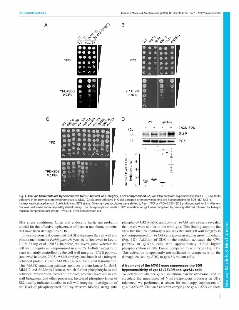
SDS stress conditions, Golgi and endocytic traffic are probablycrucial for the effective replacement of plasma membrane proteinsthat have been damaged by SDS.It was previously documented that SDS damages the cell wall and
plasma membrane in Pichia pastoris yeast cells (reviewed in Levin,2005; Zhang et al., 2015); therefore, we investigated whether thecell wall integrity is compromised in vps13Δ. Cellular integrity inyeast is mainly controlled by the cell wall integrity (CWI) pathway(reviewed in Levin, 2005), which employs one branch of a mitogen-activated protein kinase (MAPK) cascade for signal transduction.This MAPK signalling pathway involves protein kinase C, Bck1,Mkk1/2 and Slt2/Mpk1 kinase, which further phosphorylates andactivates transcription factors to produce proteins involved in cellwall biogenesis and other processes. Increased phosphorylation ofSlt2 usually indicates a defect in cell wall integrity. Investigation ofthe level of phosphorylated Slt2 by western blotting using anti-
phospho-p44/42 MAPK antibody in vps13Δ cell extracts revealedthat levels were similar to the wild type. This finding supports theview that the CWI pathway is not activated and cell wall integrity isnot compromised in vps13Δ cells grown in regular growth medium(Fig. 1D). Addition of SDS to the medium activated the CWIpathway in vps13Δ cells with approximately 5-fold higherphosphorylation of Slt2 kinase compared to wild type (Fig. 1D).This activation is apparently not sufficient to compensate for thedamage, caused by SDS, to vps13Δ mutant cells.
A fragment of the MYO3 gene suppresses the SDShypersensitivity of vps13-I2749R and vps13Δ cellsTo determine whether vps13 mutations can be overcome, and toelucidate the importance of Vps13-dependent processes in SDStolerance, we performed a screen for multicopy suppressors ofvps13-I2749R. The vps13Δ strain carrying the vps13-I2749R allele
Fig. 1. The vps13mutants are hypersensitive to SDS but cell wall integrity is not compromised. (A) vps13mutants are hypersensitive to SDS. (B) Mutantsdefective in endocytosis are hypersensitive to SDS. (C) Mutants defective in Golgi transport or endocytic sorting are hypersensitive to SDS. (D) Slt2 ishyperphosphorylated in vps13 cells following SDS stress. Overnight yeast cultures were shifted to fresh YPD or YPD+0.03% SDS and incubated for 3 h. Westernblot was performed and analysed by densitometry. The phosphorylation levels of Slt2 in relation to Pgk1 were compared by one-way ANOVA followed by Tukey’smultiple comparison test (n=3); **P<0.01. Error bars indicate s.d.
3
RESEARCH ARTICLE Disease Models & Mechanisms (2019) 12, dmm036830. doi:10.1242/dmm.036830
Disea
seModels&Mechan
isms

on a plasmid was transformed with a multicopy genomic library.Several clones, bearing various none-overlapping genomicfragments, were able to grow on SDS-containing medium, andplasmids from these clones were isolated, retested for suppressionand sequenced. One plasmid contained a fragment of the MYO3gene (MYO3-N) encoding the N-terminal 1-775 aa of type I myosin(Myo3-N), an actin cytoskeleton protein involved in endocytosis(Geli and Riezman, 1996; Goodson and Spudich, 1995), which wasresponsible for suppression. The Myo3-N fragment consists of: amotor domain (aa 36-715); a linker (aa 719-771), containing two IQmotifs, indicating ability to bind calmodulin (Ho et al., 2002); andfive amino acids of a tail lipid-binding TH1 domain (aa 771-775)(Fig. 2A). Subsequently, we found that the MYO3-N fragment alsosuppresses the vps13Δ deletion mutation (Fig. 2B). This suggeststhat the Myo3-N protein does not suppress defects by directlyinteracting with Vps13-I2479R, but instead may involve another
factor. Furthermore, we showed that a fragment of Myo3-Ncontaining the IQ motifs (aa 680-775) was necessary forsuppression (Fig. 2B), suggesting that calmodulin could be thisother factor.
To verify the hypothesis that calmodulin binding by Myo3-N isrequired for suppression, threemyo3-N alleles with one (myo3-iq1,myo3-iq2) or both (myo3-iq1/2) IQ motifs disrupted weregenerated by in vitro mutagenesis (Fig. 2A). To test theinteraction of Myo3-N and Myo3-iq1/2 with calmodulin, weused a two-hybrid system, which was previously shown to beappropriate for the detection of the interaction between Myo5 (ahomologue of Myo3) and calmodulin (Geli et al., 1998). Aspredicted, Myo3-N interacted with calmodulin but Myo3-iq1/2protein did not (Fig. 2C). Most importantly, the myo3-iq1/2 allelewas not able to suppress the SDS-hypersensitivity phenotype ofvps13Δ (Fig. 2D), although it was similarly expressed asMYO3-N,
Fig. 2. The MYO3-N fragment encoding the motor domain and calmodulin-binding motifs from Myo3 suppresses hypersensitivity of vps13Δto SDS. (A) Schematic representation of Myo3 domain structure and Myo3 variants studied. Myo3 domains and motifs are based on the Uniprot database(http://www.uniprot.org/uniprot/P36006, 02.08.2018). *, position of mutations introduced in vitro. Amino acids conforming to the consensus of the calmodulin-binding motif as defined in the SMART database (http://smart.embl-heidelberg.de, 02.08.2018). IQ motifs are underlined and amino acid substitutions areshown. (B) MYO3-N suppresses vps13Δ but MYO3-NΔ680-775 does not. (C) Myo3-N interacts with calmodulin in the two-hybrid system and mutationsin IQ motifs abolish this interaction. BD, DNA-binding domian; AD, activating domain. (D) IQ motifs of Myo3 are required for suppression of vps13Δ.
4
RESEARCH ARTICLE Disease Models & Mechanisms (2019) 12, dmm036830. doi:10.1242/dmm.036830
Disea
seModels&Mechan
isms
when the cellular levels of hemagglutinin (HA)-tagged versions ofprotein products were compared (Fig. S1). The myo3-iq1 andmyo3-iq2 mutant alleles, each with a single IQ motif removed,were less efficient in suppression than MYO3-N (Fig. 2D). Theadditive effects of IQ deletions were previously observed forMYO5 (Geli et al., 1998). Our results indicate that the mechanismby whichMYO3-N suppresses vps13Δ requires binding of Myo3-Nto calmodulin and that calmodulin signalling affects the vps13Δphenotype.
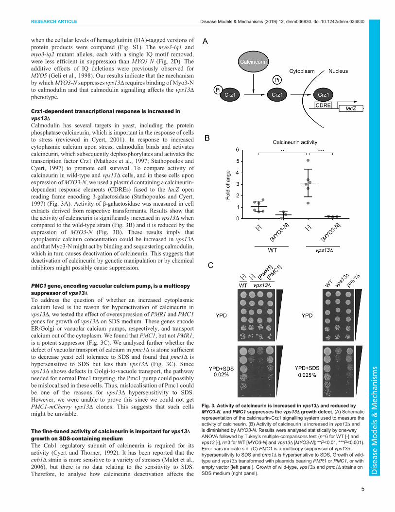
Crz1-dependent transcriptional response is increased invps13ΔCalmodulin has several targets in yeast, including the proteinphosphatase calcineurin, which is important in the response of cellsto stress (reviewed in Cyert, 2001). In response to increasedcytoplasmic calcium upon stress, calmodulin binds and activatescalcineurin, which subsequently dephosphorylates and activates thetranscription factor Crz1 (Matheos et al., 1997; Stathopoulos andCyert, 1997) to promote cell survival. To compare activity ofcalcineurin in wild-type and vps13Δ cells, and in these cells uponexpression ofMYO3-N, we used a plasmid containing a calcineurin-dependent response elements (CDREs) fused to the lacZ openreading frame encoding β-galactosidase (Stathopoulos and Cyert,1997) (Fig. 3A). Activity of β-galactosidase was measured in cellextracts derived from respective transformants. Results show thatthe activity of calcineurin is significantly increased in vps13Δ whencompared to the wild-type strain (Fig. 3B) and it is reduced by theexpression of MYO3-N (Fig. 3B). These results imply thatcytoplasmic calcium concentration could be increased in vps13Δand thatMyo3-Nmight act by binding and sequestering calmodulin,which in turn causes deactivation of calcineurin. This suggests thatdeactivation of calcineurin by genetic manipulation or by chemicalinhibitors might possibly cause suppression.
PMC1 gene, encoding vacuolar calcium pump, is amulticopysuppressor of vps13ΔTo address the question of whether an increased cytoplasmiccalcium level is the reason for hyperactivation of calcineurin invps13Δ, we tested the effect of overexpression of PMR1 and PMC1genes for growth of vps13Δ on SDS medium. These genes encodeER/Golgi or vacuolar calcium pumps, respectively, and transportcalcium out of the cytoplasm. We found that PMC1, but not PMR1,is a potent suppressor (Fig. 3C). We analysed further whether thedefect of vacuolar transport of calcium in pmc1Δ is alone sufficientto decrease yeast cell tolerance to SDS and found that pmc1Δ ishypersensitive to SDS but less than vps13Δ (Fig. 3C). Sincevps13Δ shows defects in Golgi-to-vacuole transport, the pathwayneeded for normal Pmc1 targeting, the Pmc1 pump could possiblybe mislocalised in these cells. Thus, mislocalisation of Pmc1 couldbe one of the reasons for vps13Δ hypersensitivity to SDS.However, we were unable to prove this since we could not getPMC1-mCherry vps13Δ clones. This suggests that such cellsmight be unviable.
The fine-tuned activity of calcineurin is important for vps13Δgrowth on SDS-containing mediumThe Cnb1 regulatory subunit of calcineurin is required for itsactivity (Cyert and Thorner, 1992). It has been reported that thecnb1Δ strain is more sensitive to a variety of stresses (Mulet et al.,2006), but there is no data relating to the sensitivity to SDS.Therefore, to analyse how calcineurin deactivation affects the
Fig. 3. Activity of calcineurin is increased in vps13Δ and reduced byMYO3-N, and PMC1 suppresses the vps13Δ growth defect. (A) Schematicrepresentation of the calcineurin-Crz1 signalling system used to measure theactivity of calcineurin. (B) Activity of calcineurin is increased in vps13Δ andis diminished by MYO3-N. Results were analysed statistically by one-wayANOVA followed by Tukey’s multiple-comparisons test (n=6 for WT [-] andvps13 [-], n=3 for WT [MYO3-N] and vps13Δ [MYO3-N]; **P<0.01, ***P<0.001).Error bars indicate s.d. (C) PMC1 is a multicopy suppressor of vps13Δhypersensitivity to SDS and pmc1Δ is hypersensitive to SDS. Growth of wild-type and vps13Δ transformed with plasmids bearing PMR1 or PMC1, or withempty vector (left panel). Growth of wild-type, vps13Δ and pmc1Δ strains onSDS medium (right panel).
5
RESEARCH ARTICLE Disease Models & Mechanisms (2019) 12, dmm036830. doi:10.1242/dmm.036830
Disea
seModels&Mechan
isms

response to SDS in vps13Δ cells and to establish whether activecalcineurin is required for vps13Δ suppression by MYO3-N, thegrowth of wild-type, cnb1Δ and cnb1Δ vps13Δ mutants on SDS-containing medium was compared in the presence or absence ofMYO3-N. We found that the cnb1Δ strain is more sensitive to SDSthan the wild-type strain and that cnb1Δ mutation negativelyinteracts genetically with vps13Δ (Fig. 4A), indicating that vps13Δcells rely on active calcineurin to cope with SDS stress.Interestingly, the cnb1Δ vps13Δ mutant was not suppressed byMYO3-N, indicating that Myo3-N requires the Cnb1 protein andfunctional calcineurin for vps13Δ suppression (Fig. 4A). Thus, itseems that a certain level of calcineurin activity is required fornormal growth in the presence of SDS. Sequestration of calmodulin,the activator of calcineurin, by MYO3-N results in a reduction incalcineurin activity, thus fine tuning calcineurin activity to thisrequired level.
The contribution of Crz1 to the SDS stress response and to theMYO3-N suppression of vps13Δ was also studied. Comparison ofthe growth of isogenic crz1Δ and crz1Δ vps13Δ mutants withvps13Δ and with the wild-type strain revealed that the crz1Δ strainexhibits a similar sensitivity to SDS as the wild-type strain inconditions tested. It also revealed that crz1Δ vps13Δ grows moreslowly than vps13Δ but suppression by MYO3-N is still observed(Fig. 4A). These results show that the Crz1 transcription factorpositively contributes to SDS stress tolerance in vps13Δ and, mostimportantly, that vps13Δ suppression by MYO3-N involves amechanism that is independent of Crz1 activity.
To investigate the possibility of using chemical intervention inthe vps13Δ strain, we tested the effects of: ethylene glycol tetraaceticacid (EGTA), a chelator of divalent cations that is more specific forCa2+ than for other cations; and FK506, a calcineurin inhibitor.EGTA reduces the concentration of calcium in the cytoplasm, thus
Fig. 4. A normal/intermediate activity of calmodulin-calcineurin pathway is required to cope with SDS stress in vps13Δ. (A) Growth of wild-type, vps13Δ,cnb1Δ, crz1Δ, vps13Δ cnb1Δ and vps13Δ crz1Δ cells expressing, or not, MYO3-N on YPD+SDS. (B) Addition of EGTA improves growth of vps13Δ on SDS-containing medium. (C) FK506 affects growth of vps13Δ on SDS-containing medium. Strains were grown to early exponential phase. Cells (0.05 OD) were platedon YPD+0.035% SDS, and 2 µl of 0-10 µM FK506 (HY-13756, MedChemExpress, Monmouth Junction, USA) was applied on paper discs as indicated.
6
RESEARCH ARTICLE Disease Models & Mechanisms (2019) 12, dmm036830. doi:10.1242/dmm.036830
Disea
seModels&Mechan
isms
inhibiting the Ca2+-dependent action of calmodulin and inhibitingcalcineurin. Interestingly, addition of EGTA significantly improvedgrowth of vps13Δ on SDS plates (Fig. 4B). Thus, a partial inhibitionof the calcium-dependent functions of calmodulin and the inhibitionof calcineurin is beneficial for vps13Δ cells in the presence of SDSstress. FK506 improved growth of vps13Δ at low concentrations(2.5 μM spotted onto filter paper), but higher concentrationsexhibited an adverse effect (Fig. 4C). Thus, calcineurin cannot beinhibited completely, but its downregulation can be fine-tuned inorder to improve the growth of vps13 cells. This result points toFK506 as a potential drug and to calcineurin as a useful target toselect drugs for ChAc therapy.
The cmd1-226 mutant allele, encoding a calmodulin variantwith the F92A amino acid substitution, suppresses vps13ΔThe importance of calmodulin binding by Myo3-N for thesuppression of vps13 suggests that a calmodulin sequestrationmechanism is involved. To verify this assumption, we analysedwhether expression of multiple copies of CMD1 alleles encodingwild-type or mutant calmodulin have an effect on the growth ofvps13Δ cells in the presence of SDS. The various cmd1 alleles wereselected to represent different groups of mutants with the samephenotype: defective actin cytoskeleton, altered localisation ofcalmodulin, defective karyokinetic spindle or defective budding, aspreviously described (Geli et al., 1998; Ohya and Botstein, 1994).The wild-type CMD1 negatively affected growth of vps13Δ cells,implying that the control of the calmodulin pool and/or accessibilityto different substrates is important for regulation of the cell responseto SDS stress in vps13Δ, supporting the sequestration mechanismforMYO3-N (Fig. 5A). The cmd1-231 allele, which causes a defectin budding, affected growth similarly to wild-type CMD1, while themutant cmd1-228 and cmd1-239 alleles essentially did not affectgrowth of vps13Δ. Surprisingly, the cmd1-226 allele, which resultsin the F92A substitution in calmodulin and represents a group ofmutants that show defects in the organisation of the actincytoskeleton and in endocytosis, was a potent suppressor ofvps13Δ (Fig. 5A).To test how overproduction of wild-type or mutant calmodulin
affects activity of calcineurin in vps13Δ cells, the CDRE-lacZreporter was used. Interestingly, neither wild-type nor mutantcalmodulin genes, including the cmd1-226 suppressor allele,significantly affected the increased activity of calcineurin invps13Δ cells (Fig. 5B). Since additional calmodulin did not havean effect in vps13Δ, we checked whether calcineurin could bestimulated by addition of calcium to the medium in this strain. Wild-type and vps13Δ cells showed higher activity of calcineurin aftercalcium addition, indicating that calcineurin activity in vps13Δ inregular conditions is not maximal (Fig. 5C). Moreover, in wild-typecells, the effect of calcium and expression of additional copies ofCMD1was additive. However, vps13Δ cells expressingCMD1 fromthe plasmid did not significantly increase calcineurin activity in thepresence or absence of additional calcium. Thus, even whencalcium is not a limiting factor, further activation of calcineurin byCmd1 requires Vps13. These results indicate that reduction ofcalcineurin activity is probably not a mechanism for restoringgrowth of vps13 cells on SDS plates mediated by cmd1-226. TheCmd1-226 mutant protein might target proteins other thancalcineurin. One possibility is that Cmd1-226 is different in termsof its binding with Myo3, as is the case for Myo2 (Sekiya-Kawasakiet al., 1998). Therefore, we analysed how selected cmd1 mutationsaffect the interaction of calmodulin with Myo3 using a two-hybridsystem. This analysis shows that the cmd1-226mutation, in contrast
to other mutations tested, results in total loss of interaction betweencalmodulin and Myo3-N (Fig. 5D). Thus, a complete loss ofinteraction between mutant calmodulin and Myo3 is required forcmd1-226-based suppression, the opposite to MYO3-N-basedsuppression of vps13Δ. Cmd1-226 may act by diluting wild-typecalmodulin and activating full-length Myo3, or by othermechanisms.
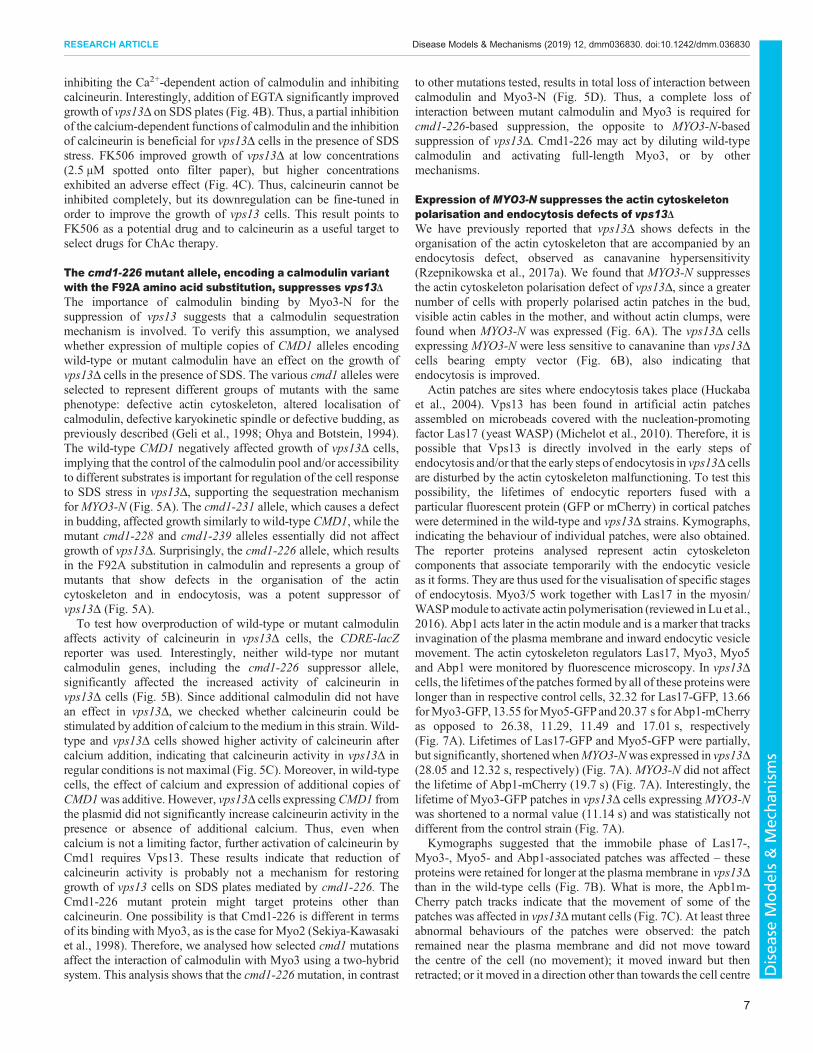
Expression of MYO3-N suppresses the actin cytoskeletonpolarisation and endocytosis defects of vps13ΔWe have previously reported that vps13Δ shows defects in theorganisation of the actin cytoskeleton that are accompanied by anendocytosis defect, observed as canavanine hypersensitivity(Rzepnikowska et al., 2017a). We found that MYO3-N suppressesthe actin cytoskeleton polarisation defect of vps13Δ, since a greaternumber of cells with properly polarised actin patches in the bud,visible actin cables in the mother, and without actin clumps, werefound when MYO3-N was expressed (Fig. 6A). The vps13Δ cellsexpressing MYO3-N were less sensitive to canavanine than vps13Δcells bearing empty vector (Fig. 6B), also indicating thatendocytosis is improved.
Actin patches are sites where endocytosis takes place (Huckabaet al., 2004). Vps13 has been found in artificial actin patchesassembled on microbeads covered with the nucleation-promotingfactor Las17 (yeast WASP) (Michelot et al., 2010). Therefore, it ispossible that Vps13 is directly involved in the early steps ofendocytosis and/or that the early steps of endocytosis in vps13Δ cellsare disturbed by the actin cytoskeleton malfunctioning. To test thispossibility, the lifetimes of endocytic reporters fused with aparticular fluorescent protein (GFP or mCherry) in cortical patcheswere determined in the wild-type and vps13Δ strains. Kymographs,indicating the behaviour of individual patches, were also obtained.The reporter proteins analysed represent actin cytoskeletoncomponents that associate temporarily with the endocytic vesicleas it forms. They are thus used for the visualisation of specific stagesof endocytosis. Myo3/5 work together with Las17 in the myosin/WASPmodule to activate actin polymerisation (reviewed inLu et al.,2016). Abp1 acts later in the actin module and is a marker that tracksinvagination of the plasma membrane and inward endocytic vesiclemovement. The actin cytoskeleton regulators Las17, Myo3, Myo5and Abp1 were monitored by fluorescence microscopy. In vps13Δcells, the lifetimes of the patches formed by all of these proteins werelonger than in respective control cells, 32.32 for Las17-GFP, 13.66forMyo3-GFP, 13.55 forMyo5-GFPand 20.37 s forAbp1-mCherryas opposed to 26.38, 11.29, 11.49 and 17.01 s, respectively(Fig. 7A). Lifetimes of Las17-GFP and Myo5-GFP were partially,but significantly, shortened whenMYO3-Nwas expressed in vps13Δ(28.05 and 12.32 s, respectively) (Fig. 7A). MYO3-N did not affectthe lifetime of Abp1-mCherry (19.7 s) (Fig. 7A). Interestingly, thelifetime of Myo3-GFP patches in vps13Δ cells expressingMYO3-Nwas shortened to a normal value (11.14 s) and was statistically notdifferent from the control strain (Fig. 7A).
Kymographs suggested that the immobile phase of Las17-,Myo3-, Myo5- and Abp1-associated patches was affected – theseproteins were retained for longer at the plasma membrane in vps13Δthan in the wild-type cells (Fig. 7B). What is more, the Apb1m-Cherry patch tracks indicate that the movement of some of thepatches was affected in vps13Δmutant cells (Fig. 7C). At least threeabnormal behaviours of the patches were observed: the patchremained near the plasma membrane and did not move towardthe centre of the cell (no movement); it moved inward but thenretracted; or it moved in a direction other than towards the cell centre
7
RESEARCH ARTICLE Disease Models & Mechanisms (2019) 12, dmm036830. doi:10.1242/dmm.036830
Disea
seModels&Mechan
isms

(chaotic movement). MYO3-N expression did not influence thedefective patch movement of vps13Δ (Fig. 7C).In summary, the results suggest that Vps13 is involved in the
early steps of endocytosis and it affects the duration of association ofseveral actin cytoskeleton proteins with endocytic sites. It alsoaffects the movement of endocytic vesicles, as observed by Abp1-mCherry, in terms of occurrence, distance travelled and direction.Production of Myo3-N in vps13Δ improves endocytosis by
correcting a number of steps in the process, in particular restoringthe functioning of full-length Myo3.
Osh2 and Osh3 proteins bridging cortical ER with plasmamembrane at endocytic sites are essential for suppressionof vps13Δ by MYO3-NWe found that the defects of vps13Δ cells – in endocytosis, in theextended lifetime of Las17-GFP and in abnormal Abp1 patch
Fig. 5. The cmd1-226 mutant allele, but not wild-type CMD1, restores growth of vps13Δ on YPD+SDS plates. (A) Different CMD1 alleles variouslyaffect the growth of vps13Δ. (B) Wild-type and mutant calmodulin do not influence calcineurin activity in vps13Δ cells. Results were analysed by one-wayANOVA (n=3); P=0.196. Error bars indicate s.d. (C) Effects of calcium addition and CMD1 overexpression on calcineurin activity in wild-type and vps13Δ cells.Results were analysed statistically by two-tailed Student’s t-test (n=6; *P<0.05, **P<0.01, ***P<0.001, ****P<0.0001. Error bars indicate s.d. (D) Cmd1-226does not interact with Myo3-N. Results from a two-hybrid experiment are shown. BD, DNA-binding domain; AD, activating domain.
8
RESEARCH ARTICLE Disease Models & Mechanisms (2019) 12, dmm036830. doi:10.1242/dmm.036830
Disea
seModels&Mechan
isms
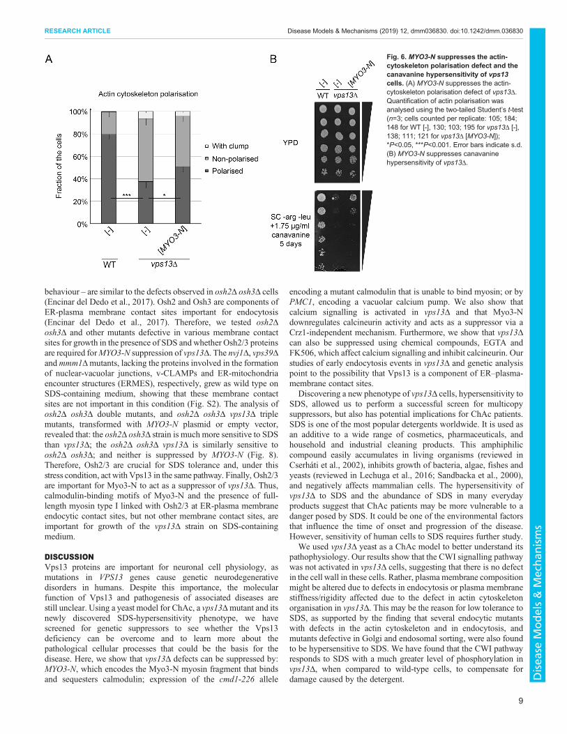
behaviour – are similar to the defects observed in osh2Δ osh3Δ cells(Encinar del Dedo et al., 2017). Osh2 and Osh3 are components ofER-plasma membrane contact sites important for endocytosis(Encinar del Dedo et al., 2017). Therefore, we tested osh2Δosh3Δ and other mutants defective in various membrane contactsites for growth in the presence of SDS and whether Osh2/3 proteinsare required forMYO3-N suppression of vps13Δ. The nvj1Δ, vps39Δandmmm1Δmutants, lacking the proteins involved in the formationof nuclear-vacuolar junctions, v-CLAMPs and ER-mitochondriaencounter structures (ERMES), respectively, grew as wild type onSDS-containing medium, showing that these membrane contactsites are not important in this condition (Fig. S2). The analysis ofosh2Δ osh3Δ double mutants, and osh2Δ osh3Δ vps13Δ triplemutants, transformed with MYO3-N plasmid or empty vector,revealed that: the osh2Δ osh3Δ strain is much more sensitive to SDSthan vps13Δ; the osh2Δ osh3Δ vps13Δ is similarly sensitive toosh2Δ osh3Δ; and neither is suppressed by MYO3-N (Fig. 8).Therefore, Osh2/3 are crucial for SDS tolerance and, under thisstress condition, act with Vps13 in the same pathway. Finally, Osh2/3are important for Myo3-N to act as a suppressor of vps13Δ. Thus,calmodulin-binding motifs of Myo3-N and the presence of full-length myosin type I linked with Osh2/3 at ER-plasma membraneendocytic contact sites, but not other membrane contact sites, areimportant for growth of the vps13Δ strain on SDS-containingmedium.
DISCUSSIONVps13 proteins are important for neuronal cell physiology, asmutations in VPS13 genes cause genetic neurodegenerativedisorders in humans. Despite this importance, the molecularfunction of Vps13 and pathogenesis of associated diseases arestill unclear. Using a yeast model for ChAc, a vps13Δmutant and itsnewly discovered SDS-hypersensitivity phenotype, we havescreened for genetic suppressors to see whether the Vps13deficiency can be overcome and to learn more about thepathological cellular processes that could be the basis for thedisease. Here, we show that vps13Δ defects can be suppressed by:MYO3-N, which encodes the Myo3-N myosin fragment that bindsand sequesters calmodulin; expression of the cmd1-226 allele
encoding a mutant calmodulin that is unable to bind myosin; or byPMC1, encoding a vacuolar calcium pump. We also show thatcalcium signalling is activated in vps13Δ and that Myo3-Ndownregulates calcineurin activity and acts as a suppressor via aCrz1-independent mechanism. Furthermore, we show that vps13Δcan also be suppressed using chemical compounds, EGTA andFK506, which affect calcium signalling and inhibit calcineurin. Ourstudies of early endocytosis events in vps13Δ and genetic analysispoint to the possibility that Vps13 is a component of ER–plasma-membrane contact sites.
Discovering a new phenotype of vps13Δ cells, hypersensitivity toSDS, allowed us to perform a successful screen for multicopysuppressors, but also has potential implications for ChAc patients.SDS is one of the most popular detergents worldwide. It is used asan additive to a wide range of cosmetics, pharmaceuticals, andhousehold and industrial cleaning products. This amphiphiliccompound easily accumulates in living organisms (reviewed inCserháti et al., 2002), inhibits growth of bacteria, algae, fishes andyeasts (reviewed in Lechuga et al., 2016; Sandbacka et al., 2000),and negatively affects mammalian cells. The hypersensitivity ofvps13Δ to SDS and the abundance of SDS in many everydayproducts suggest that ChAc patients may be more vulnerable to adanger posed by SDS. It could be one of the environmental factorsthat influence the time of onset and progression of the disease.However, sensitivity of human cells to SDS requires further study.
We used vps13Δ yeast as a ChAc model to better understand itspathophysiology. Our results show that the CWI signalling pathwaywas not activated in vps13Δ cells, suggesting that there is no defectin the cell wall in these cells. Rather, plasmamembrane compositionmight be altered due to defects in endocytosis or plasma membranestiffness/rigidity affected due to the defect in actin cytoskeletonorganisation in vps13Δ. This may be the reason for low tolerance toSDS, as supported by the finding that several endocytic mutantswith defects in the actin cytoskeleton and in endocytosis, andmutants defective in Golgi and endosomal sorting, were also foundto be hypersensitive to SDS. We have found that the CWI pathwayresponds to SDS with a much greater level of phosphorylation invps13Δ, when compared to wild-type cells, to compensate fordamage caused by the detergent.
Fig. 6. MYO3-N suppresses the actin-cytoskeleton polarisation defect and thecanavanine hypersensitivity of vps13cells. (A) MYO3-N suppresses the actin-cytoskeleton polarisation defect of vps13Δ.Quantification of actin polarisation wasanalysed using the two-tailed Student’s t-test(n=3; cells counted per replicate: 105; 184;148 for WT [-], 130; 103; 195 for vps13Δ [-],138; 111; 121 for vps13Δ [MYO3-N]);*P<0.05, ***P<0.001. Error bars indicate s.d.(B) MYO3-N suppresses canavaninehypersensitivity of vps13Δ.
9
RESEARCH ARTICLE Disease Models & Mechanisms (2019) 12, dmm036830. doi:10.1242/dmm.036830
Disea
seModels&Mechan
isms
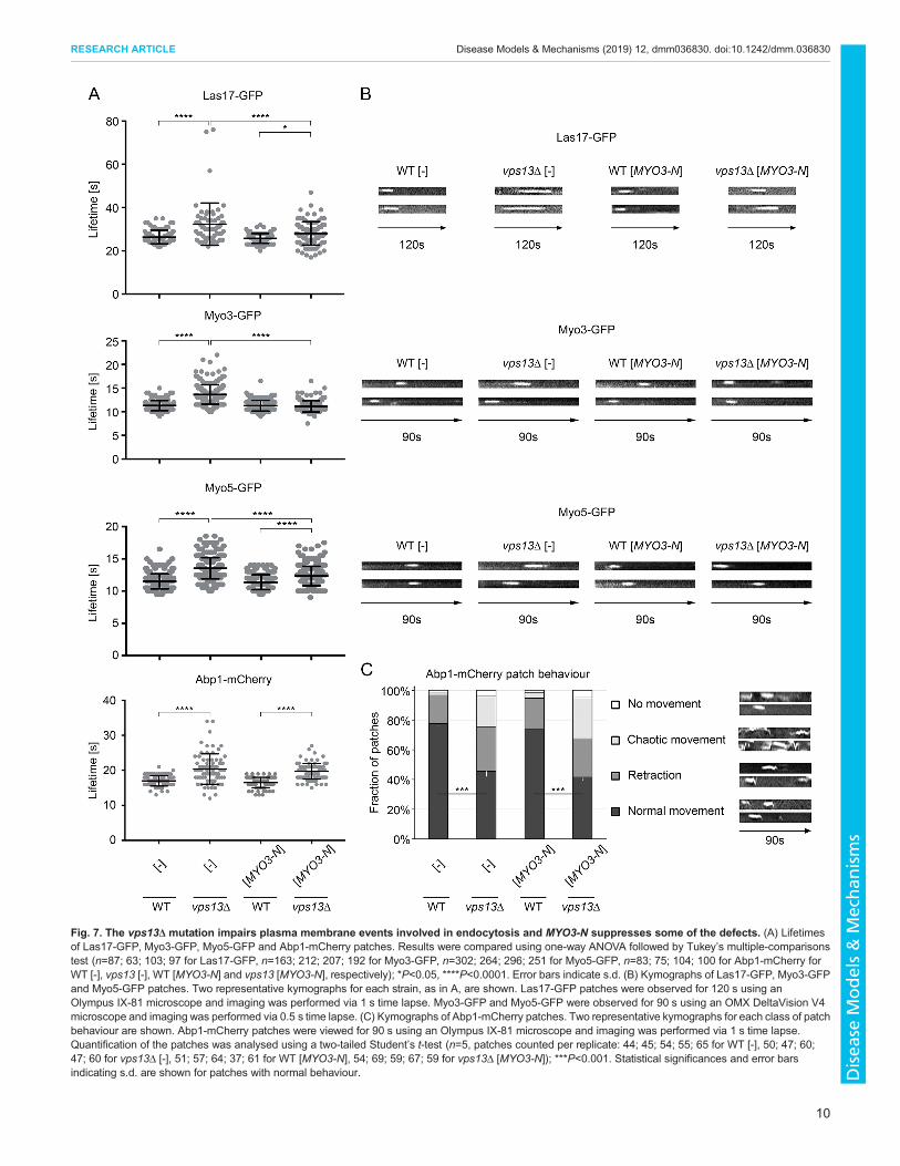
Fig. 7. The vps13Δ mutation impairs plasma membrane events involved in endocytosis and MYO3-N suppresses some of the defects. (A) Lifetimesof Las17-GFP, Myo3-GFP, Myo5-GFP and Abp1-mCherry patches. Results were compared using one-way ANOVA followed by Tukey’s multiple-comparisonstest (n=87; 63; 103; 97 for Las17-GFP, n=163; 212; 207; 192 for Myo3-GFP, n=302; 264; 296; 251 for Myo5-GFP, n=83; 75; 104; 100 for Abp1-mCherry forWT [-], vps13 [-], WT [MYO3-N] and vps13 [MYO3-N], respectively); *P<0.05, ****P<0.0001. Error bars indicate s.d. (B) Kymographs of Las17-GFP, Myo3-GFPand Myo5-GFP patches. Two representative kymographs for each strain, as in A, are shown. Las17-GFP patches were observed for 120 s using anOlympus IX-81 microscope and imaging was performed via 1 s time lapse. Myo3-GFP and Myo5-GFP were observed for 90 s using an OMX DeltaVision V4microscope and imaging was performed via 0.5 s time lapse. (C) Kymographs of Abp1-mCherry patches. Two representative kymographs for each class of patchbehaviour are shown. Abp1-mCherry patches were viewed for 90 s using an Olympus IX-81 microscope and imaging was performed via 1 s time lapse.Quantification of the patches was analysed using a two-tailed Student’s t-test (n=5, patches counted per replicate: 44; 45; 54; 55; 65 for WT [-], 50; 47; 60;47; 60 for vps13Δ [-], 51; 57; 64; 37; 61 for WT [MYO3-N], 54; 69; 59; 67; 59 for vps13Δ [MYO3-N]); ***P<0.001. Statistical significances and error barsindicating s.d. are shown for patches with normal behaviour.
10
RESEARCH ARTICLE Disease Models & Mechanisms (2019) 12, dmm036830. doi:10.1242/dmm.036830
Disea
seModels&Mechan
isms

Changes in calcineurin activity are connected with severaldiseases, including neurodegeneration. Calcineurin signalling waselevated in vps13Δ, and this is detrimental, as shown for yeastmodels for other neurodegenerative diseases (Caraveo et al., 2014).One possible explanation is that a lack of Vps13 protein causes adefect in protein trafficking that triggers a cellular responseinvolving calcineurin upregulation. Our data of vps13Δsuppression by PMC1 suggest that Pmc1 could be mislocalised invps13Δ and inefficient in pumping calcium to the vacuole, resultingin cytoplasmic calcium increase and calcineurin activation. Such aview is also supported by negative genetic interaction of vps13Δwith pmr1Δ (Costanzo et al., 2010). It is also possible that Vps13 ismore directly involved in calcium signalling, since, in high-throughput studies, Vps13 has been shown to bind calmodulin (Hoet al., 2002). However, we were not able to confirm this interactionby other methods. Although detrimental in excess, the activities ofcalcineurin and of its Crz1 effector are important for the growth ofvps13Δ cells under SDS stress, similarly to as observed for Candidaglabrata yeast (Chen et al., 2012). Calcium signalling andcalcineurin activity could be upregulated in vps13 cells by at leasttwo mechanisms: through secondary effects of disrupted transportprocesses or through the removal of potential direct interactions.We have shown that the effects of the vps13Δ mutation,
monitored through growth on SDS medium, can be overcome inseveral ways: titration of calmodulin by Myo3-N; expression ofadditional copies of PMC1; additional production of mutantcalmodulin Cmd1-226; or addition of calcineurin inhibitors.Expressed Myo3-N binds to and sequesters free calmodulin,which activates Myo3/5 and downregulates the activity ofcalcineurin. The growth defect of the cnb1Δ vps13Δ mutant is notsuppressed by MYO3-N; however, the crz1Δ vps13Δ mutant wasstill suppressed. This suggests that active calcineurin, but not Crz1,is required for the suppression and only partial downregulation isbeneficial. This effect could be also achieved by lowering
cytoplasmic calcium via overproduction of the Pmc1 vacuolarcalcium pump. This notion is further supported by the finding thatchemical calcineurin inhibitors, EGTA and FK506, suppressed thevps13Δ growth defect. FK506 suppressed vps13Δ only at a very lowconcentration, while at higher concentrations FK506 enhanced thevps13Δ hypersensitivity to SDS. Our results are in line withobservations that the moderate calcineurin activity is protective forcells, but elevated or inhibited activity is toxic (Caraveo et al., 2014,reviewed in Kipanyula et al., 2016). In agreement with acalmodulin-sequestering model, overproduction of wild-typecalmodulin is harmful to vps13Δ cells in spite of it not affectingcalcineurin activity in vps13Δ cells. The cmd1-226 also did not alterthe calcineurin activity, which suggests that the mechanism of thesuppression in this case is calcineurin independent. One possibilityis that Cmd1-226 dilutes or somehow negatively affects wild-typecalmodulin, what in turn increases the pool of the active forms ofMyo3/5, which contributes to suppression by enhancingendocytosis. The other possible explanation of cmd1-226 action isby interacting with Arc35, a subunit of the Arp2/3 complex thatactivates the polymerisation of actin filaments. Calmodulin encoded bycmd1-226 binds to Arc35 protein, while that encoded by cmd1-231,which was the most toxic to vps13Δ, does not (Schaerer-Brodbeck andRiezman, 2003). We cannot exclude the possibility that SDS stresssomehow causes cell division defects in vps13Δ andCmd1-226 helps toovercome this defect by acting on other targets, such as Spc110, aspindle-pole-body calmodulin-binding protein (Geiser et al., 1993).
As we have documented here, vps13Δ exhibits defects in an earlyendocytic processes. These defects include an extended lifetime ofseveral endocytic actin-cytoskeleton proteins and abnormal Abp1patch behaviour, which is a marker of endocytic vesicleinternalisation. Myo3-N production in vps13Δ results in restorationof the lifetime of Myo3 patches engaged in invagination and inrelease of endocytic vesicles, and in partially shortened lifetimes ofLas17 and Myo5. Myo3-N production may also have other effectsand it should also be noted that the vesicle internalisation defect ofvps13Δ is not corrected. It was recently documented that endocyticplasma membrane invaginations associate with the cortical ER andthis contact facilitates actin polymerisation and endocytic vesiclescission (Encinar del Dedo et al., 2017). Myo5 is a protein thatestablishes the contact between the ER and endocytic sites bybinding Osh2 and Osh3. Osh2/3 in turn bind Scs2 and Scs22, ERmembrane proteins, and the Osh2 sterol transfer domain is requiredfor the onset of actin polymerisation at endocytic sites (Encinar delDedo et al., 2017).Myo3 exhibits a similar domain structure toMyo5and can possibly perform similar functions at endocytic sites.Previous results are consistent with the view that type I myosin couldinduce formation of ER-plasma membrane contact sites and thiscapacity is required for myosin-Osh2/3-Scs2/22 link and the steroltransfer activity of Osh2, actin polymerisation and endocytic vesiclescission (Encinar del Dedo et al., 2017). In the osh2Δ osh3Δ andscs2Δ scs22Δ strains, the lifespan of Abp1 patches was increased,with a high fraction of patches initiating the inward movement andretracting back to the plasma membrane (Encinar del Dedo et al.,2017), similar to what we observed for vps13Δ. Our results showingthat vps13Δ defects in endocytosis are suppressed byMYO3-N in anOsh2/3-dependent way suggest that one mechanism of suppressioncould be by promoting the formation of ER-plasma membraneconnections at endocytic sites, which involve type I myosin. Also, arecent report suggests that Vps13 could be a binding partner of Osh2(Bean et al., 2018). This implies a possible Vps13 function in theformation of ER-plasma membrane contact sites, which areimportant for maintaining the lipid and protein composition of
Fig. 8. The osh2Δ osh3Δ double mutant is hypersensitive to SDS andOsh2/3 are important for vps13Δ suppression by MYO3-N.
11
RESEARCH ARTICLE Disease Models & Mechanisms (2019) 12, dmm036830. doi:10.1242/dmm.036830
Disea
seModels&Mechan
isms

plasma membrane and are a prerequisite for SDS-stress survival. Amore efficient action of myosin in promoting sterol transfer byOsh2/3 at endocytic sites helps to overcome the defects caused by thelack of Vps13. This suggests that: Vps13 may be important for thetransfer of some lipids to plasma membranes at some ER-lasmamembrane contact sites; a lack of Vps13 participation in theformation of contact sites affects endocytosis; and this defect couldbe overcome by activation of endocytic ER-plasma membranecontact sites. This model is supported by a recent study in which theN-terminal fragment of Vps13 was shown to be able to transfer thebulk of glycerolipids between liposomes (Kumar et al., 2018),implying that Vps13 might relocate lipids between membranes.Such transfer could possibly be required for growth of plasmamembrane invagination or vesicle scission during the internalisationstep of endocytosis. It requires further study to find out whether therole of Vps13 in endocytosis is direct, since the SDS growth andendocytosis defect of vps13Δ may result also from activation ofcalcium signalling. Since the growth defect of vps13Δ on SDSmedium is more severe than that of pmc1Δ, the activation of calciumsignalling is probably not the only reason and othermechanismsmaycontribute.In this study, we demonstrate the relationship between Vps13 and
calcium signalling, and the importance of Osh2/3-dependentmembrane contact sites for bypassing the vps13Δ growth defect.Our results will be helpful for studies to better understand thepathogenesis of ChAc in higher eukaryotes and in cell lines. Themulticopy suppressor that we selected can suppress the otherphenotypes of vps13Δ, not just the SDS hypersensitivity. Inaddition, chemical compounds such as EGTA or FK506 cansuppress the SDS hypersensitivity. Thus, we believe that thisphenotype is useful to learn about pathways that could be potentialtargets for chemical intervention, and could be used directly toscreen for drugs to treat ChAc.
MATERIALS AND METHODSStrains, media and growth conditionsEscherichia coli strain DH5α was used for plasmid propagation. The yeastS. cerevisiae strains used in this study are listed in Table S1. For geneticanalysis, strains BY4741 cnb1Δ vps13Δ and BY4741 crz1Δ vps13Δ wereconstructed by deletion of VPS13 using the vps13::URA3 cassette(pKA475) in the single-deletion strains BY4741 cnb1Δ and BY4741crz1Δ. Gene disruptions were performed by transformation of yeast cellswith a PCR product containing theURA3 selection marker flanked by 50 bpof homology to the 5′ and 3′ regions of VPS13. The vps13::URA3 cassettewas also used to construct strains encoding Las17-mRFP, Myo3-GFP,Myo5-GFP or Abp1-mCherry endocytic markers in the genome and devoidof Vps13 protein. Genomic integrations were confirmed by PCR ongenomic DNA.
Yeast were grown at 30°C in liquid YPD complete medium (1% yeastextract, 2% peptone, 2% glucose) or in synthetic SC medium (0.067% yeastnitrogen base without amino acids, 2% glucose) with desired supplements(adenine, uracil, amino acids) (all media ingredients: DB, Sparks, USA). Forgrowth tests, cells were grown overnight in liquid media and cultures werediluted with water to obtain a cell density equal to OD600∼1. Subsequently,aliquots of 4-fold serial dilutions of cells were spotted on plates with solidmedia. Media used for growth tests were YPD with, or without, addition ofSDS (Sigma-Aldrich, St Louis, USA), and SC-leu-arg with, or without,addition of canavanine (Sigma-Aldrich), as described in the figure legends.Plates were incubated at 30°C for 2 (YPD) or 3 (YPD+SDS) days, or asindicated. Mutant cells were very sensitive to small changes in SDSconcentration and final SDS concentration in the medium strongly dependson the lot of medium and lot of SDS stock solution, and various physicalfactors; therefore, each growth experiment was performed using severaldilutions of SDS stock solution and a representative result is shown. For
two-hybrid analysis, the PJ69-4A strain was used and respectivetransformants were grown on SC-trp-leu plates, replicated on SC-trp-leu-his supplemented with 1.5 mM aminotriazol (Sigma-Aldrich) and incubatedfor 3-5 days. Liquid cultures were inoculated with a mix of several yeastcolonies obtained after transformation. Growth tests were repeated at leasttwice. Plates were scanned by HP Scanjet G4010 or Epson Perfection 2480Photo scanner. Imagines were processed with Adobe Photoshop 6.0.
PlasmidsPlasmids used are described in Table S2. To subclone the MYO3 genefragment, the BamHI and SalI DNA fragment from the original clone fromthe genomic bank was transferred into pRS425-PGPD vector to obtainpRS425-PGPD-MYO3-N. Further transferring a 3.2 kb BamHI HindIIIfragment of pRS425-PGPD-MYO3-N to pRS425-PGPD generated pRS425-PGPD-MYO3-NΔ680-775. To destroy calmodulin-binding motifs IQ1 andIQ2 in Myo3-N, mutations were introduced in the MYO3-N fragment, aftersubcloning into pUC19 using PstI and SalI enzymes, by one-step site-directed mutagenesis. Three plasmids were obtained: pUC19-MYO3-iq1,encoding myosin fragment with I725G and Q726G substitutions; pUC19-MYO3-iq2, encoding protein with I743G and Q744G substitutions; andpUC19-MYO3-iq1/2, encoding protein with all four substitutions. Next, thePstI-SalI fragments were transferred back to create pRS425-PGPD-myo3-iq1, pRS425-PGPD-myo3-iq2 and pRS425-PGPD-myo3-iq1/2. pRS425-PGPD-MYO3-N-HA and pRS425-PGPD-myo3-iq1/2-HA were generatedby introducing the HA tag into the SalI polylinker restriction site. Sequencesof oligonucleotides used in this study are available upon request.
To construct plasmids for two-hybrid systems, the CMD1 gene andmutated alleles were amplified by PCR using YEplac181lac bearing CMD1,cmd1-226, cmd1-228, cmd1-231 or cmd1-239 alleles as the templates. PCRproducts were digested with PstI and BamHI and were cloned intopGAD424. To obtain pGBT9-MYO3-N or pGBT9-myo3-iq1/2, theappropriate fragments were amplified by PCR with pRS425-PGPD-MYO3-N or pRS425-PGPD-myo3-iq1/2 as the templates. PCR productswere cloned into EcoRI and SalI sites of pGBT9.
Screen for multicopy suppressors of vps13-I2749R mutationThe vps13Δ [vps13-I2749R] cells were transformed with pFL44-basedgenomic bank (Kabir et al., 2005), plated on SC-ura-leu plates and incubatedat 30°C for 3 days. Then, transformants were replicated on YPD plates withaddition of 0.03% SDS and incubated at 30°C for 3 days. Positive cloneswere isolated and grown in SC-ura liquid medium for plasmid isolation.Plasmids were retransformed and suppression of the vps13Δ [vps13-I2749R]growth defect was confirmed.
Actin staining and fluorescence microscopyFor actin staining, cells were grown in SC-leu medium to log-phase, fixedwith formaldehyde (Sigma-Aldrich for 2 h, washed and stained with Alexa-Fluor-546-conjugated phalloidin (Thermo Fisher Scientific, Waltham,USA). Cells were viewed with an Eclipse E800 (Nikon, Tokyo, Japan)fluorescence microscope equipped with a DS-5Mc camera (Nikon, Tokyo,Japan). Images were collected using Lucia General 5.1 software (LaboratoryImaging Ltd, Prague, Czech Republic). The same fields were viewed bydifferential interference contrast (DIC) optics. All samples in the experimentwere encoded and the microscope observations and quantifications wereperformed blindly. Charts and statistical analyses were done usingMicrosoftExcel.
For live-cell imaging, cells expressing tagged proteins were visualisedafter growing to early log-phase in synthetic medium with appropriatesupplements. Epifluorescence microscopy was performed using OlympusIX-81 inverted microscope (Olympus, Tokyo, Japan) with a DeltaVision RTRestoration Microscopy System (using a 100×/1.40 NA oil objective) andPhotometrics Coolsnap HQ camera, with imaging and image captureperformed using SoftWoRx™ image analysis and model-buildingapplication (Applied Precision Instruments, Seattle, USA). Live-cellimaging of GFP-tagged Las17 and mCherry-tagged Abp1 proteins wasperformed with 1 s time lapse. All image data sets were deconvolved, usingthe SoftWoRx application. Live-cell images of Myo3-GFP and Myo5-GFPwere acquired using OMXDeltaVision V4 microscope (GE Healthcare Life
12
RESEARCH ARTICLE Disease Models & Mechanisms (2019) 12, dmm036830. doi:10.1242/dmm.036830
Disea
seModels&Mechan
isms
Sciences, Chicago, USA) and a 60× USPLAPO (1.42 NA) objective withrefractive index 0.1514 immersion oil (Cargille, Cedar Grove, USA) with0.5 s time lapse. Samples were illuminated using Insight Solid StateIlluminator (10%), and images were taken simultaneously on separatescientific complementary metal oxide semiconductor (sCMOS) cameras(30 ms exposure). Seven 250 nm sections were acquired every 500 ms (181time points). The stacks were then deconvolved and processed, usingSoftWorx, to produce a movie composed of maximum intensity projectionsat each time point. The Myo3-/Myo5-GFP lifetime was analysed from thoseprojections. Movies and kymographs were assembled using Fiji software.Charts and statistical analyses were done in GraphPad Prism (https://www.graphpad.com/scientific-software/prism/, 02.08.2018).
Western blot analysisYeast cells were grown at 30°C in SC-ura-leu to the log-phase. Proteinextracts were prepared after disrupting cells with glass beads as described(Rzepnikowska et al., 2017a) or alkaline lysis (Yaffe and Schatz, 1984).Samples were analysed by standard SDS-PAGE followed by westernblotting using mouse monoclonal anti-Pgk1 (dilution 1:20,000, MolecularProbes Cat# A-6457, RRID:AB_221541, Thermo Fisher Scientific),monoclonal anti-HA epitope (dilution 1:2000, Cat# 901503, BioLegend,San Diego, USA), rabbit anti-phospho-p44/42 MAPK (Erk1/2) (Thr202/Thr204; dilution 1:1000, Antibody #9101, Cell Signalling Technology,Danvers, USA; validation: Pérez et al., 2016), recognising phospho-Slt2,antibodies. Secondary anti-mouse or anti-rabbit IgG horseradish peroxidase(HRP)-conjugated antibodies (dilution 1:2000, Dako, Glostrup, Denmark)were also used. Signal was detected by enhanced chemiluminescence(Millipore, Darmstadt, Germany). Densitometry was performed withImageJ (https://imagej.nih.gov/ij/, 02.08.2018). Chart and statisticalanalyses were done in GraphPad Prism.
β-galactosidase activityFor the β-galactosidase activity assay, cells were grown overnight in SC-uraor SC-ura-leu, collected and extracts prepared with glass beads. Activity ofβ-galactosidase was analysed as described (Kamin ska et al., 2005). In eachof three experiments, at least three independent transformants of respectiveyeast strains were inoculated, and their extracts were assayed in duplicates.
AcknowledgementsWe are grateful to H. Riezman (University of Geneva) and R. Kucharczyk (Instituteof Biochemistry and Biophysics PAS) for plasmids, and M. I. Geli (Institute forMolecular Biology of Barcelona) for strains.
Competing interestsThe authors declare no competing or financial interests.
Author contributionsConceptualization: J.K., T.Z.; Methodology: P.S., I. S.-de R., J.K.; Formal analysis:P.S., I. S.-de R., J.K.; Investigation: P.S., D.K., I. S.-de R., W.R., J.K.; Resources:P.S., I.S.-de R., J.K.; Writing - original draft preparation: P.S., J.K., T.Z.; Writing -review & editing: D.K., W.R., K.R.A.; Supervision: J.K., T.Z.; Project administration:T.Z.; Funding acquisition: K.R.A., T.Z.
FundingThis work was supported by the National Science Centre Poland [grant UMO-2015/19/B/NZ3/01515 to T.Z.]; and by the Biotechnology and Biological SciencesResearch Council [grant BB/N007581/1 to K.R.A.].
Supplementary informationSupplementary information available online athttp://dmm.biologists.org/lookup/doi/10.1242/dmm.036830.supplemental
ReferencesAmmons, L. M., Bingham, L. R., Callery, S., Corley, E., Katherine, A., Lipton,
J. K., Mendez, C. A., Morrison, T., Rallis, C. and Mcclellan, A. J. (2015). Yeast
require an intact tryptophan biosynthesis pathway and exogenous tryptophan for
resistance to sodium dodecyl sulfate. J. Student Res. 4, 74-82.Anderson, B. L., Boldogh, I., Evangelista, M., Boone, C., Greene, L. A. and Pon,
L. A. (1998). The Src homology domain 3 (SH3) of a yeast type I myosin, Myo5p,
binds to verprolin and is required for targeting to sites of actin polarization. J. CellBiol. 141, 1357-1370.
Bankaitis, V. A., Johnson, L. M. and Emr, S. D. (1986). Isolation of yeast mutantsdefective in protein targeting to the vacuole. Proc. Natl. Acad. Sci. USA 83,9075-9079.
Bean, B. D. M., Dziurdzik, S. K., Kolehmainen, K. L., Fowler, C. M. S., Kwong,W. K., Grad, L. I., Davey, M., Schluter, C. and Conibear, E. (2018). Competitiveorganelle-specific adaptors recruit Vps13 to membrane contact sites. J. Cell Biol.217, 3593
Bond, R., Ly, N. and Cyert, M. S. (2017). The unique C terminus of the calcineurinisoform CNAβ1 confers non-canonical regulation of enzyme activity by Ca2+ andcalmodulin. J. Biol. Chem. 292, 16709-16721.
Brickner, J. H. and Fuller, R. S. (1997). SOI1 encodes a novel, conserved proteinthat promotes TGN-endosomal cycling of Kex2p and other membrane proteins bymodulating the function of two TGN localization signals. J. Cell Biol. 139, 23-36.
Caraveo, G., Auluck, P. K., Whitesell, L., Chung, C. Y., Baru, V., Mosharov, E. V.,Yan, X., Ben-Johny, M., Soste, M., Picotti, P. et al. (2014). Calcineurindetermines toxic versus beneficial responses to -synuclein. Proc. Natl. Acad. Sci.USA 111, E3544-E3552.
Chen, Y.-L., Konieczka, J. H., Springer, D. J., Bowen, S. E., Zhang, J., Silao,F. G. S., Bungay, A. A. C., Bigol, U. G., Nicolas, M. G., Abraham, S. N. et al.(2012). Convergent evolution of calcineurin pathway roles in thermotolerance andvirulence in Candida glabrata. G3 2, 675-691.
Costanzo, M., Baryshnikova, A., Bellay, J., Kim, Y., Spear, E. D., Sevier, C. S.,Ding, H., Koh, J. L. Y., Toufighi, K., Mostafavi, S. et al. (2010). The geneticlandscape of a cell. Science 327, 425-431.
Cserhati, T., Forgacs, E. andOros, G. (2002). Biological activity and environmentalimpact of anionic surfactants. Environ. Int. 28, 337-348.
Cyert, M. S. (2001). Genetic analysis of calmodulin and its targets inSaccharomyces cerevisiae. Annu. Rev. Genet. 35, 647-672.
Cyert, M. S. and Thorner, J. (1992). Regulatory subunit (CNB1 gene product) ofyeast Ca2+/calmodulin-dependent phosphoprotein phosphatases is required foradaptation to pheromone. Mol. Cell. Biol. 12, 3460-3469.
Cyert, M. S., Kunisawa, R., Kaim, D. and Thorner, J. (1991). Yeast has homologs(CNA1 and CNA2 gene products) of mammalian calcineurin, a calmodulin-regulated phosphoprotein phosphatase. Proc. Natl. Acad. Sci. USA 88,7376-7380.
Dalton, L. E., Bean, B. D. M., Davey, M. and Conibear, E. (2017). Quantitativehigh-content imaging identifies novel regulators of Neo1 trafficking at endosomes.Mol. Biol. Cell 28, 1539-1550.
De,M., Oleskie, A. N., Ayyash, M., Dutta, S., Mancour, L., Abazeed, M. E., Brace,E. J., Skiniotis, G. and Fuller, R. S. (2017). The Vps13p-Cdc31p complex isdirectly required for TGN late endosome transport and TGN homotypic fusion.J. Cell Biol. 216, 425-439.
De Franceschi, L., Tomelleri, C., Matte, A., Brunati, A. M., Bovee-Geurts, P. H.,Bertoldi, M., Lasonder, E., Tibaldi, E., Danek, A., Walker, R. H. et al. (2011).Erythrocyte membrane changes of chorea-acanthocytosis are the result of alteredLyn kinase activity. Blood 118, 5652-5663.
Dunn, T., Gable, K. and Beeler, T. (1994). Regulation of cellular Ca2+ by yeastvacuoles. J. Biol. Chem. 269, 7273-7278.
Encinar del Dedo, J., Idrissi, F.-Z., Fernandez-Golbano, I. M., Garcia, P.,Rebollo, E., Krzyzanowski, M. K., Grotsch, H. and Geli, M. I. (2017). ORP-mediated ERcontact with endocytic sites facilitates actin polymerization.Dev. Cell43, 588-602.e6.
Fokina, A., Sokolov, S., Kang, H. A., Ter-Avanesyan, M. D. and Agaphonov, M.(2012). Inactivation of Pmc1 vacuolar Ca 2+ ATPase causes G 2 cell cycle delayin Hansenula polymorpha. Cell Cycle 11, 778-784.
Foller, M., Hermann, A., Gu, S., Alesutan, I., Qadri, S. M., Borst, O., Schmidt, E.-M., Schiele, F., Muller vom Hagen, J., Saft, C. et al. (2012). Chorein-sensitivepolymerization of cortical actin and suicidal cell death in chorea-acanthocytosis.FASEB J. 26, 1526-1534.
Fromer, M., Pocklington, A. J., Kavanagh, D. H., Williams, H. J., Dwyer, S.,Gormley, P., Georgieva, L., Rees, E., Palta, P., Ruderfer, D. M. et al. (2014). Denovo mutations in schizophrenia implicate synaptic networks. Nature 506, 1-16.
Furukawa, T., Kuboki, Y., Tanji, E., Yoshida, S., Hatori, T., Yamamoto, M.,Shibata, N., Shimizu, K., Kamatani, N. and Shiratori, K. (2011). Whole-exomesequencing uncovers frequent GNAS mutations in intraductal papillary mucinousneoplasms of the pancreas. Sci. Rep. 1, 161.
Geiser, J. R., Sundberg, H. A., Chang, B. H., Muller, E. G. D. and Davis, T. N.(1993). The essential mitotic target of calmodulin is the 110-kilodalton componentof the spindle pole body in Saccharomyces cerevisiae. Mol. Cell. Biol. 13,7913-7924.
Geli, M. I. and Riezman, H. (1996). Role of type I myosins in receptor-mediatedendocytosis in yeast. Science 272, 533-535.
Geli, M. I., Wesp, A. and Riezman, H. (1998). Distinct functions of calmodulin arerequired for the uptake step of receptor-mediated endocytosis in yeast: the type Imyosin Myo5p is one of the calmodulin targets. EMBO J. 17, 635-647.
Gietz, R. D. and Sugino, A. (1988). New yeast-Escherichia coli shuttle vectorsconstructed with in vitro mutagenized yeast genes lacking six-base pair restrictionsites. Gene 14, 527-534.
13
RESEARCH ARTICLE Disease Models & Mechanisms (2019) 12, dmm036830. doi:10.1242/dmm.036830
Disea
seModels&Mechan
isms
Goode, B. L., Eskin, J. A. and Wendland, B. (2015). Actin and endocytosis inbudding yeast. Genetics 199, 315-358.
Goodson, H. V. and Spudich, J. A. (1995). Identification and molecularcharacterization of a yeast myosin I. Cell Motil. Cytoskelet. 30, 73-84.
Goodson, H. V., Anderson, B. L., Warrick, H. M., Pon, L. A. and Spudich, J. A.(1996). Synthetic lethality screen identifies a novel yeast myosin I gene (myo5) -myosin I proteins are required for polarization of the actin cytoskeleton. J. Cell Biol.133, 1277-1291.
Grarup, N., Overvad, M., Sparsø, T., Witte, D. R., Pisinger, C., Jørgensen, T.,Yamauchi, T., Hara, K., Maeda, S., Kadowaki, T. et al. (2011). The diabetogenicVPS13C/C2CD4A/C2CD4B rs7172432 variant impairs glucose-stimulatedinsulin response in 5722 non-diabetic Danish individuals. Diabetologia 54,789-794.
Grotsch, H., Giblin, J. P., Idrissi, F.-Z., Fernandez-Golbano, I.-M., Collette, J. R.,Newpher, T. M., Robles, V., Lemmon, S. K. and Geli, M.-I. (2010). Calmodulindissociation regulates Myo5 recruitment and function at endocytic sites. EMBO J.29, 2899-2914.
Hardie, R. J., Pullon, H. W. H., Harding, A. E., Owen, J. S., Pires, M., Daniels,G. L., Imai, Y., Misra, V. P., King, R. H. M., Jacobs, J. M. et al. (1991).Neuroacanthacytosis. A clinical, haematological and pathological study of 19cases. Brain 114, 13-49.
Ho, Y., Gruhler, A., Heilbut, A., Bader, G. D., Moore, L., Adams, S.-L., Millar, A.,Taylor, P., Bennett, K., Boutilier, K. et al. (2002). Systematic identification ofprotein complexes in Saccharomyces cerevisiae by mass spectrometry. Nature415, 180-183.
Honisch, S., Fehrenbacher, B., Lebedeva, A., Alesutan, I., Castor, T., Alkahtani,S., Alarifi, S., Schaller, M., Stournaras, C. and Lang, F. (2015). Choreinsensitive dopamine release from pheochromocytoma (PC12) cells. NeuroSignals23, 1-10.
Huckaba, T. M., Gay, A. C., Pantalena, L. F., Yang, H.-C. and Pon, L. A. (2004).Live cell imaging of the assembly, disassembly, and actin cable-dependentmovement of endosomes and actin patches in the budding yeast, Saccharomycescerevisiae. J. Cell Biol. 167, 519-530.
Iida, H., Yagawa, Y. and Anraku, Y. (1990). Essential role for induced Ca2+influxfollowed by [Ca2+] i rise in maintaining viability of yeast cells late in the matingpheromone response pathway. J. Biol. Chem. 265, 13391-13399.
James, P., Halladay, J. and Craig, E. A. (1996). Genomic libraries and a host straindesigned for highly efficient two-hybrid selection in yeast. Genetics 144,1425-1436.
Kabir, M. A., Kaminska, J., Segel, G. B., Bethlendy, G., Lin, P., Della Seta, F.,Blegen, C., Swiderek, K. M., Zoładek, T., Arndt, K. T. et al. (2005). Physiologicaleffects of unassembled chaperonin Cct subunits in the yeast Saccharomycescerevisiae. Yeast 22, 219-239.
Kaminska, J., Wysocka-Kapcinska, M., Smaczynska-de Rooij, I., Rytka, J. andZoładek, T. (2005). Pan1p, an actin cytoskeleton-associated protein, is requiredfor growth of yeast on oleate medium. Exp. Cell Res. 310, 482-492.
Kipanyula, M. J., Kimaro, W. H. and Etet, P. F. S. (2016). The emerging roles of thecalcineurin-nuclear factor of activated T-lymphocytes pathway in nervous systemfunctions and diseases. J. Aging Res. 2016, 5081021.
Kolehmainen, J., Black, G. C. M., Saarinen, A., Chandler, K., Clayton-Smith, J.,Traskelin, A.-L., Perveen, R., Kivitie-Kallio, S., Norio, R., Warburg, M. et al.(2003). Cohen syndrome is caused bymutations in a novel gene, COH1, encodinga transmembrane protein with a presumed role in vesicle-mediated sorting andintracellular protein transport. Am. J. Hum. Genet. 72, 1359-1369.
Kucharczyk, R., Dupre, S., Avaro, S., Haguenauer-tsapis, R., Slonimski, P. P.and Rytka, J. (2000). The novel protein Ccz1p required for vacuolar assembly inSaccharomyces cerevisiae functions in the same transport pathway as Ypt7p.J. Cell Sci. 113, 4301-4311.
Kumar, N., Leonzino, M., Hancock-Cerutti, W., Horenkamp, F. A., Li, P. Q., Lees,J. A., Wheeler, H., Reinisch, K. M. and De Camilli, P. (2018). VPS13A andVPS13C are lipid transport proteins differentially localized at ER contact sites.J. Cell Biol. 217, 3625-3639.
Lang, A. B., Peter, A. T. J., Walter, P. and Kornmann, B. (2015). ER-mitochondrialjunctions can be bypassed by dominant mutations in the endosomal proteinVps13. J. Cell Biol. 210, 883-890.
Lechuga, M., Fernandez-Serrano, M., Jurado, E., Nun ez-Olea, J. and Rıos, F.(2016). Acute toxicity of anionic and non-ionic surfactants to aquatic organisms.Ecotoxicol. Environ. Saf. 125, 1-8.
Lesage, S., Drouet, V., Majounie, E., Deramecourt, V., Jacoupy, M., Nicolas, A.,Cormier-Dequaire, F., Hassoun, S. M., Pujol, C., Ciura, S. et al. (2016). Loss ofVPS13C function in autosomal-recessive Parkinsonism causes mitochondrialdysfunction and increases PINK1/Parkin-dependent mitophagy. Am. J. Hum.Genet. 98, 500-513.
Levin, D. E. (2005). Cell wall integrity signaling in Saccharomyces cerevisiae.Microbiol. Mol. Biol. Rev. 69, 262-291.
Liu, Y., Ishii, S., Tokai, M., Tsutsumi, H., Ohki, O., Akada, R., Tanaka, K.,Tsuchiya, E., Fukui, S. and Miyakawa, T. (1991). The Saccharomycescerevisiae genes (CMP1 and CMP2) encoding calmodulin-binding proteinshomologous to the catalytic subunit of mammalian protein phosphatase 2B. Mol.Gen. Genet. 227, 52-59.
Lu, R., Drubin, D. G. and Sun, Y. (2016). Clathrin-mediated endocytosis in buddingyeast at a glance. J. Cell Sci. 129, 1531-1536.
Luo,W.-J. andChang, A. (1997). Novel genes involved in endosomal traffic in yeastrevealed by suppression of a targeting-defective plasma membrane ATPasemutant. J. Cell Biol. 138, 731-746.
Matheos, D. P., Kingsbury, T. J., Ahsan, U. S. and Cunningham, K. W. (1997).Tcn1p/Crz1p, a calcineurin-dependent transcription factor that differentiallyregulates gene expression in Saccharomyces cerevisiae. Genes Dev. 11,3445-3458.
Michelot, A., Costanzo, M., Sarkeshik, A., Boone, C., Yates, J. R. and Drubin,D. G. (2010). Reconstitution and protein composition analysis of endocytic actinpatches. Curr. Biol. 20, 1890-1899.
Mizuguchi, T., Nakashima, M., Kato, M., Okamoto, N., Kurahashi, H.,Ekhilevitch, N., Shiina, M., Nishimura, G., Shibata, T., Matsuo, M. et al.(2018). Loss-of-function and gain-of-function mutations in PPP3CA cause twodistinct disorders. Hum. Mol. Genet. 27, 1421-1433.
Morisaki, T., Yashiro, M., Kakehashi, A., Inagaki, A., Kinoshita, H., Fukuoka, T.,Kasashima, H., Masuda, G., Sakurai, K., Kubo, N. et al. (2014). Comparativeproteomics analysis of gastric cancer stem cells. PLoS ONE 9, e110736.
Mulet, J. M., Martin, D. E., Loewith, R. and Hall, M. N. (2006). Mutual antagonismof target of rapamycin and calcineurin signaling. J. Biol. Chem. 281, 33000-33007.
Mun oz-Braceras, S., Calvo, R. and Escalante, R. (2015). TipC and the chorea-acanthocytosis protein VPS13A regulate autophagy in Dictyostelium and humanHeLa cells. Autophagy 11, 918-927.
Myers, M. D., Ryazantsev, S., Hicke, L., Payne, G. S., Myers, M. D., Ryazantsev,S., Hicke, L. and Payne, G. S. (2016). Calmodulin promotes N-BAR domain-mediated membrane constriction and endocytosis. Dev. Cell 37, 162-173.
Nakajima-Shimada, J., Iida, H., Tsuji, F. I. and Anraku, Y. (1991). Monitoring ofintracellular calcium in Saccharomyces cerevisiae with an apoaequorin cDNAexpression system. Proc. Natl. Acad. Sci. USA 88, 6878-6882.
Nakanishi, H., Suda, Y. and Neiman, A. M. (2007). Erv14 family cargo receptorsare necessary for ER exit during sporulation in Saccharomyces cerevisiae. J. CellSci. 120, 908-916.
Ohya, Y. and Botstein, D. (1994). Structure-based systematic isolation ofconditional-lethal mutations in the single yeast calmodulin gene. Genetics 138,1041-1054.
Palmer, S. E., Smaczynska-de Rooij, I. I., Marklew, C. J., Allwood, E. G., Mishra,R., Johnson, S., Goldberg, M.W. and Ayscough, K. R. (2015). A dynamin-actininteraction is required for vesicle scission during endocytosis in yeast. Curr. Biol.25, 868-878.
Park, J.-S. and Neiman, A. M. (2012). VPS13 regulates membrane morphogenesisduring sporulation in Saccharomyces cerevisiae. J. Cell Sci. 125, 3004-3011.
Park, J.-S., Halegoua, S., Kishida, S. and Neiman, A. M. (2015). A conservedfunction in phosphatidylinositol metabolism for mammalian Vps13 family proteins.PLoS ONE 10, e0124836.
Park, J.-S., Thorsness, M. K., Policastro, R., McGoldrick, L. L., Hollingsworth,N. M., Thorsness, P. E. and Neiman, A. M. (2016). Yeast Vps13 promotesmitochondrial function and is localized at membrane contact sites. Mol. Biol. Cell27, 2435-2449.
Pelzl, L., Elsir, B., Sahu, I., Bissinger, R., Singh, Y., Sukkar, B., Honisch, S.,Schoels, L., Jemaa, M., Lang, E. et al. (2017a). Lithium sensitivity of storeoperated Ca2+ entry and survival of fibroblasts isolated from chorea-acanthocytosis patients. Cell. Physiol. Biochem. 42, 2066-2077.
Pelzl, L., Hauser, S., Elsir, B., Sukkar, B., Sahu, I., Singh, Y., Hoflinger, P.,Bissinger, R., Jemaa, M., Stournaras, C. et al. (2017b). Lithium sensitiveORAI1expression, store operated Ca2+ entry and suicidal death of neurons in chorea-acanthocytosis. Sci. Rep. 7, 1-10.
Perez, J., Arcones, I., Gomez, A., Casquero, V. and Roncero, C. (2016).Phosphorylation of Bni4 by MAP kinases contributes to septum assembly duringyeast cytokinesis. FEMS Yeast Res. 16, fow060.
Pezzati, R., Bossi, M., Podini, P., Meldolesi, J. and Grohovaz, F. (1997). High-resolution calcium mapping of the endoplasmic reticulum-Golgi-exocyticmembrane system. Electron energy loss imaging analysis of quick frozen-freeze dried PC12 cells. Mol. Biol. Cell 8, 1501-1512.
Rampoldi, L., Dobson-Stone, C., Rubio, J. P., Danek, A., Chalmers, R. M.,Wood, N.W., Verellen, C., Ferrer, X., Malandrini, A., Fabrizi, G. M. et al. (2001).A conserved sorting-associated protein is mutant in chorea-acanthocytosis. Nat.Genet. 28, 119-120.
Redding, K., Brickner, J. H., Marschall, L. G., Nichols, J. W. and Fuller, R. S.(1996). Allele-specific suppression of a defective trans-Golgi network (TGN)localization signal in Kex2p identifies three genes involved in localization of TGNtransmembrane proteins. Mol. Cell. Biol. 16, 6208-6217.
Rubio, J. P., Danek, A., Stone, C., Chalmers, R., Wood, N., Verellen, C., Ferrer,X., Malandrini, A., Fabrizi, G. M., Manfredi, M. et al. (1997). Chorea-acanthocytosis: genetic linkage to chromosome 9q21. Am. J. Hum. Genet. 61,899-908.
Rzepnikowska, W., Flis, K., Kaminska, J., Grynberg, M., Urbanek, A.,Ayscough, K. R. and Zoladek, T. (2017a). Amino acid substitution equivalentto human choreaacanthocytosis I2771R in yeast Vps13 protein affects its bindingto phosphatidylinositol 3-phosphate. Hum. Mol. Genet. 26, 1497-1510.
14
RESEARCH ARTICLE Disease Models & Mechanisms (2019) 12, dmm036830. doi:10.1242/dmm.036830
Disea
seModels&Mechan
isms

Rzepnikowska, W., Flis, K., Mun oz-Braceras, S., Menezes, R., Escalante, R.and Zoladek, T. (2017b). Yeast and other lower eukaryotic organisms for studiesof Vps13 proteins in health and disease. Traffic 18, 711-719.
Sandbacka, M., Christianson, I. and Isomaa, B. (2000). The acute toxicity ofsurfactants on fish cells, Daphnia magna and fish - a comparative study. Toxicol.Vitr. 14, 61-68.
Saxena, R., Hivert, M.-F., Langenberg, C., Tanaka, T., Pankow, J. S.,Vollenweider, P., Lyssenko, V., Bouatia-Naji, N., Dupuis, J., Jackson, A. U.et al. (2010). Genetic variation in GIPR influences the glucose and insulinresponses to an oral glucose challenge. Nat. Genet. 42, 142-148.
Schaerer-Brodbeck, C. and Riezman, H. (2003). Genetic and biochemicalinteractions between the Arp2/3 complex, Cmd1p, casein kinase II, and Tub4pin yeast. FEMS Yeast Res. 4, 37-49.
Schmidt, E.-M., Schmid, E., Munzer, P., Hermann, A., Eyrich, A.-K., Russo, A.,Walker, B., Gu, S., vomHagen, J. M., Faggio, C. et al. (2013). Chorein sensitivityof cytoskeletal organization and degranulation of platelets. FASEB J. 27,2799-2806.
Sekiya-Kawasaki, M., Botstein, D. and Ohya, Y. (1998). Identification of functionalconnections between calmodulin and the yeast actin cytoskeleton. Genetics150, 43-58.
Sikorski, R. S. and Hieter, P. (1989). A system of shuttle vectors and yeast hoststrains designed for efficient manipulation of DNA in Saccharomyces cerevisiae.Genetics 122, 19-27.
Smaczynska-de Rooij, I. I., Costa, R. and Ayscough, K. R. (2008). Yeast Arf3pmodulates plasma membrane PtdIns(4,5)P2 levels to facilitate endocytosis.Traffic 9, 559-573.
Smaczynska-de Rooij, I. I., Allwood, E. G., Mishra, R., Booth, W. I.,Aghamohammadzadeh, S., Goldberg, M. W. and Ayscough, K. R. (2012).
Yeast dynamin Vps1 and amphiphysin Rvs167 function together duringendocytosis. Traffic 13, 317-328.
Stathopoulos, A. M. and Cyert, M. S. (1997). Calcineurin acts through the CRZ1/TCN1-encoded transcription factor to regulate gene expression in yeast. GenesDev. 11, 3432-3444.
Stathopoulos-Gerontides, A., Guo, J. J. and Cyert, M. S. (1999). Yeastcalcineurin regulates nuclear localization of the Crz1p transcription factorthrough dephosphorylation. Genes Dev. 13, 798-803.
Sun, Y., Martin, A. C. andDrubin, D. G. (2006). Endocytic internalization in buddingyeast requires coordinated actin nucleation and myosin motor activity. Dev. Cell11, 33-46.
Ueno, S.-I., Maruki, Y., Nakamura, M., Tomemori, Y., Kamae, K., Tanabe, H.,Yamashita, Y., Matsuda, S., Kaneko, S. and Sano, A. (2001). The geneencoding a newly discovered protein, chorein, is mutated in chorea-acanthocytosis. Nat. Genet. 28, 121-122.
Varelas, X., Stuart, D., Ellison, M. J. and Ptak, C. (2006). The Cdc34/SCFubiquitination complex mediates Saccharomyces cerevisiae cell wall integrity.Genetics 174, 1825-1839.
Velayos-Baeza, A., Vettori, A., Copley, R. R., Dobson-Stone, C. and Monaco,A. P. (2004). Analysis of the human VPS13 gene family. Genomics 84, 536-549.
Yaffe, M. P. and Schatz, G. (1984). Two nuclear mutations that block mitochondrialprotein import in yeast. Proc. Natl. Acad. Sci. USA 81, 4819-4823.
Yap, K. L., Ames, J. B., Swindells, M. B. and Ikura, M. (1999). Diversity ofconformational states and changes within the EF-hand protein superfamily.Proteins Struct. Funct. Genet. 37, 499-507.
Zhang, M., Liu, Z., Yu, Q., Mao, J., Zhang, B., Xing, L. and Li, M. (2015). Deletionof genes encoding fatty acid desaturases leads to alterations in stress sensitivity inPichia pastoris. FEMS Yeast Res. 15, fov020.
15
RESEARCH ARTICLE Disease Models & Mechanisms (2019) 12, dmm036830. doi:10.1242/dmm.036830
Disea
seModels&Mechan
isms