Received: Rounded atelectasis of the lung: A … · Rounded atelectasis of the lung: ... a history...
7
Rounded atelectasis of the lung: A pictorial review Magdalena Sobocińska 1 ABCDEFG, Bartosz Sobociński 2 BCDE, Agnieszka Jarzemska 3 ACDE, Zbigniew Serafin 2 CDEF 1 Department of Radiology and Imaging Diagnostics, Kuyavian and Pomeranian Pulmonology Centre, Bydgoszcz, Poland 2 Department and Chair of Radiology and Imaging Diagnostics, Nicolaus Copernicus University in Toruń, Bydgoszcz, Poland 3 Clinical Department of Pulmonary Diseases, Tuberculosis and Cancer, Kuyavian and Pomeranian Pulmonology Centre, Bydgoszcz, Poland Author’s address: Magdalena Sobocińska, Department of Radiology and Imaging Diagnostics, Kuyavian and Pomeranian Pulmonology Centre in Bydgoszcz, Seminaryjna 1 Str., 85-326 Bydgoszcz, Poland, e-mail: [email protected] Summary Rounded atelectasis of the lung is well described in medical literature, but still difficult to diagnose. Since lesions give no clinical symptoms in patients, radiologists are often the first to recognize the round lesion in an X-ray picture or a CT scan. Rounded atelectasis is an atypical form of lung collapse that usually occurs adjacent to scarred pleura and can be mistaken for lung cancer. Patients with rounded atelectasis have a history of asbestos exposure or pleural effusion due to various causes. When characteristic imaging findings are present, the diagnosis is rarely dubious and no further investigation is necessary. However, differential diagnosis of rounded atelectasis poses a challenge to pulmonary specialists and radiologists. Keywords: Rounded Atelectasis • Computed Tomography • Lung Cancer PDF file: http://www.polradiol.com/abstract/index/idArt/889983 Received: 2013.11.03 Accepted: 2013.12.10 Published: 2014.07.15 Background Rounded atelectasis is a rare form of peripheral alveolar collapse that develops as a result of pleural diseases [1–4]. The lesion is always adherent to the surface of the pleura and may suggest a carcinoma of the lung. Due to incom- mensurability between the radiological image and clinical presentation, the lesion may lead to unnecessary exten- sion of diagnostic scope, exposure of the patient to invasive diagnostic techniques and oncological anxiety and unneces- sary costs to health care units. Rounded atelectasis has been described many times in the literature [5]; however, it may still be difficult to diagnose, particularly by radiologists not experienced in everyday pneumological diagnostics and not able of consulting pneu- mologists directly. The goal of this work is to present the image of rounded atelectasis in CT scans and X-rays, along with basic clinical data and differentiation. Etiology and Clinical Presentation Rounded atelectasis is more common in men (80%) than in women. The most common cause of rounded atelectasis is occupational exposure to mineral dusts: asbestosis, pneumoconiosis, inhalation of mixed mineral dusts [1,6]. Frequent incidence of rounded atelectasis was also demon- strated in exudative pleuritis due to various causes: in the course of tuberculosis, hemothorax, following cardiosurgi- cal procedures, in chronically dialyzed patients. Rounded atelectasis is less common in pulmonary diseases directly affecting pleura, such as in legionellosis, histoplasmosis and in patients with end stage renal disease. Atelectasis may also occur in the course of sarcoidosis and in young adults without history of pulmonary diseases [1,7,8] The direct mechanism for the development of rounded atelectasis has not been fully explained. According to one of the theories, pleural fluid causes local atelectasis due to the pressure on the adjacent lung. If the rate of fluid pleu- ral accumulation exceeds the absorptive capacity of adjacent alveoli, visceral pleura damage occurs with formation of a fissure and translocation of the lung towards that fissure. As a result of this process, the lung folds in a concentric shape maintained by developing adhesions. When the effusion is resorbed, the lung fills in the space around rounded atelecta- sis. According to another theory, the lesions are initiated by local pleuritis due to irritant agents, such as asbestos. Local Authors’ Contribution: A Study Design B Data Collection C Statistical Analysis D Data Interpretation E Manuscript Preparation F Literature Search G Funds Collection Signature: © Pol J Radiol, 2014; 79: 203-209 DOI: 10.12659/PJR.889983 203 REVIEW ARTICLE
Transcript of Received: Rounded atelectasis of the lung: A … · Rounded atelectasis of the lung: ... a history...
Rounded atelectasis of the lung: A pictorial reviewMagdalena Sobocińska1
ABCDEFG, Bartosz Sobociński2BCDE, Agnieszka Jarzemska3ACDE,
Zbigniew Serafin2CDEF
1 Department of Radiology and Imaging Diagnostics, Kuyavian and Pomeranian Pulmonology Centre, Bydgoszcz, Poland2 Department and Chair of Radiology and Imaging Diagnostics, Nicolaus Copernicus University in Toruń, Bydgoszcz, Poland3 Clinical Department of Pulmonary Diseases, Tuberculosis and Cancer, Kuyavian and Pomeranian Pulmonology Centre,
Bydgoszcz, Poland
Author’s address: Magdalena Sobocińska, Department of Radiology and Imaging Diagnostics, Kuyavian and Pomeranian Pulmonology Centre in Bydgoszcz, Seminaryjna 1 Str., 85-326 Bydgoszcz, Poland, e-mail: [email protected]
Summary Rounded atelectasis of the lung is well described in medical literature, but still difficult to diagnose.
Since lesions give no clinical symptoms in patients, radiologists are often the first to recognize the round lesion in an X-ray picture or a CT scan. Rounded atelectasis is an atypical form of lung collapse that usually occurs adjacent to scarred pleura and can be mistaken for lung cancer. Patients with rounded atelectasis have a history of asbestos exposure or pleural effusion due to various causes. When characteristic imaging findings are present, the diagnosis is rarely dubious and no further investigation is necessary. However, differential diagnosis of rounded atelectasis poses a challenge to pulmonary specialists and radiologists.
Keywords: Rounded Atelectasis • Computed Tomography • Lung Cancer
PDF fi le: http://www.polradiol.com/abstract/index/idArt/889983
Received: 2013.11.03 Accepted: 2013.12.10 Published: 2014.07.15
Background
Rounded atelectasis is a rare form of peripheral alveolar collapse that develops as a result of pleural diseases [1–4]. The lesion is always adherent to the surface of the pleura and may suggest a carcinoma of the lung. Due to incom-mensurability between the radiological image and clinical presentation, the lesion may lead to unnecessary exten-sion of diagnostic scope, exposure of the patient to invasive diagnostic techniques and oncological anxiety and unneces-sary costs to health care units.
Rounded atelectasis has been described many times in the literature [5]; however, it may still be difficult to diagnose, particularly by radiologists not experienced in everyday pneumological diagnostics and not able of consulting pneu-mologists directly. The goal of this work is to present the image of rounded atelectasis in CT scans and X-rays, along with basic clinical data and differentiation.
Etiology and Clinical Presentation
Rounded atelectasis is more common in men (80%) than in women. The most common cause of rounded atelectasis
is occupational exposure to mineral dusts: asbestosis, pneumoconiosis, inhalation of mixed mineral dusts [1,6]. Frequent incidence of rounded atelectasis was also demon-strated in exudative pleuritis due to various causes: in the course of tuberculosis, hemothorax, following cardiosurgi-cal procedures, in chronically dialyzed patients. Rounded atelectasis is less common in pulmonary diseases directly affecting pleura, such as in legionellosis, histoplasmosis and in patients with end stage renal disease. Atelectasis may also occur in the course of sarcoidosis and in young adults without history of pulmonary diseases [1,7,8]
The direct mechanism for the development of rounded atelectasis has not been fully explained. According to one of the theories, pleural fluid causes local atelectasis due to the pressure on the adjacent lung. If the rate of fluid pleu-ral accumulation exceeds the absorptive capacity of adjacent alveoli, visceral pleura damage occurs with formation of a fissure and translocation of the lung towards that fissure. As a result of this process, the lung folds in a concentric shape maintained by developing adhesions. When the effusion is resorbed, the lung fills in the space around rounded atelecta-sis. According to another theory, the lesions are initiated by local pleuritis due to irritant agents, such as asbestos. Local
Authors’ Contribution: A Study Design B Data Collection C Statistical Analysis D Data Interpretation E Manuscript Preparation F Literature Search G Funds Collection
Signature: © Pol J Radiol, 2014; 79: 203-209DOI: 10.12659/PJR.889983
203
R E V I E W A R T I C L E
accumulation of pleural fluid or irritant dusts in the course of asbestosis, leads to shrinkage and thickening of pleura. The adjacent lung also shrinks and rounded atelectasis devel-ops [9]. This theory, albeit most popular at this time, does not account for all cases of rounded atelectasis, and therefore multifactorial etiology is often mentioned [2,4,10,11].
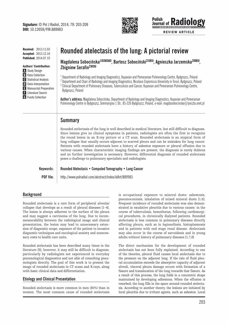
In most cases, rounded atelectasis is asymptomatic [1,8,11] and clinicians are usually surprised by incommensurabil-ity between clinical presentation and radiological lesions. Some patients may complain of chest pains, dyspnoea or cough [1,8]. Symptoms are more common in atelectasis associated with asbestosis is more common, although they are probably due to the underlying disease. Physical exami-nations reveal no abnormalities or minimum abnormali-ties (crackling in the affected region upon deep inhalation). Rounded atelectasis without concomitant pleural lesions shows no changes in functional lungs tests [1,8].Figure 1. Posteroanterior chest radiography: peripheral, well-defined
opacity (round atelectasis).
Figure 2. CT examination of the chest after administration of contrast medium (pulmonary and mediastinal window) in the patient shown in Figure 1. Oval soft tissue density in both lower lobes. Lesions adhere to the pleura, forming acute angles. “Comet tail” sign better visible on the right side (arrow head). The volume of both lower lobes is reduced, both oblique fissures are displaced (arrows). Bilateral pleural effusion.
Figure 3. Rounded atelectasis in the left lower lobe, the volume of the lobe is reduced and the left major fissure is displaced (arrow).
Review Article
204
© Pol J Radiol, 2014; 79: 230-209
Radiological Image
A major part of lesions is detected accidentally in X-rays taken for other reasons [1,2,3,8] (Figure 1). Rounded atelec-tasis is usually an isolated lesions, also cases of multiple lesions are possible (Figure 2). It is typically found in lower lung lobes, particularly in posterior regions. Usually, ate-lectasis has the form of peripheral round, oval or fusiform lesion of diffuse outline. according to the literature, the size of the lesion ranges from 2.5 to 8 cm. Usually, the lesion does not adhere to the diaphragm, being separated by pulmonary parenchyma. Pleural thickening at the level of atelectasis is typical. The volume of the affected lobe is reduced (Figure 3).
In computed tomography scans, rounded atelectasis is visu-alized as a mass with a soft tissue density, usually with air bronchogram in the centre or in the proximal part [1,6–11] (Figure 3). Contrast enhancement is not typical, although
higher density of lesion periphery may sometimes be observed as compared to the lesion centre. The lesion is also characterized in that it is never completely surrounded by pulmonary parenchyma, forming an acute angle with the pleura (Figure 4). Vessels and bronchi entering the mass are compressed and bent – this is the so-called “comet tail” or “crab nippers: symptom characteristic for rounded atelectasis and observed in most cases (Figure 5 and 6B). Pleural thickening is common at the level of the mass, although this is not a pathognomic symptom, particularly in patients with history of asbestos exposure; pleural fluid may also be observed. Typical features of rounded atelecta-sis are summarized in Table 1.
Distant (up to 1 year) follow up usually reveals no change in the nature of the lesion or an increase in its mass; less commonly, partial regression may be observed [1,2,6,10] (Figure 6). Rounded atelectasis associated with asbestosis
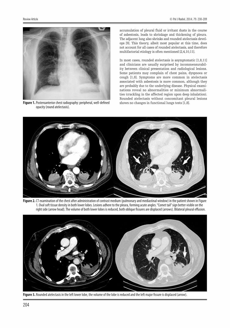
Figure 4. Pleural effusion and rounded atelectasis affecting most of the lower right lobe and adjacent to the distorted and displaced oblique fissure. Air bronchogram in proximal part of RA is also visible.
Figure 5. Sagittal projections. Round atelectasis with “comet tail” signs visible on both sides. Displacement of the oblique fissure on both sides and reduced volume of the lower lobes.
© Pol J Radiol, 2014; 79: 203-209 Sobocińska M. et al. – Rounded atelectasis of the lung: A pictorial review
205
most commonly adheres to pleural thickenings, has len-tiform or cuneiform shape with a “comet tail” sign and leads to a reduction in the volume of the affected lobe [1]. If the lesion is concomitant to pneumoconiosis, pneumo-coniotic nodules may be visible within the mass contour [1]. Enlargement of hilar lymph nodes should always rise suspicion of cancer [1]
Differentiation
Rounded atelectasis requires differentiation from sub-pleural rounded masses. Of highest importance is differen-tiation of rounded atelectasis from lung carcinoma or lung metastasis [1,2,3,6].
Typical features of lung carcinoma that differentiate the disease from rounded carcinoma include poorly defined edges, polycyclic or nodular outline and spicules referred to as corona radiata or radial bands overlapping the pleura (pleural tail) (Figures 7 and 8). Some forms of cancer may be accompanied by the air bronchogram symptom [8]. In larger lesions, ground-glass or air regions may occur in lesion centre as symptoms of central necrosis. The volume of cancer-affected lung is unchanged; the volume may be changed if the neoplastic lesion is accompanied by atelec-tatic lesions associated with bronchial obstruction. If the pathological mass is accompanied by inflammatory lesions (with or without atelectasis), the volume of the lobe may be increased. Cancer lesions are usually accompanied by enlarged lymph nodes within the hila and/or the mediasti-num [12,13]. Differentiation between atelectasis and car-cinoid is facilitated by clinical symptoms associated with hormonal release. In addition, carcinoids differ from round-ed lung lesions by intense enhancement following con-trast administration. A similar radiological image may be
A
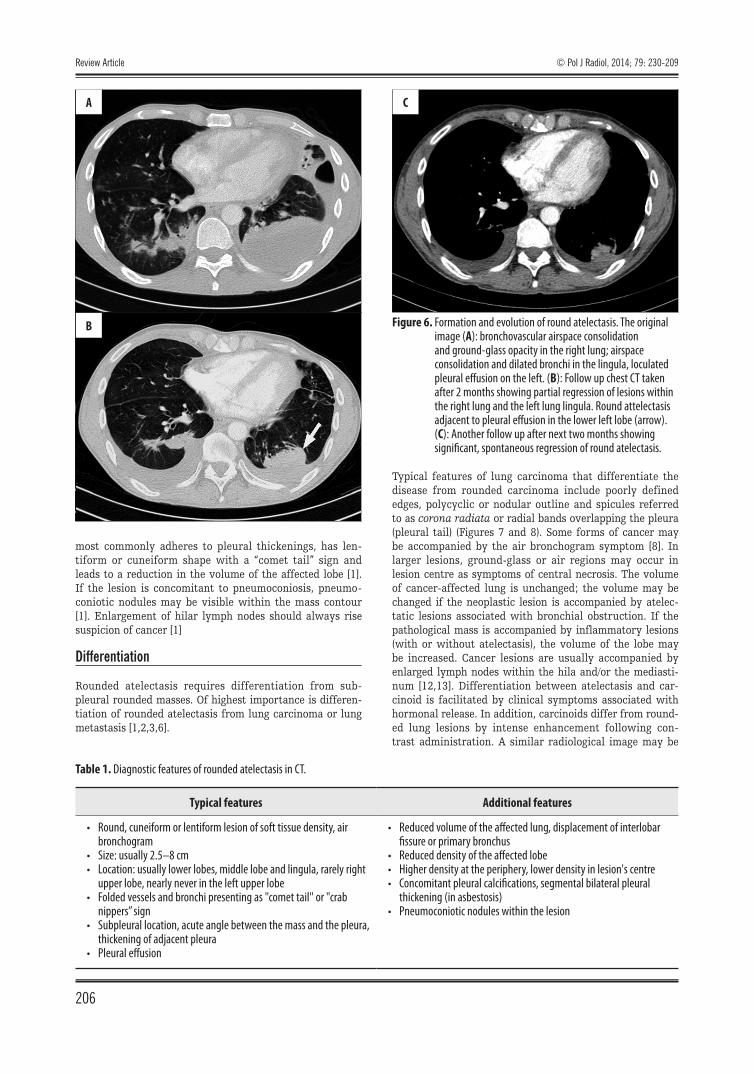
Figure 6. Formation and evolution of round atelectasis. The original image (A): bronchovascular airspace consolidation and ground-glass opacity in the right lung; airspace consolidation and dilated bronchi in the lingula, loculated pleural effusion on the left. (B): Follow up chest CT taken after 2 months showing partial regression of lesions within the right lung and the left lung lingula. Round attelectasis adjacent to pleural effusion in the lower left lobe (arrow). (C): Another follow up after next two months showing significant, spontaneous regression of round atelectasis.
C
B
Typical features Additional features
• Round, cuneiform or lentiform lesion of soft tissue density, air bronchogram
• Size: usually 2.5–8 cm• Location: usually lower lobes, middle lobe and lingula, rarely right
upper lobe, nearly never in the left upper lobe• Folded vessels and bronchi presenting as "comet tail" or "crab
nippers” sign• Subpleural location, acute angle between the mass and the pleura,
thickening of adjacent pleura• Pleural effusion
• Reduced volume of the affected lung, displacement of interlobar fissure or primary bronchus
• Reduced density of the affected lobe• Higher density at the periphery, lower density in lesion's centre• Concomitant pleural calcifications, segmental bilateral pleural
thickening (in asbestosis)• Pneumoconiotic nodules within the lesion
Table 1. Diagnostic features of rounded atelectasis in CT.
Review Article
206
© Pol J Radiol, 2014; 79: 230-209
observed in round pneumonia, Wegener’s granulomatosis, pulmonary abscess as well as in fungal or mycobacterial lung diseases – in such cases, the medical history and clini-cal symptoms are decisive in differentiation [1].
Lung metastases are usually multiple and bilateral; how-ever, isolated lesions may occur in ca. 5% of patients. Metastases are usually smoothly defined and located most-ly in peripheral region of the lungs. A blood vessel enter-ing the lesion – known as the supplying vessel symptom – is visible in 40% of cases. Most common tumors with lung
metastases are melanoma, sarcoma and testicular cancer [12]. In case of a single subpleural lung carcinoma, patients usually present with clinical symptoms of the underly-ing disease and differentiation is made on the basis of investigations.
Calcifications within rounded atelectasis may be present in patients with pneumoconiosis; however, in such cases, multiple nodules with calcifications are present in entire lungs. Basically, calcifications within the rounded lesion suggest its benign nature. However, such calcification must be located centrally or for a ring; in case of multiple calci-fications, they should be uniformly distributed in the lesion – these are the features of granuloma (Figure 9). Nodular calcifications with accompanying adipose tissue are sug-gestive of hamartoma [12,13]. Round lesions in Wegener’s granulomatosis may present with spicules or supply-ing vessels; most commonly, however, these are multiple lesions (Figure 10). In the active phase, these are accom-panied by ground glass symptoms corresponding to alveo-lar bleeding; in chronic disease, these are accompanied by
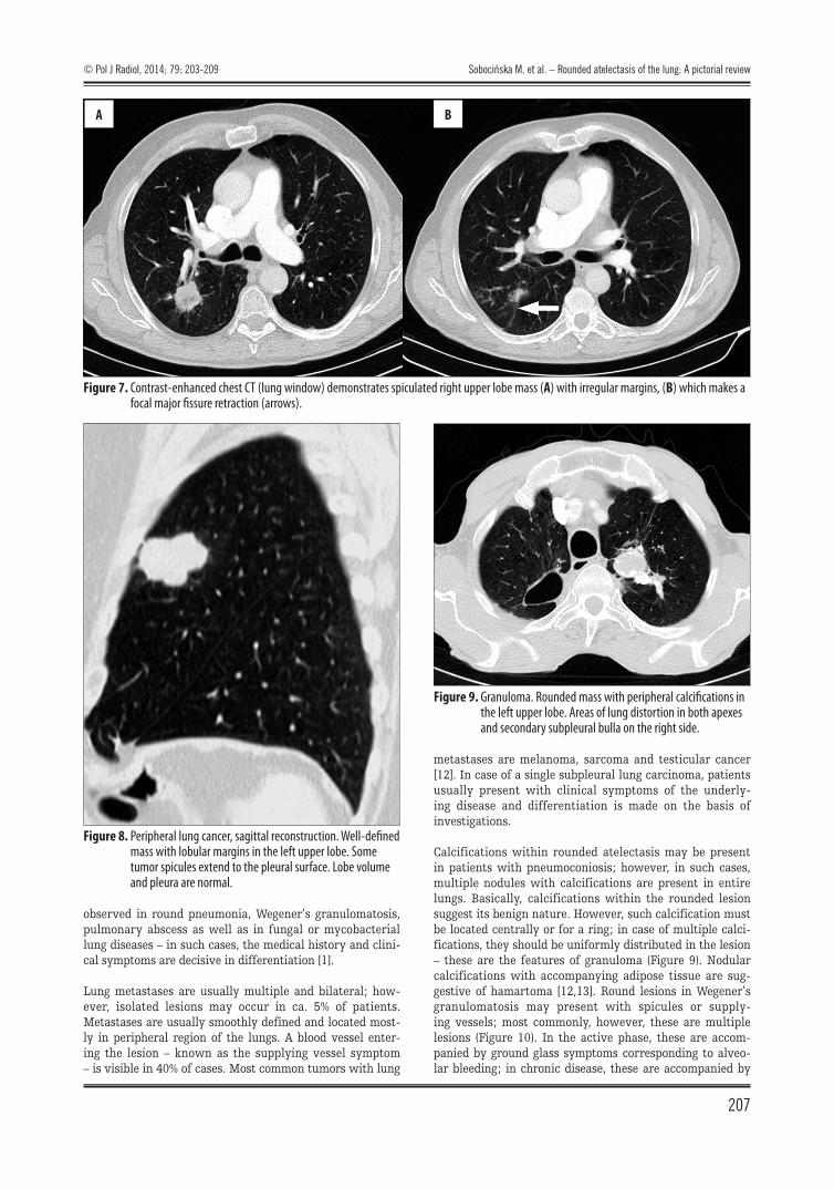
Figure 8. Peripheral lung cancer, sagittal reconstruction. Well-defined mass with lobular margins in the left upper lobe. Some tumor spicules extend to the pleural surface. Lobe volume and pleura are normal.
Figure 9. Granuloma. Rounded mass with peripheral calcifications in the left upper lobe. Areas of lung distortion in both apexes and secondary subpleural bulla on the right side.
Figure 7. Contrast-enhanced chest CT (lung window) demonstrates spiculated right upper lobe mass (A) with irregular margins, (B) which makes a focal major fissure retraction (arrows).
A B
© Pol J Radiol, 2014; 79: 203-209 Sobocińska M. et al. – Rounded atelectasis of the lung: A pictorial review
207
the thickening of interlobar septa [14]; such lesions are not observed in rounded atelectasis.
Pulmonary abscess may be mistaken for rounded atelectasis only in the infiltration phase. A mature abscess is encap-sulated in a wall with the thickness not exceeding 15 mm. Walls having the thickness of up to 4 mm suggest benign lesions; walls having the thickness of 15 mm suggest malig-nant lesions with the intermediate values being non-spe-cific [12]. Rounded atelectasis has also to be differentiated from round pneumonia, which occurs mainly in children and is very rare in adults. Round pneumonia has the form of round, spiculated mass accompanied by pleural thick-ening and satellite lesions. Air bronchogram image may be observed. Clinical symptoms of pneumonia facilitate dif-ferentiation of the disease from rounded atelectasis [15,16]. The lesions resolve following anti-inflammatory treatment.
Organizing pneumonia (OP) should also be taken into account when making the differential diagnosis of rounded atelec-tasis. Nodular densities of different sizes are observed in 30–50% of OP cases; these sometimes occur as isolated find-ings (Figure 11). The lesions may be of peribronchial or sub-pleural location. Air bronchogram image is typical. In 30% of cases, lesions are spiculated; small amounts of pleural fluid or pleural thickening are possible. Differentiation is facili-tated by concomitant presence of parenchymal densities and
ground glass in both lungs. Patients with organizing pneu-monia present with respiratory tract symptoms [14].
Invasive Diagnostics and Treatment
Most experts believe that computed tomography is an ideal tool for making the correct diagnosis and avoiding exploratory thoracotomy or surgical resection of the lesion [1,5,6,11]. It should be kept in mind that cytological confir-mation is not required in case of radiological diagnosis of rounded atelectasis [1].
Therefore, in most cases of typical tomographic picture of rounded atelectasis, transbronchial or transthoracic thin-needle biopsy is inexpedient. Biopsy should only be consid-ered when radiological criteria for diagnosis are not met or when radiological image does not allow for exclusion of malignant growth [1,11]. Negative result of lesion biop-sy is of little value as it does not rule out growth process and requires further, more detailed diagnostics [1,6,10]. Therefore, in cases when rounded atelectasis does not pre-sent with typical picture, surgical videothoracoscopy (VATS) is the method of choice in patients at high risk of cancer. VATS permits the correct diagnosis and is associated with less complications than open lung biopsy. In patients with respiratory or cardiac insufficiency, thoracoscopy is recom-mended as being less invasive than VATS [1,8].
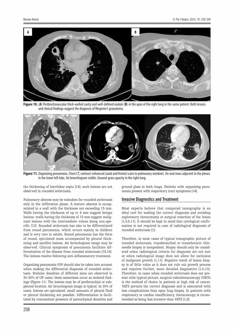
Figure 11. Organizing pneumonia. Chest CT, contrast-enhanced (axial and frontal scans in pulmonary window). An oval mass adjacent to the pleura in the lower left lobe. Air bronchogram visible. Ground-grass opacity in the right lung.
Figure 10. (A) Peribrochovascular thick-walled cavity and well-defined nodule (B) in the apex of the right lung in the same patient. Both lesions and clinical findings suggest the diagnosis of Wegener’s granuloma.
A B
Review Article
208
© Pol J Radiol, 2014; 79: 230-209
No method for the treatment of rounded atelectasis is available [1,10,11]. Surgical intervention should be con-sidered only in cases of deep lung function impairment or when concomitant malignant growth is suspected [1].
Radiological Practice
Rounded atelectasis is not a rare finding in chest CT scans; however, it is often not properly diagnosed by radiologists. In our review of examinations consulted in the Kuyavian and Pomeranian Pulmonology Centre in Bydgoszcz over the period of last 3 years (6 cases), the predominant descrip-tions included: “solid lesion of unknown nature”, “lesion for further diagnostics”, “contrast-enhanced lesion” or “regress-ing inflammatory lesion”.
Each of these radiological reports forces the pneumolo-gist to take up a specific diagnostic route, most typically transthoracic biopsy. In histopathological examination of rounded atelectasis, the biopsy result does not reveal tumor cells; however, the clinician should not rely on such result as there always exist a risk that tumor cells have not been properly collected. This in turn may lead to repeated biopsy or other invasive examinations (e.g. VATS or thoracotomy). Thus, the radiologist contributes to unnecessary extension of diagnostic procedures, increasing costs and potential risks to the patient.
On the other hand, when radiological summary descrip-tion reads: “regressing inflammatory lesions”, there is a discrepancy between negative history, clinical presentation and lack of abnormalities in physical examination and the radiological picture; this requires the clinician to consider
initiation of antibiotic treatment which is not necessary. The treatment does not lead to regression in follow up examination or the regression is in fact spontaneous.
One could suppose that cautious descriptions of rounded atelectasis as e.g. solid lesion for further diagnostics is due on one hand to the realities of radiologists’ work, as the radiologists must usually be specialists in all areas of medicine, and on the other hand, to frequent lack of suffi-cient clinical data. In addition, there is an increasing anxi-ety among physicians regarding possible medical errors. However, in case of typical radiological signs of rounded atelectasis, particularly in computed tomography scans, the summary should always include a suggestion of such diagnosis. This is particularly the case when the lesion is accompanied by pleural fluid, when the patient has pos-itive history of exposure to asbestos or other dusts or in case when patient presents with no clinical symptoms and the lesion is “incidentally” detected in the X-ray image. Clinical diagnosis and further management of patient is at the discretion of the clinician who can base their decisions on the interview and physical examinations as well as lab-oratory investigations and should accordingly make their decisions regarding further diagnostics or discontinuation of diagnostic procedures on the basis of radiological image.
Conclusions
Thanks to its typical radiological features, rounded atelec-tasis may be in most cases diagnosed on the basis of com-puted tomography scan without the need of a biopsy. CT differentiation of rounded atelectasis should include in par-ticular peripheral lung cancer, metastases, granulomas and organizing pneumonia.
1. Stathopoulos GT, Karamessini MT, Sotiriadi AE, Pastromas VG: Rounded atelectasis of the lung. Respir Med, 2005; 99(5): 615–23
2. Batra P, Brown K, Hayashi K et al: Rounded atelectasis. J Thorac Imaging, 1996; 11(3): 187–97
3. Partap VA: The comet tail sign. Radiology, 1999; 213: 553–54 4. Woodring JH, Reed JC: Types and mechanisms of pulmonary
atelectasis. J Thorac Imaging, 1996; 11(2): 92–108 5.ProkopM,GalanskiM:Spiralnaiwielorzędowatomografia
komputerowaczłowieka.2nd edition (Polish). Medipage, Warsaw, 2010; 320 [in Polish]
6. Doyle TC, Lawler GA: CT Features of rounded atelectasis of the lung. Am J Roentgenol, 1984; 143(2): 225–28
7. Thachil RT, Krishnan P, Lapidus C: Rounded atelectasis. Clin Pulmon Med, 2002; 9(1): 66–67
8. Tetikkurt C, Tetikkurt S, Ozdemir I, Bayar N: Round atelectasis in sarcoidosis. Multidiscip Respir Med, 2011; 6(3): 180–82
9. Woodring JH: Pleural effusion is a cause of round atelectasis of the lung. J Ky Med Assoc, 2000; 98(12): 527–32
10. Szydlowski GW, Cohn HE, Steiner RM, Edie RN: Rounded atelectasis: a pulmonary pseudotumor. Ann Thorac Surg, 1992; 53(5): 817–21
References:
11. Younis WG, Dernaika TA, Jones KR et al: Round atelectasis with atypical computed tomography findings. Respir Care, 2006; 51(7): 761–63
12.KowalewskiJ,DancewiczM:Pojedynczycieńokrągływpłucu–ciągleaktualnyproblemdiagnostyczny.PneumonolAlergolPol,2005;73(2): 198–201 [in Polish]
13.KuziemskiK:Cieńkrągłypojedynczywpłucu.Podejściepraktycznew podstawowej opiece zdrowotnej. Forum Medycyny Rodzinnej, 2011; 5(3): 217–21 [in Polish]
14. Bestry I, Oniszh K, Pawlicka L, Dobkowski P: Rola metod obrazowychwdiagnostycechoróbśródmiąższowychchoróbpłuc.In:WiatrE,Rowińska-ZakrzewskaE,PirożyńskiM(eds.):Chorobyśródmiąższowepłuc.AlfaMedicaPress,Warsaw,2012;62–89[inPolish]
15. Wagner AL, Szabunio M, Hazlett KS, Wagner SG: Radiologic manifestations of round pneumonia in adults. Am J Roentgenol, 1998; 170(3): 723–26
16.HalkiewiczF,KozielskiJ,Witkowska-KlinkeJetal:Pojedynczycieńkrągływmiąższupłucnym:rakpłucaczyokrągłezapaleniepłuc?–problemdiagnostyczny,różnicowanie.KardiochirTorakochiPol,2012; 9(4): 477–80 [in Polish]
© Pol J Radiol, 2014; 79: 203-209 Sobocińska M. et al. – Rounded atelectasis of the lung: A pictorial review
209