Peng et al
4
Scientific Research and Essays Vol. 6(14), pp. 2976-2979, 18 July, 2011 Available online at http://www.academicjournals.org/SRE DOI: 10.5897/SRE11.079 ISSN 1992-2248 ©2011 Academic Journals Full Length Research Paper Endometriosis induced acute intestinal obstruction: A case report and literature review Baoan Peng*, Jianhua Qian, Xiaoyun Wan and Chang Su Women's Hospital, School of Medicine, Zhejiang University, Hangzhou 31006, China. Accepted 6 April, 2011 Intestinal obstruction caused by the invasion of endometriosis on intestinal walls is rarely reported, and it is difficult to make timely and accurate diagnosis in clinical practice. Herein, we report a rare case of acute intestinal obstruction caused by endometriosis and make a review on related literature. Digestive tract endometriosis should be considered if the patient had a long history of constipation, abdominal distension or incomplete intestinal obstruction, especially when these symptoms were closely related with the menstrual cycle. Key words: Endometriosis, invasion, intestinal obstruction. INTRODUCTION Endometriosis (EMT) is a gynecological medical condition in which endometrial-like cells appear and flourish in areas outside the uterine cavity, most commonly on the ovaries. Ectopic endometrium may invade any part of the body, frequently in ovaries, uterosacral ligament, etc. However, intestinal obstruction caused by the invasion of endometriosis on intestinal walls is rare in clinical practice, and it is easily misdiagnosed or missed diagnosed. Now, we report a rare case of acute intestinal obstruction due to endometriosis. CLINICAL DATA A 22-year-old married female, G0P0, was hospitalized because of hypogastralgia for 1 month, abdominal distension, no defecation, and fever for 3 days. The menstrual cycle of the patient used to be regular, and she had an 8-year history of dysmenorrhea. One year before hospitalization, B-type ultrasonography showed right sactosalpinx and left ovarian cystic mass (3.1 × 2.6 cm in size), but she did not receive any treatment. Four months earlier, she received artificial insemination because of infertility for 2 years. One month ago, she w as *Corresponding author. E-mail: [email protected] hospitalized again because of hypogastralgia, and B-type ultrasonography showed a 7.8 × 6.3 × 6.4 cm cystic mass in the right attachment area and a 5.9 × 5.6 × 5.8 cm cystic mass in the left ovary, and there were many septations within cystic masses and blood flow in septations, with a resistance index of 0.54. The primary diagnosis was bilateral ovarian endometriosis cyst accompanied with infection and bilateral hydrosalpinx. After two weeks of anti-infection treatment, abdominal pain obviously alleviated, and B-type ultrasonography revealed that the cystic masses were slightly smaller. Then, the patient discharged and prepared to receive tylectomy at the end of the next menstrual cycle. Three days ago, the patient suffered from lower abdominal distension and the symptoms gradually aggravated, and subsequently there was nausea and vomiting with gastric contents for several times, followed by no defecation, less and less fart and lower fever near 38.0 . Urine acetone bodies were “+++”. Plain abdominal radiography revealed that the ascending colon and splenic flexure of the colon were significantly expanded, so she was hospitalized on October 12, 2009 with the initial diagnosis of incomplete intestinal obstruction. Gynecologic examination showed anteversion of uterus; the size was normal, without tenderness and less activity; there was a cystic mass 6 ~ 8 cm in diameter in left and right attachment area, with high tension and less activity and obvious tenderness. B-type ultrasonography showed that: there was unclear boundary between uterus
-
Upload
lediayukime -
Category
Documents
-
view
221 -
download
0
Transcript of Peng et al
8/12/2019 Peng et al
http://slidepdf.com/reader/full/peng-et-al 1/4
Scientific Research and Essays Vol. 6(14), pp. 2976-2979, 18 July, 2011Available online at http://www.academicjournals.org/SRE DOI: 10.5897/SRE11.079ISSN 1992-2248 ©2011 Academic Journals
Full Length Research Paper
Endometriosis induced acute intestinal obstruction: Acase report and literature review
Baoan Peng*, Jianhua Qian, Xiaoyun Wan and Chang Su
Women's Hospital, School of Medicine, Zhejiang University, Hangzhou 31006, China.
Accepted 6 April, 2011
Intestinal obstruction caused by the invasion of endometriosis on intestinal walls is rarely reported, andit is difficult to make timely and accurate diagnosis in clinical practice. Herein, we report a rare case ofacute intestinal obstruction caused by endometriosis and make a review on related literature. Digestive
tract endometriosis should be considered if the patient had a long history of constipation, abdominaldistension or incomplete intestinal obstruction, especially when these symptoms were closely relatedwith the menstrual cycle.
Key words: Endometriosis, invasion, intestinal obstruction.
INTRODUCTION
Endometriosis (EMT) is a gynecological medicalcondition in which endometrial-like cells appear andflourish in areas outside the uterine cavity, mostcommonly on the ovaries. Ectopic endometrium may
invade any part of the body, frequently in ovaries,uterosacral ligament, etc. However, intestinal obstructioncaused by the invasion of endometriosis on intestinalwalls is rare in clinical practice, and it is easilymisdiagnosed or missed diagnosed. Now, we report arare case of acute intestinal obstruction due toendometriosis.
CLINICAL DATA
A 22-year-old married female, G0P0, was hospitalizedbecause of hypogastralgia for 1 month, abdominaldistension, no defecation, and fever for 3 days. Themenstrual cycle of the patient used to be regular, and shehad an 8-year history of dysmenorrhea. One year beforehospitalization, B-type ultrasonography showed rightsactosalpinx and left ovarian cystic mass (3.1 × 2.6 cm insize), but she did not receive any treatment. Four monthsearlier, she received artificial insemination because ofinfertility for 2 years. One month ago, she was
*Corresponding author. E-mail: [email protected]
hospitalized again because of hypogastralgia, and B-typeultrasonography showed a 7.8 × 6.3 × 6.4 cm cystic massin the right attachment area and a 5.9 × 5.6 × 5.8 cmcystic mass in the left ovary, and there were many
septations within cystic masses and blood flow inseptations, with a resistance index of 0.54. The primarydiagnosis was bilateral ovarian endometriosis cysaccompanied with infection and bilateral hydrosalpinxAfter two weeks of anti-infection treatment, abdominapain obviously alleviated, and B-type ultrasonographyrevealed that the cystic masses were slightly smallerThen, the patient discharged and prepared to receivetylectomy at the end of the next menstrual cycle. Threedays ago, the patient suffered from lower abdominadistension and the symptoms gradually aggravated, andsubsequently there was nausea and vomiting with gastriccontents for several times, followed by no defecationless and less fart and lower fever near 38.0 . Urineacetone bodies were “+++”. Plain abdominal radiographyrevealed that the ascending colon and splenic flexure ofthe colon were significantly expanded, so she washospitalized on October 12, 2009 with the initial diagnosisof incomplete intestinal obstruction.
Gynecologic examination showed anteversion outerus; the size was normal, without tenderness and lessactivity; there was a cystic mass 6 ~ 8 cm in diameter inleft and right attachment area, with high tension and lessactivity and obvious tenderness. B-type ultrasonographyshowed that: there was unclear boundary between uterus
8/12/2019 Peng et al
http://slidepdf.com/reader/full/peng-et-al 2/4
Peng et al. 2977
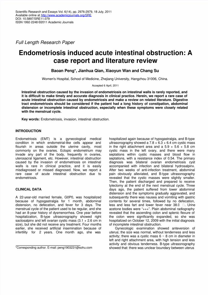
Figure 1. Intraopeative findings showed that the proximal end of the junction of sigmoid colon and rectum was
significantly enlarged, and the intestine was thin-walled and high-tensioned, and closely adhered to neighboringtissues.
and neighboring tissues; the size of right ovary wasnormal; a 8.0 × 5.7 × 6.7 cm sausage-shaped thick-walled cystic mass was found near the right ovary, and a4.5 × 4.3 × 3.1 cm cystic mass was found in the leftovary, presenting flocculent echo, and a 3.3 × 3.3 × 1.7cm sausage-shaped thick-walled cystic mass was foundnear the left ovary. Preoperative diagnosis wasendometriosis of bilateral ovaries and adhesive intestinalobstruction secondary to pelvic inflammatory disease.
The patient received anti-infection treatment withcefoperazone sodium, sulbactam sodium and ornidazolefor 2 days, but symptoms above-mentioned were notimproved. Abdominal distention aggravated, and bowel
sounds were decreased. The patient underwentexploratory laparotomy on October 14, 2009, and it wasfound that there was about 200 ml yellow inflammatoryexudate in the abdominal and pelvic cavities, and theproximal end of junction of sigmoid colon and rectum wassignificantly enlarged (Figure 1), and the mass was 10cm in diameter, thin-walled and high-tensioned, and theintestinal wall was congestive and edematous, closelyadhered to anterior and posterior walls of uterus, leftattachment, left pelvic wall and gastrocolic omentum.Uterus rectum lacunae were completely closed. The size
of left ovary was normal, and left fallopian tube wasenlarged to 3 cm in diameter due to inflammation andclosely adhered to neighboring tissues. The rightattachment was enlarged to 10 cm in diameter due to thecystis, and it also closely adhered to neighboring tissuesand there were some septations in the cystic massThere was thick liquid in one cavity and brown thin liquidin another cavity. Frozen section examination of the rightattachment revealed right chronic salpingitis, righhydrosalpinx, and endometriosis of serosal muscleAdhesions were isolated to restore normal anatomicstructure, but the sigmoid colon was still inflated. Thecolorectal junction was narrowed and intestinal walls
were rigid, and thus it was recognized as the site ofincomplete intestinal obstruction, but it could not beexcluded that obstruction caused by tumors. Narrowedrectum 3 cm and sigmoid colon 12 cm near colorectaobstruction ring were removed, and sigmoid colostomy inthe left lower abdominal wall was performed.
Gross observation of specimen showed that: thelength of colorectal obstruction segment was 1 cm, andintestinal wall was fibrous ring-like thickened, with smoothmucous membrane, and only little fingertip could passthrough the lumen of colorectal obstruction ring. Frozen
8/12/2019 Peng et al
http://slidepdf.com/reader/full/peng-et-al 3/4
2978 Sci. Res. Essays
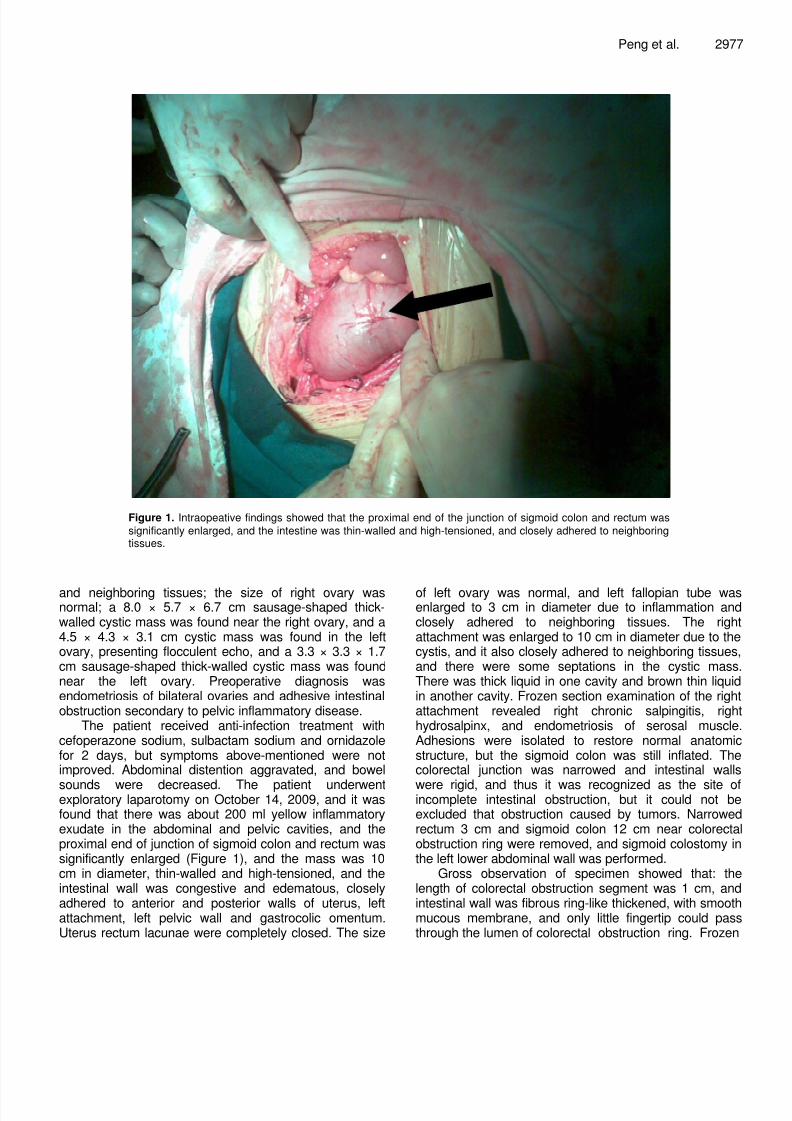
Figure 2. Postoperative pathological findings. There was endometrial gland and interstitial substance
accompanied with hemorrhage in the smooth muscle layer of rectum, and no intestinal mucosa was invaded(H&E×100).
section examination showed rectosigmoid endometriosis,which was consistent with the postoperative pathological
diagnosis (Figure 2). Postoperative diagnoses werepelvic endometriosis (IV period), incomplete intestinalobstruction and acute exacerbation of chronic pelvicinflammation. Good postoperative recovery was obtainedin this patient, and intestinal anastomosis was performed3 months later. Defecation function of the patientrecovered well. Treatment for endometriosis withgonadotropin-releasing hormone agonist (GnRH-a) wasperformed for 6 months.
DISCUSSION
EMT is a common disease of reproductive women, butthe pathological factors are still now unknown.Uterosacral ligament, rectouterine pouch and lowerposterior wall of uterus locate at the lower part of pelviccavity, and endometrial debris in endometrial debrisusually pollutes theses places, and thus these sites arethe predilection sites of EMT. Digestive tract EMTaccounts for 5% of all, and rectum and colon are thepredilection sites of digestive tract EMT, accounting for70%. But, small intestine EMT almost is confined to theterminal ileum (1 - 7%) and that in the proximal ileum and
jejunum is rarely reported, but no EMT in stomach has
been reported. The exact incidence of intestinaobstruction caused by digestive tract EMT is unknown
and complete intestinal obstruction is less than 1%. Distaileal obstruction caused by EMT is rare and only accountsfor 7 - 23% of EMT intestinal obstruction. Due to intestinaobstruction, about 0.7% patients with endometriosis inabdominal and pelvic cavities receive enterectomy (Riazand Khurshaidi, 2007; Sun et al., 2006). Jubanyik et al(1997) reviewed about 1000 patients who once receivedgynecological operations, and there were 181(18%digestive tract EMT, but only one patient had smalintestine lesion. It is also reported that pelvic cavityparacolic lymph nodes and para-aortic lymph nodes canbe invaded by EMT. Lymph node EMT usually co-existedwith EMT of intestinal walls, and lymph node EMT mayresult from the lymphatic metastasis of EMT of intestinawalls (Cameron et al., 1995).
Because digestive tract EMT usually has no typicasymptoms of gynecological EMT, it is difficult to maketimely and accurate diagnosis of EMT. The symptomsinclude recurrent abdominal pain, abdominal distension,tenesmus, constipation, diarrhea, hemafecia anddefecation pain, but these symptoms are not EMTspecific. Digestive tract EMT should be identified withirritable bowel syndrome, infectious diarrhea, ischemicbowel disease, inflammatory bowel disease and tumorPatient history still plays important roles in diagnosis, and
8/12/2019 Peng et al
http://slidepdf.com/reader/full/peng-et-al 4/4
the symptoms of some patients may be closelyassociated with menstrual cycle (Scarmato et al., 2000).50% of digestive tract EMT usually is accompanied withpelvic EMT, and small intestine EMT should beconsidered in young unfertile women with abdominal painand intestinal obstruction. Mussa et al. (2001) once
reported a case of small intestine EMT accompanied withintestinal obstruction who mainly presentedhypoproteinemia and anasarca. Although colonoscopycan diagnose colon EMT, it is observed by colonoscopythat colonic mucosa usually is normal or mildly chronicinflammatory, and EMT lesions and fibrous stenosismainly locate in placenta percreta and muscular layer ofintestinal canal. There is no history of rectal bleeding inthis patient, and repeated stool examinations are normal,and digital rectal examination shows that intestinalmucosa is smooth. Rectal bleeding may result fromenlarged EMT lesions in intestinal walls and more narrowof intestinal canal during menstruation, and stool injurescongestive mucosa when stool passes through narrowintestinal canal. Interestingly, colon mucosa rapidlyrestores normal after menstruation, and no abnormalitiesare found under coloscope. EMT generally only invadesdeep layers of intestinal walls, and there are no specialchanges in intestinal mucosa. However, the pathologicalexaminations by colonoscopy are usually negativebecause the specimen tissues are not enough deep.Double contrast barium enema shows mass due toexternal pressure and serrated plicas. Although CT canclearly detect colorectal tumors, CT is not the first choiceof intestinal tract EMT (Takeuchi et al., 2005). Magneticresonance image (MRI) has a high sensitivity (77 - 93%)in the diagnosis of intestinal canal EMT (Takeuchi et al.,
2005), but it is difficult to determine the invasion depth ofrectum wall EMT. Recently, the combination of MRI andendoscopic ultrasound can increase the accuracy rate ofdiagnosis. Serum markers have poor accuracy in thediagnosis of digestive tract EMT, but tumor marker CA-125 can indicate the progress of EMT. CA19-9 has lowersensitivity than CA-125, but cytokine interleukin-6 hashigher sensitivity and specificity than CA-125 (Mounseyet al., 2006).
Most EMT patients with intestinal obstruction mustreceive surgical treatments, but occasional EMT patientswithout intestinal obstruction can take hormones such asdanazol and GnRH-a, and they must receive surgical
treatments if they suffered from the change of bowelhabits, abdominal pain, hemafecia, or even intestinalobstruction. If the lesion is located in small intestine, drugtreatment is only temporary, and EMT segments of smallintestine must be resected to obtain good prognosis.Intestinal EMT is still active after menostasis, so intestinalEMT should also be considered if these patientsexperienced intestinal obstruction and can not beneglected (De Ceglie et al., 2008).. After operation, thispatient received anti-endometriosis treatment withgonadotropin-releasing hormone agonist (GnRH-a) for 6
Peng et al. 2979
months, and intestinal anastomosis was performed 3months later, good bowel function was obtained, and upto now (1 year) no evidence of EMT recurrence wasfound.
This paper reports a rare case of digestive tract EMTwith sudden intestinal obstruction. EMT lesion in the
muscular layer of colon resulted in fibrous ringconstriction and oppressed intestinal canal, and then thepatient presented the symptoms of intestinal obstructionPreoperative auxiliary examinations and intraoperativefrozen pathological section are conducive to finadiagnosis, which can effectively decrease surgical areaand corresponding adverse complications. In clinicapractice, it is difficult to diagnose digestive tract EMTwhose symptoms are independent with menstrual cycleFor patients with menorrhalgia and chocolate cyst ofovary, digestive tract EMT should be considered if thereis a hard mass around rectum and colon and incompleteintestinal obstruction. These patients should receivesurgical treatment as early as possible, and EMT tissuesinvading intestinal canal should be removed. Afteoperation, the patients must receive anti-EMT treatmentsto decrease the recurrence rate. The long history ofconstipation in this patient indicates that digestive tractEMT should be considered if patients have a long historyof constipation, abdominal distension and incompleteintestinal obstruction, especially when these symptomsare closely related with the menstrual cycle.
REFERENCES
Cameron IC, Rogers S, Collins MC, Reed MW (1995). Intestinaendometriosis: presentation, investigation, and surgical managementInt. J. Colorectal. Dis., 10: 83-86.
De Ceglie A, Bilardi C, Blanchi S, Picasso M, Di Muzio M, Trimarchi AConio M (2008). Acute small bowel obstruction caused byendometriosis: A case report and review of the literature. World. JGastroenterol., 14: 3430-3434.
Jubanyik KJ, Comite F (1997). Extrapelvic endometriosis. ObstetGynecol. Clin. North. Am., 24: 411-440.
Mounsey AL, Wilgus A, Slawson DC (2006). Diagnosis andmanagement of endometriosis. Am. Fam. Phys., 74: 594-600.
Mussa FF, Younes Z, Tihan T, Lacy BE (2001). Anasarca and smalbowel obstruction secondary to endometriosis. J. Clin. Gastroenterol.32: 167-171.
Riaz N, Khurshaidi N (2007). Acute small bowel obstruction secondaryto ileal endometrioma. J. Coll. Physicians. Surg. Pak., 17: 228-229.
Scarmato VJ, Levine MS, Herlinger H, Wickstrom M, Furth EE, TureckRW (2000). Ileal endometriosis: radiographic findings in five cases
Radiology, 214: 509-512.Sun Mei-Ling, Li Yu, Zhang Yin-Min (2006). Clinical analysis of 12cases of endometriosis of rectum . Matern. Child. Health. Care. Chin.12: 2233-2234.
Takeuchi H, Kuwatsuru R, Kitade M, Sakurai A, Kikuchi I, Shimanuki HKinoshita K (2005). A novel technique using magnetic resonanceimaging jelly for evaluation of rectovaginal endometriosis. FertilSteril., 83: 442-447.


![[B4] PENG Yan](https://static.fdocuments.pl/doc/165x107/577ce47b1a28abf1038e744b/b4-peng-yan.jpg)