Nanoparticles as drug delivery systems - Polish Academy...
18
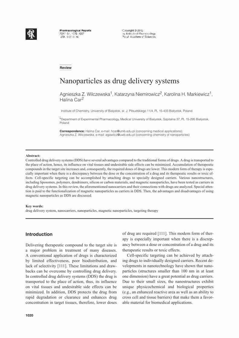
Review Nanoparticles as drug delivery systems Agnieszka Z. Wilczewska 1 , Katarzyna Niemirowicz 2 , Karolina H. Markiewicz 1 , Halina Car 2 Institute of Chemistry, University of Bialystok, al. J. Pilsudskiego 11/4, PL 15-433 Bia³ystok, Poland Department of Experimental Pharmacology, Medical University of Bialystok, Szpitalna 37, PL 15-295 Bia³ystok, Poland Correspondence: Halina Car, e-mail: [email protected] (concerning medical applications); Agnieszka Z. Wilczewska, e-mail: [email protected] (concerning chemistry of nanoparticles) Abstract: Controlled drug delivery systems (DDS) have several advantages compared to the traditional forms of drugs. A drug is transported to the place of action, hence, its influence on vital tissues and undesirable side effects can be minimized. Accumulation of therapeutic compounds in the target site increases and, consequently, the required doses of drugs are lower. This modern form of therapy is espe- cially important when there is a discrepancy between the dose or the concentration of a drug and its therapeutic results or toxic ef- fects. Cell-specific targeting can be accomplished by attaching drugs to specially designed carriers. Various nanostructures, including liposomes, polymers, dendrimers, silicon or carbon materials, and magnetic nanoparticles, have been tested as carriers in drug delivery systems. In this review, the aforementioned nanocarriers and their connections with drugs are analyzed. Special atten- tion is paid to the functionalization of magnetic nanoparticles as carriers in DDS. Then, the advantages and disadvantages of using magnetic nanoparticles as DDS are discussed. Key words: drug delivery system, nanocarriers, nanoparticles, magnetic nanoparticles, targeting therapy Introduction Delivering therapeutic compound to the target site is a major problem in treatment of many diseases. A conventional application of drugs is characterized by limited effectiveness, poor biodistribution, and lack of selectivity [111]. These limitations and draw- backs can be overcome by controlling drug delivery. In controlled drug delivery systems (DDS) the drug is transported to the place of action, thus, its influence on vital tissues and undesirable side effects can be minimized. In addition, DDS protects the drug from rapid degradation or clearance and enhances drug concentration in target tissues, therefore, lower doses of drug are required [111]. This modern form of ther- apy is especially important when there is a discrep- ancy between a dose or concentration of a drug and its therapeutic results or toxic effects. Cell-specific targeting can be achieved by attach- ing drugs to individually designed carriers. Recent de- velopments in nanotechnology have shown that nano- particles (structures smaller than 100 nm in at least one dimension) have a great potential as drug carriers. Due to their small sizes, the nanostructures exhibit unique physicochemical and biological properties (e.g., an enhanced reactive area as well as an ability to cross cell and tissue barriers) that make them a favor- able material for biomedical applications. 1020
Transcript of Nanoparticles as drug delivery systems - Polish Academy...
Review
Nanoparticles as drug delivery systems
Agnieszka Z. Wilczewska1, Katarzyna Niemirowicz2, Karolina H. Markiewicz1,
Halina Car2
1Institute of Chemistry, University of Bialystok, al. J. Pilsudskiego 11/4, PL 15-433 Bia³ystok, Poland
2Department of Experimental Pharmacology, Medical University of Bialystok, Szpitalna 37, PL 15-295 Bia³ystok,
Poland
Correspondence: Halina Car, e-mail: [email protected] (concerning medical applications);
Agnieszka Z. Wilczewska, e-mail: [email protected] (concerning chemistry of nanoparticles)
Abstract:
Controlled drug delivery systems (DDS) have several advantages compared to the traditional forms of drugs. A drug is transported tothe place of action, hence, its influence on vital tissues and undesirable side effects can be minimized. Accumulation of therapeuticcompounds in the target site increases and, consequently, the required doses of drugs are lower. This modern form of therapy is espe-cially important when there is a discrepancy between the dose or the concentration of a drug and its therapeutic results or toxic ef-fects. Cell-specific targeting can be accomplished by attaching drugs to specially designed carriers. Various nanostructures,including liposomes, polymers, dendrimers, silicon or carbon materials, and magnetic nanoparticles, have been tested as carriers indrug delivery systems. In this review, the aforementioned nanocarriers and their connections with drugs are analyzed. Special atten-tion is paid to the functionalization of magnetic nanoparticles as carriers in DDS. Then, the advantages and disadvantages of usingmagnetic nanoparticles as DDS are discussed.
Key words:
drug delivery system, nanocarriers, nanoparticles, magnetic nanoparticles, targeting therapy
Introduction
Delivering therapeutic compound to the target site isa major problem in treatment of many diseases.A conventional application of drugs is characterizedby limited effectiveness, poor biodistribution, andlack of selectivity [111]. These limitations and draw-backs can be overcome by controlling drug delivery.In controlled drug delivery systems (DDS) the drug istransported to the place of action, thus, its influenceon vital tissues and undesirable side effects can beminimized. In addition, DDS protects the drug fromrapid degradation or clearance and enhances drugconcentration in target tissues, therefore, lower doses
of drug are required [111]. This modern form of ther-
apy is especially important when there is a discrep-
ancy between a dose or concentration of a drug and its
therapeutic results or toxic effects.Cell-specific targeting can be achieved by attach-
ing drugs to individually designed carriers. Recent de-velopments in nanotechnology have shown that nano-particles (structures smaller than 100 nm in at leastone dimension) have a great potential as drug carriers.Due to their small sizes, the nanostructures exhibitunique physicochemical and biological properties(e.g., an enhanced reactive area as well as an ability tocross cell and tissue barriers) that make them a favor-able material for biomedical applications.
1020 Pharmacological Reports, 2012, 64, 1020�1037
Pharmacological Reports2012, 64, 1020�1037ISSN 1734-1140
Copyright © 2012by Institute of PharmacologyPolish Academy of Sciences
Nanocarriers used in drug delivery system
According to the definition from NNI (National
Nanotechnology Initiative), nanoparticles are struc-
tures of sizes ranging from 1 to 100 nm in at least one
dimension. However, the prefix “nano” is commonly
used for particles that are up to several hundred
nanometers in size.Nanocarriers with optimized physicochemical and
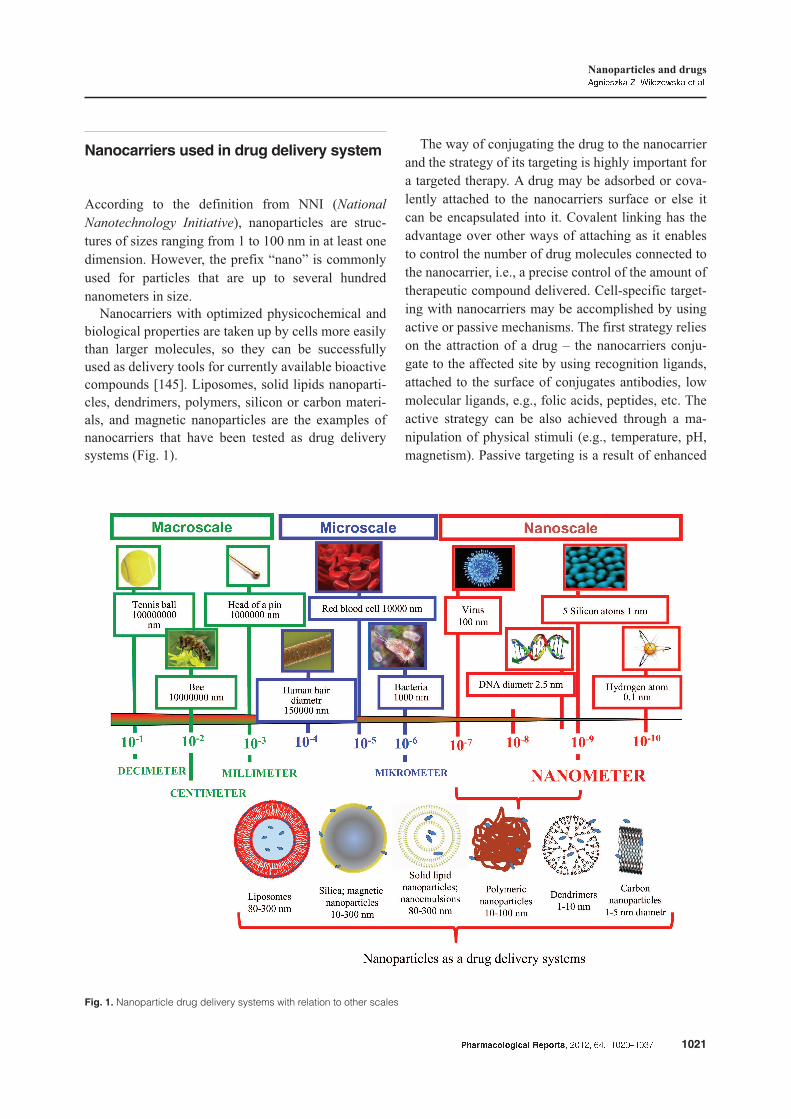
biological properties are taken up by cells more easilythan larger molecules, so they can be successfullyused as delivery tools for currently available bioactivecompounds [145]. Liposomes, solid lipids nanoparti-cles, dendrimers, polymers, silicon or carbon materi-als, and magnetic nanoparticles are the examples ofnanocarriers that have been tested as drug deliverysystems (Fig. 1).
The way of conjugating the drug to the nanocarrierand the strategy of its targeting is highly important fora targeted therapy. A drug may be adsorbed or cova-lently attached to the nanocarriers surface or else itcan be encapsulated into it. Covalent linking has theadvantage over other ways of attaching as it enablesto control the number of drug molecules connected tothe nanocarrier, i.e., a precise control of the amount oftherapeutic compound delivered. Cell-specific target-ing with nanocarriers may be accomplished by usingactive or passive mechanisms. The first strategy relieson the attraction of a drug – the nanocarriers conju-gate to the affected site by using recognition ligands,attached to the surface of conjugates antibodies, lowmolecular ligands, e.g., folic acids, peptides, etc. Theactive strategy can be also achieved through a ma-nipulation of physical stimuli (e.g., temperature, pH,magnetism). Passive targeting is a result of enhanced
Pharmacological Reports, 2012, 64, 1020�1037 1021
Nanoparticles and drugsAgnieszka Z. Wilczewska et al.
Fig. 1. Nanoparticle drug delivery systems with relation to other scales
vascular permeability and retention (EPR) which ischaracteristic of leaky tissues of tumors [111].
Once the drug-nanocarrier conjugates reach thediseased tissues, the therapeutic agents are released.A controlled release of drugs from nanocarriers canbe achieved through changes in physiological envi-ronment such as temperature, pH, osmolality, or via
an enzymatic activity.Nanocarriers used for medical applications have to
be biocompatible (able to integrate with a biologicalsystem without eliciting immune response or anynegative effects) and nontoxic (harmless to a givenbiological system). Undesirable effects of nanoparti-cles strongly depend on their hydrodynamic size,shape, amount, surface chemistry, the route of admini-stration, reaction of the immune system (especiallya route of the uptake by macrophages and granulo-cytes) and residence time in the bloodstream. Due toa number of factors which may affect the toxicity ofnanoparticles, their estimation is rather difficult and,thus, toxicological studies of each new DDS formula-tion are needed. However, with respect to their size,one can make some generalizations – smaller particleshave a greater surface area, thus, they are more reac-tive and, in consequence, more toxic [5]. It is gener-ally accepted that nanoparticles with a hydrodynamicdiameter of 10–100 nm have optimal pharmacokineticproperties for in vivo applications. Smaller nanoparti-cles are subjects to tissue extravasations and renalclearance whereas larger are quickly opsonized andremoved from the bloodstream via the macrophagesof the reticuloendothelial system [35].
Liposomes
Liposomes have been the first to be investigated asdrug carriers. They are nano/micro-particular or col-loidal carriers, usually with 80–300 nm size range[144]. They are spherical vesicles composed of phos-pholipids and steroids (e.g., cholesterol), bilayers, orother surfactants and form spontaneously when cer-tain lipids are dispersed in aqueous media where lipo-somes can be prepared, e.g., by sonication [139].Liposomes have been reported to increase the solubil-ity of drugs and improve their pharmacokinetic prop-erties, such as the therapeutic index of chemothera-peutic agents, rapid metabolism, reduction of harmfulside effects and increase of in vitro and in vivo anti-
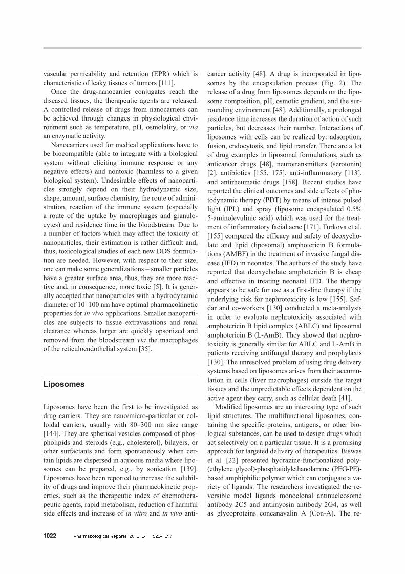
cancer activity [48]. A drug is incorporated in lipo-somes by the encapsulation process (Fig. 2). Therelease of a drug from liposomes depends on the lipo-some composition, pH, osmotic gradient, and the sur-rounding environment [48]. Additionally, a prolongedresidence time increases the duration of action of suchparticles, but decreases their number. Interactions ofliposomes with cells can be realized by: adsorption,fusion, endocytosis, and lipid transfer. There are a lotof drug examples in liposomal formulations, such asanticancer drugs [48], neurotransmitters (serotonin)[2], antibiotics [155, 175], anti-inflammatory [113],and antirheumatic drugs [158]. Recent studies havereported the clinical outcomes and side effects of pho-todynamic therapy (PDT) by means of intense pulsedlight (IPL) and spray (liposome encapsulated 0.5%5-aminolevulinic acid) which was used for the treat-ment of inflammatory facial acne [171]. Turkova et al.[155] compared the efficacy and safety of deoxycho-late and lipid (liposomal) amphotericin B formula-tions (AMBF) in the treatment of invasive fungal dis-ease (IFD) in neonates. The authors of the study havereported that deoxycholate amphotericin B is cheapand effective in treating neonatal IFD. The therapyappears to be safe for use as a first-line therapy if theunderlying risk for nephrotoxicity is low [155]. Saf-dar and co-workers [130] conducted a meta-analysisin order to evaluate nephrotoxicity associated withamphotericin B lipid complex (ABLC) and liposomalamphotericin B (L-AmB). They showed that nephro-toxicity is generally similar for ABLC and L-AmB inpatients receiving antifungal therapy and prophylaxis[130]. The unresolved problem of using drug deliverysystems based on liposomes arises from their accumu-lation in cells (liver macrophages) outside the targettissues and the unpredictable effects dependent on theactive agent they carry, such as cellular death [41].
Modified liposomes are an interesting type of suchlipid structures. The multifunctional liposomes, con-taining the specific proteins, antigens, or other bio-logical substances, can be used to design drugs whichact selectively on a particular tissue. It is a promisingapproach for targeted delivery of therapeutics. Biswaset al. [22] presented hydrazine-functionalized poly-(ethylene glycol)-phosphatidylethanolamine (PEG-PE)-based amphiphilic polymer which can conjugate a va-riety of ligands. The researchers investigated the re-versible model ligands monoclonal antinucleosomeantibody 2C5 and antimyosin antibody 2G4, as wellas glycoproteins concanavalin A (Con-A). The re-
1022 Pharmacological Reports, 2012, 64, 1020�1037
versible attachment of homing devices is useful espe-cially in modified liposomal systems, whereafter theysuccessfully perform the function of targeting at thespecific site. Ligands, such as antibodies, are cleavedoff in response to an environmental stimulus, e.g., pH[22]. In addition, cationic liposomes (CLs) can beused as a gene delivery carrier [167]. They are betterthan natural or anionic liposomes for gene transfer[167]. Kim and co-workers [72] studied modifiedcationic liposomes either by polyethylene glycol(PEG)-grafting or PEG-adding methods as transfec-tion complexes of plasmid DNA. In a recent study,Biswas et al. [21] have examined polyethyleneglycol-phosphatidylethanolamine (PEG-PE) conju-gate with the TPP group as drug carriers. They usedpaclitaxel (PTX) as a model drug and studied them fortheir toxicity, mitochondrial targeting, and efficacy indelivering. As a result, they suggested that TPP-PEG-PE can be used as non-toxic, mitochondria-targeted drug delivery systems [21].
Nanoparticles based on solid lipids
SLN (solid lipid nanoparticles), NLC (nanostructuredlipid carriers) and LDC (lipid drug conjugates) aretypes of carrier systems based on solid lipid matrix,
i.e., lipids solid at the body temperature [165]. Theyhave been exploited for the dermal [1], peroral [100],parenteral [109], ocular [13], plumonary [83], andrectal [147] delivery.
SLN are particles made of solid lipids, e.g., highlypurified triglycerides, complex glyceride mixtures orwaxes stabilized by various surfactants [76]. Themain characteristics of SLN include a good physicalstability, protection of incorporated drugs from degra-dation, controlled drug release, and good tolerability.Additionally, some disadvantages have been ob-served, such as low loading capacity (limited by thesolubility of drug in the lipid and the structure andpolymorphic state of the lipid matrix), drug expulsionafter crystallization, and a relatively high water con-tent of the dispersions [104, 165].
NLC and LDC are modifications of lipid basednanoparticles that have been developed to overcomelimitations of conventional SLN. NLC are producedby mixing solid lipids with liquid lipids, which leadsto special nanostructure with increased payload andprevented drug expulsion. Three types of NLC havebeen introduced: imperfect type NLC (general imper-fections in the matrix nanostructure form free spacesfor the accommodation of the guest molecules), mul-tiple type NLC (drugs are solved in oils and protectedfrom degradation by the surrounding solid lipid) andamorphous type NLC (the crystallization that causesdrug expulsion is avoided) [157]. NLC are mainly ex-
Pharmacological Reports, 2012, 64, 1020�1037 1023
Nanoparticles and drugsAgnieszka Z. Wilczewska et al.
Fig. 2. Drug delivery by liposomes
ploited for dermal applications [103, 125]. LDC weredeveloped in order to expand applicability of lipidbased carriers to lipophobic drug molecules. These in-soluble drug-lipid conjugates can be prepared by saltformation or by covalent linking followed by homog-enization [102, 165].
Polymeric nanoparticles
Polymeric nanoparticles (PNPs) are structures witha diameter ranging from 10 to 100 nm. The PNPs areobtained from synthetic polymers, such as poly-e-caprolactone [20], polyacrylamide [14] and poly-acrylate [156], or natural polymers, e.g., albumin[94], DNA [93], chitosan [93, 128] gelatin [131].Based on in vivo behavior, PNPs may be classified asbiodegradable, i.e., poly(L-lactide) (PLA) [91], poly-glycolide (PGA) [118], and non-biodegradable, e.g.,polyurethane [55].
PNPs are usually coated with nonionic surfactantsin order to reduce immunological interactions (e.g.,opsonization or presentation PNPs to CD8 T-lympho-cytes) as well as intermolecular interactions betweenthe surface chemical groups of PNPs (e.g., van derWaals forces, hydrophobic interaction or hydrogenbonding) [152].
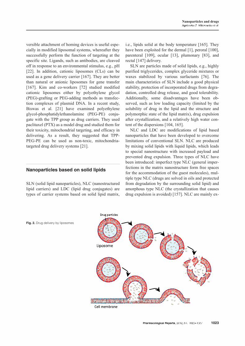
Drugs can be immobilized on PNPs surface aftera polymerization reaction [87] or can be encapsulatedon PNP structure during a polymerization step [99].Moreover, drugs may be released by desorption,diffusion, or nanoparticle erosion in target tissue[152]. The examples of drug-polymeric nanocarrierconjugates used as drug delivery systems are shownin Table 1.
Among the aforementioned applications the onethat is particularly interesting is the immobilization ofretinyl acetate (RA) on ethyl cellulose (EC), whichimproves aqueous stability and photostability ofa drug. In ex vivo tests on a skin tissue of mice,a 100% absorption of RA after 24 h has been demon-strated [8]. It is also worth to point out the biodegrad-
1024 Pharmacological Reports, 2012, 64, 1020�1037
Tab. 1. Polymer nanocarriers as DDS
Drug Therapeutic activity Nanocarrier Ref.
Carboplatin Antineoplastic drug, ovarian, head, neck and lung cancer Sodium alginate 106
5-Fluorouracil (5-FU) Anticancer drug, colon cancer CS-g-poly(N-vinylcaprolactam)
128
Doxorubicin Antineoplastic agent, wide spectrum of tumors PEGylatedPLGA 118
Capecitabine Pro-drug of fluorouracil, metastatic colorectal and breastcancer
CS-poly(ethyleneoxide-g-acrylamide)
3
Mitomycin C Chemotherapeutic agent, bladder tumors CS, CS-PCL, PLL-PCL 20
Rifampicin Antitubercular drugs, latent M. tuberculosis infectionin adults
Gelatin 131
Cyclosporine A Cyclic polypeptide, immunosuppressant GMO/poloxamer 407 cubicnanoparticles
79
Lamivudine Anti-HIV drug PLA/CS 43
Tacrine Anti-Alzheimer drug CS 164
Retinyl acetate Photoaging, severe acne and skin inflammation EC 8
Analogue of b-lactam Antibiotic, infection G(+)bacteria Polyacrylate 156
Clotrimazole Antifungal drug PLA-co-PLG 115
CS – chitosan; PEGylatedPLGA – PEGylated poly(lactic-co-glycolic acid); PCL – polycaprolactone; PLL – poly(L-lysine); GMO – glycerylmonooleate; EC – ethyl cellulose, PLA – polylactide; PLG – polyglycolide
able thermo-responsive chitosan-g-poly(N-vinylcapro-lactam)-biopolymer used for the delivery of 5-fluoro-uracil to cancer cells. The hypothesized mechanism of5-FU controlled release from this polymeric nanocar-rier is swelling followed by conformational changesduring a LCST (lower critical solution temperature)transition. The in vitro drug release showed a signifi-cant release above LCST. The high toxicity to cancercells, comparatively lower to the normal ones, wasobserved [128].
The application of biodegradable nanosystems forthe development of nanomedicines is one of the mostsuccessful ideas. Nanocarriers composed of biode-gradable polymers undergo hydrolysis in the body,producing biodegradable metabolite monomers, suchas lactic acid and glycolic acid. Kumari et al. [78] re-ported a minimal systemic toxicity associated with us-ing of PLGA for drug delivery or biomaterial applica-tions. Such nanoparticles are biocompatible with tis-sue and cells [116]. Drug-biodegradable polymericnanocarrier conjugates used for drug delivery are sta-ble in blood, non-toxic, and non-thrombogenic. Theyare also non-immunogenic as well as non-proinflam-matory, and they neither activate neutrophils nor af-fect reticuloendothelial system [42].
Dendrimer nanocarriers
Dendrimers are unique polymers with well-definedsize and structure. Dendritic architecture is one of themost popular structures observed throughout all bio-logical systems. Some of the examples of nanometricmolecules possessing dendritic structure include: gly-cogen, amylopectin, and proteoglycans [146].
In the structure of dendrimer, in contrast to the lin-ear polymer, the following elements can be distin-guished: a core, dendrons, and surface active groups.The core is a single atom or molecule (only if it has atleast two identical functional groups) that dendronsare attached to. The dendrons (dendrimer arms) aremolecules of monomer linked with the core, forminglayers and building successive generations (theirgrowth is spatially limited). Biocompatibility andphysicochemical properties of dendrimers are deter-mined by surface functional groups [23].
Selection of a core, type of a monomer and surfacefunctional groups determine the usability of den-drimers in medical applications. Cytotoxicity of den-
drimers and their so-called polyvalence is particularlyrelevant for biomedical purposes. Dendrimers cyto-toxicity depends on the core material and is stronglyinfluenced by the nature of the dendrimers surface.For example, changing the surface amine groups intohydroxyl ones may result in lower levels of cytotoxic-ity. The term polyvalence defines the number of activegroups on a dendrimers surface. The presence of sev-eral surface functional groups enables a sumultanoeusinteraction with a number of receptors, thus, it en-hances biological activity.
There are a few ways of connecting biologicallyactive compounds to dendrimers. The drug may beencapsulated in the internal structure of dendrimers[40] or it can be chemically attached or physically ad-sorbed on dendrimers surface [97]. The choice of theimmobilization method depends on the drug proper-ties. Encapsulation is used when drugs are labile,toxic, or poorly soluble. In turn, chemical attachmentprovides the possibility to control quantity of drugs ondendrimers surface by controlling the number of co-valent bonds [140]. The selectivity of both methodsmay be enhanced by attaching on the dendrimers sur-face such targeting agents as folic acid [40, 140] orepidermal growth factor [173].
The surface of dendrimers provides an excellentplatform for an attachment of specific ligands, whichmay include folic acid [140], antibodies [161], cyclictargeting peptides – arginine-glycine-aspartic acid(RGD) [159], selective A3 adenosine receptor [153],silver salts complexes antimicrobial agents [16], orpoly(ethylene glycol) (PEG) [85]. The attached com-pounds can improve surface activity as well as thebiological and physical properties of dendrimers.
Poly(amido amide) (PAMAM) is a dendrimerwhich is frequently used in biomedical applications.Both the structure of PAMAM dendrimers and thedistribution of drugs [135] or genes [173] inside thesemolecules have been intensively investigated. PAMAMdendrimers grow through generations G = 1–10 andtheir size increases from 1.1 to 12.4 nm. The size ofthe respective generations of PAMAM is comparableto the size of selected proteins, e.g., dendrimer G3with insulin and G7 with hemerythrin [150]. An ex-ample of a drug immobilization in PAMAM dendri-mer is cisplatin. This complex compared with freecisplatin exhibits several advantages, such as slowerrate of drug release, higher accumulation of the drugin solid tumors, and lower toxicity in all organs[40, 92]. Further examples of drug incorporation in
Pharmacological Reports, 2012, 64, 1020�1037 1025
Nanoparticles and drugsAgnieszka Z. Wilczewska et al.
PAMAM dendrimers are anticancer drugs, includingmethotrexate [108], doxorubicin [59], 5-FU [140],and anti-inflammatory drugs, e.g., ibuprofen [149],piroxicam [122], or indomethacin [26].
The size and charge of PAMAM dendrimers influ-ence their cytotoxicity. The higher-generation (G4-G8) PAMAM dendrimers exhibit toxicity due to theirhigh cationic charge density [134]. Comparative tox-icity studies on cationic and anionic dendrimers usingCaco-2 cells have shown a significantly lower cyto-toxicity of anionic compounds in comparison with thecationic ones [74]. Positively charged dendrimers in-troduced into the systemic circulation interact withblood components causing destabilization of cellmembranes and cell lysis [63]. Roberts and co-workers [129] observed that cationic PAMAM den-drimers caused a decrease in cell viability, however,they found no evidence of their immunogenicity inrabbits. Additionally, they studied the toxicity of cati-onic PAMAM Starburst® in mice and they suggestedthat even high doses of low generation cationic den-drimers do not cause side effects. Recent studies haveshown that G4 dendrimers, which have amino termi-nal groups, are toxic and impair the growth and devel-opment of zebrafish embryos. In turn, dendrimerswith carboxylic acid functional groups did not exertadverse effect on zebrafish embryos [61]. Dendrimerscan modulate cytokine and chemokine release. Thisproperty turned out to be helpful in therapy, but it canalso cause serious toxic effects [49]. The PAMAMgeneration of 3,5-glucosamine dendrimers inducesa synthesis of pro-inflammatory chemokines, such asMIP-1a, MIP-1h, and cytokines TNF-a, IL-1h, IL-6,IL-8 in human dendritic cells and macrophages,which exhibits an immunomodulatory effect. Chau-han et al. [27] studied in vivo toxicity profile ofPAMAM dendrimers in mice. The researchers as-sessed the following parameters: the animal behavior,feed intake, body weight, carbohydrate, lipid and pro-tein metabolism, hematological parameters, histopa-thology, and cell viability. They observed no effect onother hematological (excluding red blood cells, hema-tocrit value and hemoglobin) and biochemical pa-rameters (excluding the decrease of glucose levels inthe high-NH2 dose) as well as on feed intake, bodyand organ weights. In addition, the histopathologyshowed a toxic effect on liver and kidneys [27]. PE-Gylation of dendritic systems is a way of loweringgeneral toxicity. This process enables a long-lasting
blood circulation and an avoidance of dendrimers ac-cumulation in normal organs, such as kidneys andliver [75].
Silica materials
Silica materials used in controlled drug delivery sys-tems are classified as xerogels [36] and mesoporoussilica nanoparticles (MSNs), e.g., MCM-41 (MobilComposition of Matter) and SBA-15 (Santa BarbaraUniversity mesoporous silica material) [163]. They ex-hibit several advantages as carrier systems, includingbiocompatibility, highly porous framework and an easein terms of functionalization [7]. Among inorganicnanoparticles, silica materials are the carriers whichmost often are chosen for biological purposes [141].
Silica xerogels possess an amorphous structure withhigh porosity and enormous surface area. A porousstructure (shape and pore dimensions) depends on syn-thesis parameters [50]. Sol-gel technique is frequentlyused to form silica xerogels loaded with drugs.A modification of the synthesis conditions, such as theratio of reagents, temperature, concentration of thecatalyst, and pressure of drying, allows to alter proper-ties of xerogels used in controlled drug release [36,126]. Phenytoin [52], doxorubicin [124], cisplatin [38],metronidazole [39], nifedipine [95], diclofenac [37],and heparin [4] are examples of drugs which have beenloaded into xerogels using this technique.
The best known types of mesoporous silica nano-materials are MCM-41 with a hexagonal arrangementof the mesopores and SBA-15 with a well-orderedhexagonal connected system of pores [163]. TheMSNs, in comparison with xerogels, possess morehomogenous structure, lower polydispersity andhigher surface area for adsorption of therapeutic or di-agnostic agents [46]. The mechanism of drug loadinginto mesoporous silica material is a chemical orphysical adsorption [46]. By these processes, diversetypes of drugs, including anticancer drugs [46, 60],antibiotics [81], and heart disease drugs [121], havebeen embedded into MNSs. The drug release is usu-ally controlled by diffusion [81]. The silicalites andmesoporous silica nanoparticles potential applicationin photodynamic therapy has been also studied [62].
The MSNs properties make them an excellent ma-terial for various pharmaceutical and biomedical ap-
1026 Pharmacological Reports, 2012, 64, 1020�1037
plications. The structure of MSNs enables the incor-poration of both small [46] and large molecules [73],adsorption of DNA, and gene transfer [142]. Thisgives a possibility of using these nanomaterials ina combined therapy [60].
Some data indicate that nano-sized silica particles(SNPs) are biocompatible and have a great potentialfor a variety of diagnostic and therapeutic applica-tions in medicine. However, recent studies have re-vealed in vitro and in vivo toxicity and certain hazardsof using nanosilica. Most of the in vitro studies ofsilica nanoparticles show the adverse effect in investi-gated cells. The described effect depends on the celltype and nanoparticle size. Silica nanoparticles havean impact on a generation of oxidative stress in cellsvia formation of reactive oxygen species [84], ele-vated production of malondialdehyde [82], decreasingglutathione level [174], and induction of antioxidantenzymes, including superoxide dismutase (SOD) andheme oxygenase 1 (OH-1) [117]. All of these eventsare responsible for lipid peroxidation and cell mem-brane damage [82]. Previous work showed that expo-sure to silica nanoparticles at high concentrationscaused activation of NF-kB in endothelial cells [84]or Nrf-2-ERK MAP kinase signaling pathway in hu-man bronchial epithelial cells [51]. Pro-inflammatoryresponse resulted in an induction of various chemoki-nes (MCP-1 and MIP-2) [33, 84] and cytokines, suchas interleukins IL-1 [121], IL-8, IL-6 [32, 87] andTNF-a [33, 121], CD54 and CD62E [32, 162]. Otherresearchers also reported that silica nanoparticles in-duced apoptosis via JNK/p53-dependent, mitochon-drial pathways [84]. Chen et al. [29] suggested an ab-sorption of SNPs in the nucleus generated aberrantclusters of topoisomerase I and protein aggregates inthe nucleoplasm. The formation of nucleoplasmic ag-gregates impairs such nuclear functions as inhibitingreplication and transcription [29]. Yang and co-workers implied that perturbation of intracellular freecalcium homeostasis may be responsible for cytotoxiceffect of silica nanoparticles [169]. A recent work byZhao et al. [178] have demonstrated the effects ofnanoparticles (depending on the surface properties,structure and size) on human red blood cells (RBCs).The uptake of large silica nanoparticles by RBCsshowed a strong local membrane deformation leadingto a spiculation of RBCs, internalization of the parti-cles, and eventual hemolysis. On the contrary, adsorp-tion of small particles occurred without affectingmembrane or morphology of RBCs [178].
The size of nanoparticles plays a key role in theirtoxicity. Cho et al. [32] investigated the effect of theparticle size on the pharmacokinetic parameters, suchas tissue distribution and excretion via intravenouslyinjected silica particles of different sizes in mice.They observed an occurrence of inflammatory re-sponse of the liver within 12 h after injection of 200and 100 nm silica nanoparticles. However, this effectwas not reported for the smaller particles (50 nm). Alltypes of particles were excreted via urine and bile.These three types of nanoparticles were accumulatedby macrophage in liver and spleen and remained thereup to 4 weeks after the first injection of a single dose[32]. The authors then presented the effect of amor-phous silica by intratracheal instillation. They ob-served significant increase of lung weights, totalnumber of BAL cells and proteins concentration. Thehistopathological evaluation showed an inflammation,characterized by late neutrophils infiltration which re-sulted in the occurrence of symptoms of chronicgranulomatous inflammation of the lung [33]. Otherresearchers reported that inhalation of silica nanopar-ticles caused the inflammation of pulmonary tractmyocardial ischemic damage and atrio-ventricularblockage. In addition, they observed an increase of fi-brinogen concentration in blood [30]. Although or-ganically modified silica nanoparticles accumulate inall organs, there were no inducted signs of organ tox-icity [77]. Nishimori et al. [112] evaluating the acutetoxicity of amorphous silica particles observed a sig-nificant hepatotoxicity. During chronic administrationnano-size materials cause liver fibrosis in mice [112].
Carbon nanomaterials
Carbon nanocarriers used in DDS are differentiatedinto nanotubes (CNTs) and nanohorns (CNH).
CNTs are characterized by unique architectureformed by rolling of single (SWNCTs – single walledcarbon nanotubes) or multi (MWCNTs – multi walledcarbon nanotubes) layers of graphite with an enor-mous surface area and an excellent electronic andthermal conductivity [17]. Biocompatibility of nano-tubes may be improved by chemical modification oftheir surface [54]. Such adjustment can be imple-mented by covalent anchoring of PAMAM den-drimers [176], amphiphilic diblock copolymers [45],
Pharmacological Reports, 2012, 64, 1020�1037 1027
Nanoparticles and drugsAgnieszka Z. Wilczewska et al.
or PEG layers [18] on CNTs surface or dispersionwithin a hyaluronic acid matrix [137]. Due to theirmechanical strength, SWCNTs have been used asa support to improve properties of other carriers, e.g.,polymeric or non-polymeric composites [137].
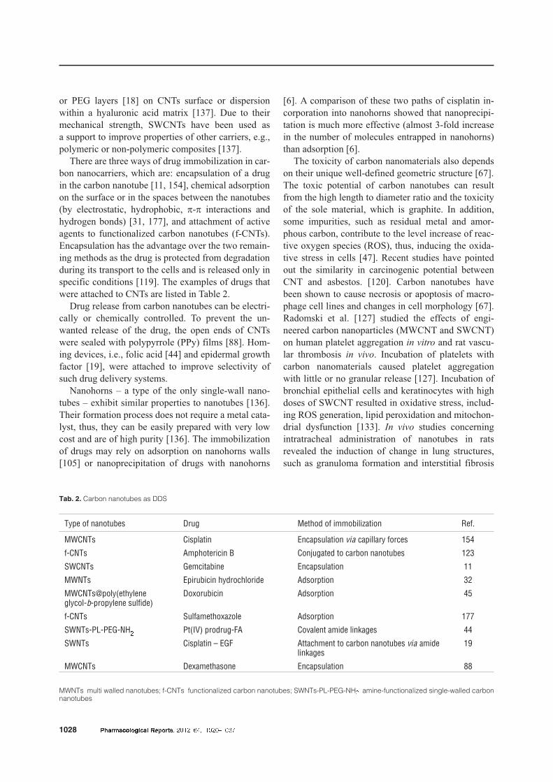
There are three ways of drug immobilization in car-bon nanocarriers, which are: encapsulation of a drugin the carbon nanotube [11, 154], chemical adsorptionon the surface or in the spaces between the nanotubes(by electrostatic, hydrophobic, p-p interactions andhydrogen bonds) [31, 177], and attachment of activeagents to functionalized carbon nanotubes (f-CNTs).Encapsulation has the advantage over the two remain-ing methods as the drug is protected from degradationduring its transport to the cells and is released only inspecific conditions [119]. The examples of drugs thatwere attached to CNTs are listed in Table 2.
Drug release from carbon nanotubes can be electri-cally or chemically controlled. To prevent the un-wanted release of the drug, the open ends of CNTswere sealed with polypyrrole (PPy) films [88]. Hom-ing devices, i.e., folic acid [44] and epidermal growthfactor [19], were attached to improve selectivity ofsuch drug delivery systems.
Nanohorns – a type of the only single-wall nano-tubes – exhibit similar properties to nanotubes [136].Their formation process does not require a metal cata-lyst, thus, they can be easily prepared with very lowcost and are of high purity [136]. The immobilizationof drugs may rely on adsorption on nanohorns walls[105] or nanoprecipitation of drugs with nanohorns
[6]. A comparison of these two paths of cisplatin in-corporation into nanohorns showed that nanoprecipi-tation is much more effective (almost 3-fold increasein the number of molecules entrapped in nanohorns)than adsorption [6].
The toxicity of carbon nanomaterials also dependson their unique well-defined geometric structure [67].The toxic potential of carbon nanotubes can resultfrom the high length to diameter ratio and the toxicityof the sole material, which is graphite. In addition,some impurities, such as residual metal and amor-phous carbon, contribute to the level increase of reac-tive oxygen species (ROS), thus, inducing the oxida-tive stress in cells [47]. Recent studies have pointedout the similarity in carcinogenic potential betweenCNT and asbestos. [120]. Carbon nanotubes havebeen shown to cause necrosis or apoptosis of macro-phage cell lines and changes in cell morphology [67].Radomski et al. [127] studied the effects of engi-neered carbon nanoparticles (MWCNT and SWCNT)on human platelet aggregation in vitro and rat vascu-lar thrombosis in vivo. Incubation of platelets withcarbon nanomaterials caused platelet aggregationwith little or no granular release [127]. Incubation ofbronchial epithelial cells and keratinocytes with highdoses of SWCNT resulted in oxidative stress, includ-ing ROS generation, lipid peroxidation and mitochon-drial dysfunction [133]. In vivo studies concerningintratracheal administration of nanotubes in ratsrevealed the induction of change in lung structures,such as granuloma formation and interstitial fibrosis
1028 Pharmacological Reports, 2012, 64, 1020�1037
Tab. 2. Carbon nanotubes as DDS
Type of nanotubes Drug Method of immobilization Ref.
MWCNTs Cisplatin Encapsulation via capillary forces 154
f-CNTs Amphotericin B Conjugated to carbon nanotubes 123
SWCNTs Gemcitabine Encapsulation 11
MWNTs Epirubicin hydrochloride Adsorption 32
MWCNTs@poly(ethyleneglycol-b-propylene sulfide)
Doxorubicin Adsorption 45
f-CNTs Sulfamethoxazole Adsorption 177
SWNTs-PL-PEG-NH2 Pt(IV) prodrug-FA Covalent amide linkages 44
SWNTs Cisplatin – EGF Attachment to carbon nanotubes via amidelinkages
19
MWCNTs Dexamethasone Encapsulation 88
MWNTs multi walled nanotubes; f-CNTs functionalized carbon nanotubes; SWNTs-PL-PEG-NH2 amine-functionalized single-walled carbonnanotubes
[80, 101]. Unmodified MWCNT caused pro-inflam-matory response in keratinocytes cell lines, e.g., re-lease of cytokine interleukin 8 and formation of cyto-plasmic vacuoles [98].
Magnetic nanoparticles
Magnetic nanoparticles exhibit a wide variety of at-tributes, which make them highly promising carriersfor drug delivery. In particular, these are: easy han-dling with the aid of an external magnetic field, thepossibility of using passive and active drug deliverystrategies, the ability of visualization (MNPs are usedin MRI), and enhanced uptake by the target tissue re-sulting in effective treatment at the therapeutically op-timal doses [10].
However, in most of the cases where magneticnanocarriers have been used, difficulties in achievingthese objectives appeared. It is most likely associatedwith inappropriate features of magnetic nanoparticlesor inadequate magnet system. Magnetic nanoparti-cles, for instance, tend to aggregate into larger clus-ters loosing the specific properties connected withtheir small dimensions and making physical handlingdifficult. In turn, magnetic force may not be strongenough to overcome the force of blood flow and toaccumulate magnetic drugs only at target site [110].
Therefore, designing magnetic drug delivery systemsrequires taking into consideration many factors, e.g.,magnetic properties and size of particles, strength of mag-netic field, drug loading capacity, the place of accessibil-ity of target tissue, or the rate of blood flow [24].
Depending on magnetic properties, MNPs can bedivided into pure metals (such as cobalt [96], nickel[69], manganese [132], and iron [143]), their alloysand oxides. However, narrowing the area of MNPsapplications only to biomedicine reduces significantlythe choice of magnetic material. Such a restriction re-sults from the lack of knowledge of the negative ef-fects which the majority of these nanomaterials haveon the human body.
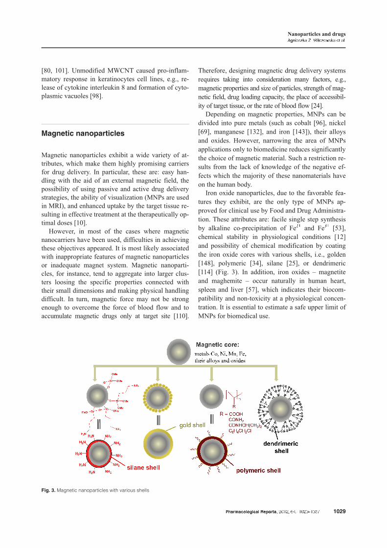
Iron oxide nanoparticles, due to the favorable fea-tures they exhibit, are the only type of MNPs ap-proved for clinical use by Food and Drug Administra-tion. These attributes are: facile single step synthesisby alkaline co-precipitation of Fe2+ and Fe3+ [53],chemical stability in physiological conditions [12]and possibility of chemical modification by coatingthe iron oxide cores with various shells, i.e., golden[148], polymeric [34], silane [25], or dendrimeric[114] (Fig. 3). In addition, iron oxides – magnetiteand maghemite – occur naturally in human heart,spleen and liver [57], which indicates their biocom-patibility and non-toxicity at a physiological concen-tration. It is essential to estimate a safe upper limit ofMNPs for biomedical use.
Pharmacological Reports, 2012, 64, 1020�1037 1029
Nanoparticles and drugsAgnieszka Z. Wilczewska et al.
Fig. 3. Magnetic nanoparticles with various shells
Connecting a drug with MNP may be achieved bycovalent binding [53], electrostatic interactions [53],adsorption [168], or encapsulation process [166]. Tar-geting of drug-MNPs conjugates to diseased tissues(magnetic targeted drug delivery systems – MTDDS),depending on their size and surface chemistry, can becarried out by passive or active mechanism. Passivetargeting is a result of enhanced vascular permeabilityand retention (EPR) of tumor tissues. Active strategyrelies on the attraction of nanoparticle to the affectedsite by using recognition ligands (e.g., antibodies) at-tached to the surface of MNPs and by handling of anexternal magnetic field [53].
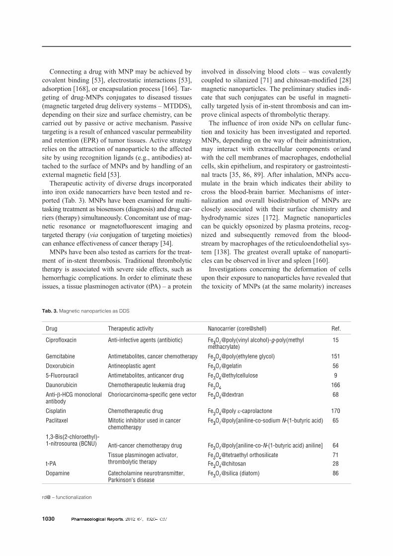
Therapeutic activity of diverse drugs incorporatedinto iron oxide nanocarriers have been tested and re-ported (Tab. 3). MNPs have been examined for multi-tasking treatment as biosensors (diagnosis) and drug car-riers (therapy) simultaneously. Concomitant use of mag-netic resonance or magnetofluorescent imaging andtargeted therapy (via conjugation of targeting moieties)can enhance effectiveness of cancer therapy [34].
MNPs have been also tested as carriers for the treat-ment of in-stent thrombosis. Traditional thrombolytictherapy is associated with severe side effects, such ashemorrhagic complications. In order to eliminate theseissues, a tissue plasminogen activator (tPA) – a protein
involved in dissolving blood clots – was covalentlycoupled to silanized [71] and chitosan-modified [28]magnetic nanoparticles. The preliminary studies indi-cate that such conjugates can be useful in magneti-cally targeted lysis of in-stent thrombosis and can im-prove clinical aspects of thrombolytic therapy.
The influence of iron oxide NPs on cellular func-tion and toxicity has been investigated and reported.MNPs, depending on the way of their administration,may interact with extracellular components or/andwith the cell membranes of macrophages, endothelialcells, skin epithelium, and respiratory or gastrointesti-nal tracts [35, 86, 89]. After inhalation, MNPs accu-mulate in the brain which indicates their ability tocross the blood-brain barrier. Mechanisms of inter-nalization and overall biodistribution of MNPs areclosely associated with their surface chemistry andhydrodynamic sizes [172]. Magnetic nanoparticlescan be quickly opsonized by plasma proteins, recog-nized and subsequently removed from the blood-stream by macrophages of the reticuloendothelial sys-tem [138]. The greatest overall uptake of nanoparti-cles can be observed in liver and spleen [160].
Investigations concerning the deformation of cellsupon their exposure to nanoparticles have revealed thatthe toxicity of MNPs (at the same molarity) increases
1030 Pharmacological Reports, 2012, 64, 1020�1037
Tab. 3. Magnetic nanoparticles as DDS
Drug Therapeutic activity Nanocarrier (core@shell) Ref.
Ciprofloxacin Anti-infective agents (antibiotic) Fe3O4@poly(vinyl alcohol)-g-poly(methylmethacrylate)
15
Gemcitabine Antimetabolites, cancer chemotherapy Fe3O4@poly(ethylene glycol) 151
Doxorubicin Antineoplastic agent Fe3O4@gelatin 56
5-Fluorouracil Antimetabolites, anticancer drug Fe3O4@ethylcellulose 9
Daunorubicin Chemotherapeutic leukemia drug Fe3O4 166
Anti-b-HCG monoclonalantibody
Choriocarcinoma-specific gene vector Fe3O4@dextran 68
Cisplatin Chemotherapeutic drug Fe3O4@poly e-caprolactone 170
Paclitaxel Mitotic inhibitor used in cancerchemotherapy
Fe3O4@poly[aniline-co-sodium N-(1-butyric acid) 65
1,3-Bis(2-chloroethyl)-1-nitrosourea (BCNU) Anti-cancer chemotherapy drug Fe3O4@poly[aniline-co-N-(1-butyric acid) aniline] 64
t-PA
Tissue plasminogen activator,thrombolytic therapy
Fe3O4@tetraethyl orthosilicate
Fe3O4@chitosan
71
28
Dopamine Catecholamine neurotransmitter,Parkinson’s disease
Fe3O4@silica (diatom) 86
rd@ – functionalization
in the following order: from nanobeads, to nanowormsand nanospheres [90]. MNPs coated with long-chainpolymers are less cell toxic (slightly lower cell viabilityin relation to the control probe in MTT assay) thanMNPs coated with shorter polymer chain of the sametype (with a significantly reduced viability) [58].
Magnetic nanoparticles promote activation ofphagocytotic and cytokine-release functions of macro-phages [47]. Gas vesicles were observed in MNPs-treated cells with increased granularity of the cells [89].Magnetite nanoparticles have the potency of causingoxidative DNA lesions in cultured A549 cells (the hu-man lung epithelial cell line) [70]. They also causetransient increase of ALT (serum alanine aminotransfe-rase), AST (aspartate aminotransferase), and ALP (al-kaline phosphatase) levels [66]. In several reports ironoxide was found to accumulate in tissues but withoutsignificant histological changes in vital organs [166].
It is known that iron oxide NPs can induce oxida-tive stress by excess of ROS production. The responseof defense elements leads to increased expression ofantioxidant enzymes, such as heme oxygenase 1 andsuperoxide dismutase. This stage is followed by pro-inflammatory response, i.e., cytokine, chemokine, andmatrix metalloproteinase (MMP) release, leading toapoptosis and mutagenesis. An increase in the activityof MMP in the nervous system can enlarge BBB per-meability and cause neuronal damages. Oxidativestress-induced cell injury and death may be avoidedusing appropriate dose of MNPs [107].
Conclusion
Nanocarriers as drug delivery systems are designed toimprove the pharmacological and therapeutic proper-ties of conventional drugs. The incorporation of drugmolecules into nanocarrier can protect a drug againstdegradation as well as offers possibilities of targetingand controlled release. Due to small dimensions,nanocarriers are able to cross the blood-brain-barrier(BBB) and operate on cellular level. In comparisonwith the traditional form of drugs, nanocarrier-drugconjugates are more effective and selective. They canreduce the toxicity and other adverse side effects innormal tissues by accumulating drugs in target sites.In consequence, the required doses of drugs are lower.
Although there are several nanoparticle-basedtherapeutic agents which are currently being devel-
oped and are under preclinical evaluation, onlya handful of nanoparticle drugs are available on thepharmaceutical market, e.g., liposomal conjugates:Doxil® (doxorubicin) or DaunoXome® (daunorubi-cin). It is due to the fact that nanoparticle based drugdelivery systems do have a lot of drawbacks and limi-tations. Some of them arise from scaling up problems.For instance, small size and large surface area ofnanoparticle-based targeting system can lead to an ag-gregation, making physical handling difficult. Nano-carrier-drug conjugates can be phagocytosed by cellswhereas their intracellular degradation may cause cy-totoxic effects. Other issues include low drug loadingcapacity, low loading efficiency, and poor ability tocontrol the size distribution of carriers. Furthermore,there is a lack of technological methods, which willlead to nanodevices of approvable quality.
Despite all the limitations and shortcomings, nano-particle DDS which respond to slight changes in thelocal cellular environment have a potential to resolvemany of the current drug delivery problems. How-ever, before the ongoing research will bring a clini-cally useful drug delivery system, challenges whichinclude developing toxicity testing protocols, improv-ing biocompatibility, drug loading, targeting, trans-port and release, controlling interaction with biologi-cal barriers, detecting and monitoring exposure leveland assessing the impact on the environment have tobe met.
Due to a number of functional groups on the sur-face of nanoparticles, the drug can be attached to thecarrier only in a stoichiometric ratio. The oxidativestress and inflammation in different cell types havebeen often reported as toxic mechanisms of varioustypes of nanoparticles. Nanoparticles of diameter10 nm can remain in cells and induct chronic inflam-matory response and fibrosis of tissue. An additionalproblem is the lack of knowledge concerning the dis-tribution of drug carriers and the unpredictability ofthe process. Thus, in our opinion, the magnetic tar-geted drug delivery system is one of the most attrac-tive strategy target therapy. Magnetic nanoparticleshave their unique magnetic properties and they can beattracted by magnetic fields, thus, acting as drug carri-ers in a target therapy. In addition, inorganic magneticnanoparticles containing the iron and gadoliniumserve as an excellent contrast enhancing agents inMRI (approved by FDA – Food and Drug Administra-tion).
Pharmacological Reports, 2012, 64, 1020�1037 1031
Nanoparticles and drugsAgnieszka Z. Wilczewska et al.
A real therapeutic breakthrough can be achievedsolely by carrying out painstaking studies in the fieldof nano-therapy. Using nanosystems in therapies ofdiseases may contribute to achieving an effective can-cer treatment. Moreover, immobilization of homingdevices, such as folic acid, epidermal growth factor orantibodies, to the surface of nanoparticles, improvesselectivity of drug carriers. The key applications ofnanoparticles in medicine are diagnosis and targettherapy, however, their wider use is still the future.
References:
1. Abdel-Mottaleb MMA, Neumann D, Lamprecht A:Lipid nanocapsules for dermal application: A compara-tive study of lipid-based versus polymer-based nanocar-riers. Eur J Pharm Biopharm, 2011, 79, 36–42.
2. Afergan E, Epstein H, Dahan R, Koroukhov N, RohekarK, Danenberg HD, Golomb G: Delivery of serotonin tothe brain by monocytes following phagocytosis of lipo-somes. J Control Release, 2008, 132, 84–90.
3. Agnihotri SA, Aminabhavi TM: Novel interpenetratingnetwork chitosan-poly(ethylene oxide-g-acrylamide)hy-drogel microspheres for the controlled release of cape-citabine. Int J Pharm, 2006, 324, 103–115.
4. Ahola MS, Säilynoja ES, Raitavuo MH, Vaahtio MM,Salonen JI, Yli-Urpo AU: In vitro release of heparinfrom silica xerogels. Biomaterials, 2001, 22, 2163–2170.
5. Ai J, Biazar E, Montazeri M, Majdi A, Aminifard S,Safari M, Akbari HR: Nanotoxicology and nanoparticlesafety in biomedical designs. Int J Nanomedicine, 2011,6, 1117–1127.
6. Ajima K, Murakami T, Mizoguchi Y, Tsuchida K, Ichi-hashi T, Iijima S, Yudasaka M: Enhancement of in vivoanticancer effects of cisplatin by incorporation inside single-wall carbon nanohorns. ACS Nano, 2008, 2, 2057–2064.
7. Amato G: Silica-encapsulated efficient and stable siquantum dots with high biocompatibility. Nanoscale ResLett, 2010, 5, 1156–1160.
8. Arayachukeat S, Wanichwecharungruang SP, Tree-UdomT: Retinyl acetate-loaded nanoparticles: Dermal penetra-tion and release of the retinyl acetale. Int J Pharm, 2011,404, 281–288.
9. Arias JL, López-Viota M, Delgado AV, Ruiz MA:Iron/ethylcellulose (core/shell) nanoplatform loaded with5-fluorouracil for cancer targeting. Colloids Surf B Bio-interfaces, 2010, 77, 111–116.
10. Arruebo M, Fernández-Pacheco R, Ibarra, MR, Santa-maría J: Magnetic nanoparticles for drug delivery. NanoToday, 2007, 2, 22–32.
11. Arsawang U, Saengsawang O, Rungrotmongkol T,Sornmee P, Wittayanarakul K, Remsungnen T,Hannongbua S: How do carbon nanotubes serve ascarriers for gemcitabine transport in a drug delivery sys-tem? J Mol Graph Model, 2011, 29, 591–596.
12. Asmatulu R, Zalich MA, Claus RO, Riffle J: Synthesis,characterization and targeting of biodegradable magneticnanocomposite particles by external magnetic fields.J Magn Magn Mater, 2005, 292, 108–119.
13. Attama AA, Schicke BC, Paepenmüller T, Müller-Goymann CC: Solid lipid nanodispersions containingmixed lipid core and a polar heterolipid: characteriza-tion. Eur J Pharm Biopharm, 2007, 67, 48–57.
14. Bai J, Li Y, Du J, Wang S, Zheng J, Yang O, Chen X:One-pot synthesis of polyacrylamide-gold nanocompos-ite. Mater Chem Phys, 2007, 106, 412–415.
15. Bajpai AK, Gupta R: Magnetically mediated release ofciprofloxacin from polyvinyl alcohol based superpara-magnetic nanocomposites. J Mater Sci Mater Med, 2011,22, 357–369.
16. Balogh L, Swanson DR, Tomalia DA, Hagnauer GL,McManus AT: Dendrimer-silver complexes and nano-composites as antimicrobial agents. Nano Lett, 2001, 1,18–21.
17. Beg S, Rizwan M, Sheikh AM, Hasnain MS, Anwer K,Kohli K: Advancement in carbon nanotubes: basics, bio-medical applications and toxicity. J Pharm Pharmacol,2011, 63, 141–163.
18. Bhirde AA, Patel S, Sousa AA, Patel V, Molinolo AA,Ji Y, Leapman RD et al.: Distribution and clearance ofPEG-single-walled carbon nanotube cancer drug deliveryvehicles in mice. Nanomedicine, 2010, 5, 1535–1546.
19. Bhirde AA, Patel V, Gavard J, Zhang G, Sousa AA,Masedunskas A, Leapman RD et al.: Targeted killing ofcancer cells in vivo and in vitro with EGF-directed car-bon nanotube-based drug delivery. ACS Nano, 2009, 3,307–316.
20. Bilensoy E, Sarisozen C, Esendagl G, Dogan LA, AktasY, Sen M, Mangan AN: Intravesical cationic nanoparti-cles of chitosan and polycaprolactone for the delivery ofMitomycin C to bladder tumors. Int J Pharm, 2009, 371,170–176.
21. Biswas S, Dodwadkar NS, Deshpande PP, Torchilin VP:Liposomes loaded with paclitaxel and modified withnovel triphenylphosphonium-PEG-PE conjugate possesslow toxicity, target mitochondria and demonstrate en-hanced antitumor effects in vitro and in vivo. J ControlRelease, 2012, 159, 393–402.
22. Biswas S, Dodwadkar NS, Sawant RR, Torchilin VP:Development of the novel PEG-PE-based polymer forthe reversible attachment of specific ligands to lipo-somes: synthesis and in vitro characterization. BioconjugChem, 2011, 22, 2005–2013.
23. Caminade AM, Laurent R, Majoral JP: Characterizationof dendrimers. Adv Drug Deliv Rev, 2005, 57, 2130– 2146.
24. Cao Q, Han X, Li L: Enhancement of the efficiency ofmagnetic targeting for drug delivery: Development andevaluation of magnet system. J Magn Magn Mater, 2011,323, 1919–1924.
25. Chang JH, Kang KH, Choi J, Jeong YK: High efficiencyprotein separation with organosilane assembled silicacoated magnetic nanoparticles. Superlattice Microst,2008, 44, 442–448.
26. Chauhan AS, Diwan PV, Jain NK, Tomalia DA: Unex-pected in vivo anti-inflammatory activity observed for
1032 Pharmacological Reports, 2012, 64, 1020�1037
simple, surface functionalized poly(amidoamine) den-drimers. Biomacromolecules, 2009, 10, 1195–1202.
27. Chauhan AS, Jain NK, Diwan PV: Pre-clinical and be-havioural toxicity profile of PAMAM dendrimers inmice. Proc R Soc A, 2010, 466, 1535–1550.
28. Chen JP, Yang PC, Ma YH, Wu T: Characterization ofchitosan magnetic nanoparticles for in situ delivery oftissue plasminogen activator. Carbohydr Polym, 2011,84, 364–372.
29. Chen M, von Mikecz A: Formation of nucleoplasmicprotein aggregates impairs nuclear function in responseto SiO2 nanoparticles. Exp Cell Res, 2005, 305, 51–62.
30. Chen Z, Meng H, Xing G, Yuan H, Zhao F, Liu R,Chang X et al.: Age-related differences in pulmonaryand cardiovascular responses to SiO2 nanoparticle inha-lation: nanotoxicity has susceptible population. EnvironSci Technol, 2008, 42, 8985–8992.
31. Chen Z, Pierre D, He H, Tan S, Pham-Huy C, Hong H,Huang J: Adsorption behavior of epirubicin hydrochlo-ride on carboxylated carbon nanotubes. Int J Pharm,2011, 28, 405, 153–161.
32. Cho M, Cho WS, Choi M, Kim SJ, Han BS, Kim SH,Kim HO et al.: The impact of size on tissue distributionand elimination by single intravenous injection of silicananoparticles. Toxicol Lett, 2009, 189, 177–183.
33. Cho WS, Choi M, Han BS, Cho M, Oh J, Park K, KimSJ et al.: Inflammatory mediators induced by intratra-cheal instillation of ultrafine amorphous silica particles.Toxicol Lett, 2007, 175, 24–33.
34. Chomoucka J, Drbohlavova J, Huska D, Adam V, KizekR, Hubalem J: Magnetic nanoparticles and targeted drugdelivering. Pharm Res, 2010, 62, 144–149.
35. Cole AJ, Yang VC, David AE: Cancer theranostics: therise of targeted magnetic nanoparticles. Trends Biotech-nol, 2011, 29, 323–332.
36. Czarnobaj K: Preparation and characterization of silica xe-rogels as carriers for drugs. Drug Deliv, 2008, 15, 485–492.
37. Czarnobaj K, Czarnobaj J: Sol-gel processed porous sil-ica carriers for the controlled release of diclofenac dieth-ylamine. J Biomed Mater Res B Appl Biomater, 2008¸87, 114–120.
38. Czarnobaj K, Lukasiak J: In vitro release of cisplatinfrom sol-gel processed organically modified silica xero-gels. J Mater Sci Mate Med, 2007, 18, 2041–2044.
39. Czarnobaj K, Sawicki W: The sol-gel preparedSiO2-CaO-P2O5 composites doped with metronidazolefor application in local delivery systems. Pharm DevTechnol, 2011, (doi: 10.3109/10837450.2011.572894).
40. D’Emanuele A, Attwood D: Dendrimer-drug interac-tions. Adv Drug Deliv Rev, 2005, 57, 2147–2162.
41. Daemen T, Hofstede G, Ten Kate MT, Bakker-WoudenbergIA, Scherphof GL: Liposomal doxorubicin-induced tox-icity: depletion and impairment of phagocytic activity ofliver macrophages. Int J Cancer, 1995, 61, 666–671.
42. des Rieux A, Fievez V, Garinot M, Schneider YJ,Préat V: Nanoparticles as potential oral delivery systemsof proteins and vaccines: a mechanistic approach. J Con-trol Release, 2006,116, 1–27.
43. Dev A, Binulal NS, Anita A, Nair SV, Furuike T, TamuraH, Jayakumar R: Preparation of poly(lactic acid)/chito-
san nanoparticles for anti-HIV drug delivery applica-tions. Carbohydr Polym, 2010, 80, 833–838.
44. Dhar S, Liu Z, Thomale J, Dai H, Lippard SJ: Targetedsingle-wall carbon nanotube-mediated Pt(IV) prodrugdelivery using folate as a homing device. J Am ChemSoc, 2008, 27, 130, 11467–11476.
45. Di Crescenzo A, Velluto D, Hubbell JA, Fontana A:Biocompatible dispersions of carbon nanotubes: a poten-tial tool for intracellular transport of anticancer drugs.Nanoscale, 2011, 3, 925–928.
46. Di Pasqua AJ, Wallner S, Kerwood DJ, Dabrowiak JC:Adsorption of the Pt(II) anticancer drug carboplatin bymesoporous silica. Chem Biodiv, 2009, 6, 1343–1349.
47. Dobrovolskaia MA, McNeil SE: Immunological proper-ties of engineered nanomaterials. Nat Nano, 2007, 2,469–478.
48. dos Santos Giuberti C, de Oliveira Reis EC, RibeiroRocha TG, Leite EA, Lacerda RG, Ramaldes GA,de Oliveira MC: Study of the pilot production processof long-circulating and pH-sensitive liposomes contain-ing cisplatin. J Liposome Res, 2011, 21, 60–69.
49. Duncan R, Izzo L: Dendrimer biocompatibility and tox-icity. Adv Drug Deliv Rev, 2005, 57, 2215–2237.
50. Echeverría JC, Estella J, Barbería V, Musgo J, GarridoJJ: Synthesis and characterization of ultramicroporoussilica xerogels. J Non-Cryst Solids, 2010, 356, 378–382.
51. Eom HJ, Choi J: Oxidative stress of silica nanoparticlesin human bronchial epithelial cell, Beas-2B. Toxicol InVitro, 2009, 23, 1326–1332.
52. Fidalgo A, Lopez TM , Ilharco LM: Wet sol–gel silicamatrices as delivery devices for phenytoin. J Sol-Gel SciTechnol, 2009, 49, 320–328.
53. Figuerola A, Di Corato R, Manna L, Pellegrino T: Fromiron oxide nanoparticles towards advanced iron-based in-organic materials designed for biomedical applications.Pharmacol Res, 2010, 62, 126–143.
54. Foldvari M, Bagonluri M: Carbon nanotubes as func-tional excipients for nanomedicines: I. Pharmaceuticalproperties. Nanomedicine, 2008, 4, 173–182.
55. Fritzen-Garcia MB, Zanetti-Ramos BG, Schweitzer deOliveira C, Soldi V, Pasa AA, Creczynski-Pasa TB:Atomic force microscopy imaging of polyurethane nano-particles onto different solid. Mater Sci Eng C, 2009, 29,405–409.
56. Gaihre B, Khil MS, Lee DR, Kim HY: Gelatin-coatedmagnetic iron oxide nanoparticles as carrier system: drugloading and in vitro drug release study. Int J Pharm,2009, 365, 180–189.
57. Grassi-Schultheiss PP, Heller F, Dobson J: Analysis ofmagnetic material in the human heart, spleen and liver.Biometals, 1997, 10, 351–355.
58. Hafeli UO, Riffle JS, Harris-Shekhawat L, Carmichael-Baranauskas A, Mark F, Dailey JP, Bardenstein D: Celluptake and in vitro toxicity of magnetic nanoparticles suit-able for drug delivery. Mol Pharm, 2009, 6, 1417–1428.
59. Han L, Huang R, Liu S, Huang S, Jiang Ch: Peptide-conjugated PAMAM for targeted doxorubicin delivery totransferrin receptor overexpressed tumors. Mol Pharm,2010, 7, 2156–2165.
60. He Q, Gao Y, Zhang L, Zhang Z, Gao F, Ji X, Li Y,Shi J: A pH-responsive mesoporous silica nanoparticles-
Pharmacological Reports, 2012, 64, 1020�1037 1033
Nanoparticles and drugsAgnieszka Z. Wilczewska et al.
based multi-drug delivery system for overcoming multi-drug resistance. Biomaterials, 2011, 32, 7711–7720.
61. Heiden TCK, Dengler E, Kao WJ, Heideman W, Peter-son DE: Developmental toxicity of low generationPAMAM dendrimers in zebrafish. Toxicol Appl Pharma-col, 2007, 225, 70–79.
62. Hocine O, Gary-Bobo M, Brevet D, Maynadier M,Fontanel S, Raehm L, Richeter S et al.: Silicalites andmesoporous silica nanoparticles for photodynamictherapy. Int J Pharm, 2010, 402, 221–230.
63. Hong S, Bielinska AU, Mecke A, Keszler B, Beals JL,Shi X, Balogh L et al.: Interaction of poly(amidoamine)dendrimers with supported lipid bilayers and cells: holeformation and the relation to transport. Bioconjug Chem,2004, 15, 774–782.
64. Hua MY, Liu HL, Yang HW, Chen PY, Tsai RY, HuangCY, Tseng IC et al.: The effectiveness of a magneticnanoparticle-based delivery system for BCNU in thetreatment of gliomas. Biomaterials, 2011, 32, 516–527.
65. Hua MY, Yang HW, Chuang CK, Tsai RY, Chen WJ,Chuang KL, Chang YH et al.: Magnetic-nanoparticle-modified paclitaxel for targeted therapy for prostate can-cer. Biomaterials, 2010, 31, 7355–7363.
66. Jain TK, Reddy MK, Morales MA, Leslie-Pelecky DL,Labhasetwar V: Biodistribution, clearance, and biocom-patibility of iron oxide magnetic nanoparticles in rats.Mol Pharm, 2007, 5, 316–327.
67. Jia G, Wang H, Yan L, Wang X, Pei R, Yan T, Zhao Y,Guo X: Cytotoxicity of carbon nanomaterials: single-wall nanotube, multi-wall nanotube, and fullerene. Envi-ron Sci Technol, 2005, 39, 1378–1383.
68. Jingting C, Huining L, Yi Z: Preparation and characteri-zation of magnetic nanoparticles containingFe3O4-dextran-anti-b-human chorionic gonadotropin,a new generation choriocarcinoma-specific gene vector.Int J Nanomedicine, 2011, 6, 285–294.
69. Kale SN, Jadhav AD, Verma S, Koppikar SJ, Kaul-Ghanekar R, Dhole SD, Ogale SB: Characterization ofbiocompatible NiCo2O4 nanoparticles for applications inhyperthermia and drug delivery. Nanomedicine, 2012, 8,452–459.
70. Karlsson HL, Cronholm P, Gustafsson J, Moller L:Copper oxide nanoparticles are highly toxic: a compari-son between metal oxide nanoparticles and carbon nano-tubes. Chem Res Toxicol, 2008, 21, 1726–1732.
71. Kempe H, Kempe M: The use of magnetite nanoparticlesfor implant-assisted magnetic drug targeting in throm-bolytic therapy. Biomaterials, 2010, 31, 9499–9510.
72. Kim JK, Choi SH, Kim CO, Park JS, Ahn WS, Kim CK:Enhancement of polyethylene glycol (PEG)-modifiedcationic liposome-mediated gene deliveries: effects onserum stability and transfection efficiency. J Pharm Phar-macol, 2003, 55, 453–460.
73. Kim TW, Slowing II, Chung PW, Lin VS: Orderedmesoporous polymer-silica hybrid nanoparticles as vehi-cles for the intracellular controlled release of macro-molecules. ACS Nano, 2011, 5, 360–366.
74. Kitchens KM, Kolhatkar RB, Swaan PW, Eddington ND,Ghandehari H.: Transport of poly(amidoamine) den-drimers across Caco-2 cell monolayers: influence of size,
charge and fluorescent labeling. Pharm Res, 2006, 23,2818–2826.
75. Kojima C, Regino C, Umeda Y, Kobayashi H, Kono K:Influence of dendrimer generation and polyethylene gly-col length on the biodistribution of PEGylated den-drimers. Int J Pharm, 2010, 383, 293–296.
76. Kovacevic A, Savic S, Vuleta G, Müller RH, Keck CM:Polyhydroxy surfactants for the formulation of lipidnanoparticles (SLN and NLC): Effects on size, physicalstability and particle matrix structure. Int J Pharm, 2011,406, 163–172.
77. Kumar R, Roy I, Ohulchanskky TY, Vathy LA, BergeyEJ, Sajjad M, Prasad PN: In vivo biodistribution andclearance studies using multimodal organically modifiedsilica nanoparticles. ACS Nano, 2010, 4, 699–708.
78. Kumari A, Yadav SK, Yadav SC: Biodegradable poly-meric nanoparticles based drug delivery systems. Col-loids Surf B Biointerfaces, 2010, 75, 1–18.
79. Lai J, Lu Y, Yin Z, Hu F, Wu W: Pharmacokinetics andenhanced oral bioavailability in beagle dogs of cyclo-sporine A encapsulated in glyceryl monooleate/polox-amer 407 cubic nanoparticles. Int J Nanomedicine, 2010,5, 13–23.
80. Lam CW, James JT, McCluskey R, Hunter RL: Pulmo-nary toxicity of single-wall carbon nanotubes in mice 7and 90 days after intratracheal instillation. Toxicol Sci,2004, 77, 126–134.
81. Li Z, Su K, Cheng B, Deng Y: Organically modifiedMCM-type material preparation and its usage in con-trolled amoxicillin delivery. J Colloid Interface Sci,2010, 342, 607 –613.
82. Lin W, Huang YW, Zhou XD, Ma Y: In vitro toxicity ofsilica nanoparticles in human lung cancer cells. ToxicolAppl Pharmacol, 2006, 217, 252–259.
83. Liu J, Gong T, Fu H, Wang C, Wang X, Chen Q, Zhang Q,He Q, Zhang Z: Solid lipid nanoparticles for pulmonarydelivery of insulin, Int J Pharm, 2008, 356, 333–344.
84. Liu X, Sun J: Endothelial cells dysfunction induced bysilica nanoparticles through oxidative stress via JNK/P53and NF-kB pathways. Biomaterials, 2010, 31, 8198– 8209.
85. Lope AL, Reins RY, McDermott AM, Trautner BW,Cai C: Antibacterial activity and cytotoxicity ofPEGylated poly(amidoamine) dendrimers. Mol Biosyst,2009, 5, 1148 –1156.
86. Losic D, Yu Y, Aw MS, Simovic S, Thierry B, Addai-Mensah J: Surface functionalisation of diatoms with do-pamine modified iron-oxide nanoparticles: toward mag-netically guided drug microcarriers with biologically de-rived morphologies. Chem Commun (Camb), 2010, 46,6323–6325.
87. Luo G, Yu X, Jin C, Yang F, Fu D, Long J, Xu J et al.:LyP-1-conjugated nanoparticles for targeting drug deliv-ery to lymphatic metastatic tumors. Int J Pharm, 2010,385, 150–156.
88. Luo X, Matranga C, Tan S, Alba N, Cui XT: Carbonnanotube nanoreservior for controlled release of anti-inflammatory dexamethasone. Biomaterials, 2011, 32,6316–6323.
89. Mahmoudi M, Simchi A, Imani M, Shokrgozar MA,Milani AS, Hafeli U, Stroeve P: A new approach forthe in vitro identification of the cytotoxicity of super-
1034 Pharmacological Reports, 2012, 64, 1020�1037
paramagnetic iron oxide nanoparticles. Colloids Surf BBiointerfaces, 2010, 75, 300–309.
90. Mahmoudi M, Simchi A, Milani AS, Stroeve P: Cell tox-icity of superparamagnetic iron oxide nanoparticles.J Colloid Interface Sci, 2009, 336, 510–518.
91. Mainardes RM, Khalil NM, Gremião MPD: Intranasaldelivery of zidovudine by PLA and PLA–PEG blendnanoparticles. Int J Pharm, 2010, 395, 266–271.
92. Malik N, Evagorou EG, Duncan R: Dendrimer-platinate:a novel approach to cancer chemotherapy. Anti-CancerDrugs Des, 1999, 10, 767–776.
93. Mao HQ, Roy K, Troung-Le VL, Janes KA, Lin KY,Wang J, August JT, Leong KW: Chitosan-DNA nanopar-ticles as gene carriers: synthesis, characterization andtransfection efficiency. J Control Release, 2001, 70,399–421.
94. Martinem A, Iglesias I, Lozano R, Teijon JM, BlancoMD: Synthesis and characterization of thiolatedalginate-albumin nanoparticles stabilized by disulfidebonds. Evaluation as drug delivery systems. CarbohydrPolym, 2011, 83, 1311–1321.
95. Maver U, Godec A, Bele M, Planinšek O, Gaberšèek M,Srèiè S, Jamnik J: Novel hybrid silica xerogels for stabi-lization and controlled release of drug. Int J Pharm,2007, 330,164–174.
96. Meng X, Seton HC, Lu le T, Prior IA, Thanh NT, SongB: Magnetic CoPt nanoparticles as MRI contrast agentfor transplanted neural stem cells detection. Nanoscale,2011, 3, 977–984.
97. Menjoge AR, Kannan RM, Tomalia DA: Dendrimer-based drug and imaging conjugates: design considera-tions for nanomedical applications. Drug Discov Today,2010, 15, 171–187.
98. Monteiro-Riviere NA, Nemanich RJ, Inman AO, WangYY, Riviere JE: Multi-walled carbon nanotube interac-tions with human epidermal keratinocytes. Toxicol Lett,2005, 155, 377–384.
99. Mora-Huertas CE, Fessi H, Elaissari A: Polymer-basednanocapsules for drug delivery. Int J Pharm, 2010, 385,113–142.
100. Muchow M, Maincent P, Müller RH: Lipid nanoparticleswith a solid matrix (SLN, NLC LDC) for oral drug deliv-ery. Drug Dev Ind Pharm, 2008, 34, 1394–1405.
101. Muller J, Huaux F, Moreau N, Misson P, Heilier JF, De-los M, Arras M et al.: Respiratory toxicity of multi-wallcarbon nanotubes. Toxicol Appl Pharmacol, 2005, 207,221–231.
102. Müller RH, Keck CM: Challenges and solutions for thedelivery of biotech drugs – a review of drug nanocrystaltechnology and lipid nanoparticles. J Biotechnol, 2004,113, 151–170.
103. Müller RH, Petersen RD, Hommoss A, Pardeike J:Nanostructured lipid carriers (NLC) in cosmetic dermalproducts. Adv Drug Deliv Rev, 2007, 59, 522–530.
104. Müller RH, Radtke M, Wissing SA: Nanostructured lipidmatrices for improved microencapsulation of drugs, IntJ Pharm, 2002, 242, 121–128.
105. Murakami T, Sawada H, Tamura G, Yudasaka M, IijimaS, Tsuchida K: Water-dispersed single-wall carbon nano-horns as drug carriers for local cancer chemotherapy.Nanomedicine, 2008, 3, 453–463.
106. Nanjwade BK, Singh J, Parikh KA, Manvi FV: Prepara-tion and evaluation of carboplatin biodegradable poly-meric nanoparticles. Int J Pharm, 2010, 385, 176–180.
107. Naqvi S, Samim M, Abdin MZ, Ahmed FJ, Maitra AN,Prashant CK, Dinda AK: Concentration-dependent toxic-ity of iron oxide nanoparticles mediated by increasedoxidative stress. Int J Nanomedicine, 2010, 5, 983–989.
108. Natali S, Mijovic J: Dendrimers as drug carriers: dynam-ics of PEGylated and methotrexate-loaded dendrimers inaqueous solution. Macromolecules, 2010, 43, 3011–3017.
109. Nayak AP, Tiyaboonchai W, Patankar S, MadhusudhanB, Souto EB: Curcuminoids-loaded lipid nanoparticles:novel approach towards malaria treatment. ColloidsSurf B Biointerfaces, 2010, 81, 263–273.
110. Neuberger T, Schopf B, Hofmann H, Hofmann M, vonRechenberg B: Superpara-magnetic nanoparticles forbiomedical applications: possibilities and limitations ofa new drug delivery system. J Magn Magn Mater, 2005,293, 483–496.
111. Nevozhay D, Kañska U, Budzyñska R, Boratyñski J:Current status of research on conjugates and related drugdelivery systems in the treatment of cancer and other dis-eases (Polish). Postepy Hig Med Dosw, 2007, 61, 350–360.
112. Nishimori H, Kondoh M, Isoda K, Tsunoda S, TsutsumiY, Yagi K: Silica nanoparticles as hepatotoxicants. EurJ Pharm Biopharm, 2009, 72, 496–501.
113. Paavola A, Kilpeläinen I, Yliruusi J, Rosenberg P: Con-trolled release injectable liposomal gel of ibuprofen forepidural analgesia. Int J Pharm, 2000, 199, 85–93.
114. Pan BF, Gao F, Gu HC: Dendrimer modified magnetitenanoparticles for protein immobilization. J Colloid Inter-face Sci, 2005, 284, 1–6.
115. Pandey R, Ahmad Z, Sharma S, Khuller GK: Nano-encapsulation of azole antifungals: Potential applicationsto improve oral drug delivery. Int J Pharm, 2005, 301,268–276.
116. Panyam J, Labhasetwar V: Biodegradable nanoparticlesfor drug and gene delivery to cells and tissue. Adv DrugDeliv Rev, 2003, 55, 329–347.
117. Park EJ, Park K: Oxidative stress and pro-inflammatoryresponses induced by silica nanoparticles in vivo and invitro. Toxicol Lett, 2009, 184, 18–25.
118. Park J, Fong PM, Lu J, Russell KS, Booth KJ, SaltzmanWM, Fahmy TM: PEGylated PLGA nanoparticles forthe improved delivery of doxorubicin. Nanomedicine,2009, 5, 410-418.
119. Perry JL, Martin CR, Stewart JD: Drug-delivery strate-gies by using template-synthesized nanotubes. Chemis-try, 2011, 17, 6296–6302.
120. Poland CA, Duffin R, Kinloch I, Maynard A, WallaceWA, Seaton A, Stone V et al.: Carbon nanotubes intro-duced into the abdominal cavity of mice show asbestos-like pathogenicity in a pilot study. Nat Nanotechnol,2008, 3, 423–428.
121. Popovici RF, Seftel EM, Mihai GD, Popovici E, VoicuVA: Controlled drug delivery system based on orderedmesoporous silica matrices of captopril as angiotensin-converting enzyme inhibitor drug. J Pharm Sci, 2011,100, 704–714.
122. Prajapati RN, Tekade RK, Gupta U, Gajbhiye V, JainNK: Dendimer-mediated solubilization, formulation de-
Pharmacological Reports, 2012, 64, 1020�1037 1035
Nanoparticles and drugsAgnieszka Z. Wilczewska et al.
velopment and in vitro-in vivo assessment of piroxicam.Mol Pharm, 2009, 6, 940–950.
123. Prajapati VK, Awasthi K, Gautam S, Yadav TP, Rai M,Srivastava ON, Sundar S et al.: Targeted killing of Leish-mania donovani in vivo and in vitro with amphotericin Battached to functionalized carbon nanotubes. J Antimi-crob Chemother, 2011, 66, 874 –879.
124. Prokopowicz M: Synthesis and in vitro characterizationof freeze-dried doxorubicin-loaded silica xerogels.J Sol-Gel Sci Technol, 2010, 53, 525–533.
125. Puglia C, Blasi P, Rizza L, Schoubben A, Bonina F,Rossi C, Ricci M: Lipid nanoparticles for prolongedtopical delivery: an in vitro and in vivo investigation.Int J Pharm, 2008, 357, 295–304.
126. Quintanar-Guerrero D, Ganem-Quintanar A, Nava-Arzaluz MG, Piñón-Segundo E: Silica xerogels as phar-maceutical drug carriers. Expert Opin Drug Deliv, 2009,6, 485–498.
127. Radomski A, Jurasz P, Alonso-Escolano D, Drews M,Morandi M, Malinski T, Radomski MW: Nanoparticle-induced platelet aggregation and vascular thrombosis.Br J Pharmacol, 2005, 146, 882–893.
128. Rejinold NS, Chennazhi KP, Nair SV, Tamura H,Jayakumar R: Biodegradable and thermo-sensitivechitosan-g-poly(N-vinylcaprolactam) nanoparticles asa 5-fluorouracil carrier. Carbohydr Polym, 2011, 83,776–786.
129. Roberts JC, Bhalgat MK, Zera RT: Preliminary biologi-cal evaluation of polyamidoamine (PAMAM) Starburstdendrimers. J Biomed Mater Res, 1996, 30, 53–65.
130. Safdar A, Ma J, Saliba F, Dupont B, Wingard JR,Hachem RY, Mattiuzzi GN et al.: Drug-induced nephro-toxicity caused by amphotericin B lipid complex and li-posomal amphotericin B: a review and meta-analysis.Medicine (Baltimore), 2010, 89, 236–244.
131. Saraog GK, Gupta P, Gupta UD, Jain NK, Agrawal GP:Gelatin nanocarriers as potential vectors for effectivemanagement of tuberculosis. Int J Pharm, 2010, 385,143–149.
132. Sayed FN, Jayakumar OD, Sudakar C, Naik R,Tyagi AK: Possible weak ferromagnetism in pure andM (Mn, Cu, Co, Fe and Tb) doped NiGa2O4 nanoparti-cles. J Nanosci Nanotechnol, 2011, 11, 3363–3369.
133. Sayes CM, Liang F, Hudson JL, Mendez J, Guo W,Beach JM, Moore VC et al.: Functionalization densitydependence of single-walled carbon nanotubes cytotox-icity in vitro. Toxicol Lett, 2006, 161, 135–142.
134. Shah N, Steptoe RJ, Parekh HS: Low-generation asym-metric dendrimers exhibit minimal toxicity and effec-tively complex DANN. J Pept Sci, 2011, 17, 470–478.
135. Shi L, Fleming CJ, Riechers SL, Yin N-N, Luo J,Lam KS, Liu GY: High-resolution imaging of dendrimersused in drug delivery via scanning probe microscopy.J Drug Deliv, 2011, (doi:10.1155/2011/254095).
136. Shiba K, Yudasaka M, Iijima S: Carbon nanohorns asa novel drug carrier. Nihon Rinsho, 2006, 64, 239–246.
137. Shin US, Yoon IK, Lee GS, Jang WC, Knowles JC,Kim HW: Carbon nanotubes in nanocomposites and hy-brids with hydroxyapatite for bone replacements. J Tis-sue Eng, 2011, (doi:10.4061/2011/674287).
138. Shubayev VI, Pisanic TR 2nd, Jin S: Magnetic nanopar-ticles for theragnostics. Adv Drug Deliv Rev, 2009, 61,467–477.
139. Silva R, Ferreira H, Cavaco-Paulo A: Sonoproductionof liposomes and protein particles as templates for deliv-ery purposes. Biomacromolecules, 2011, 12, 3353–3368.
140. Singh P, Gupta U, Asthana A, Jain NK: Folate andFolate-PEG-PAMAM dendrimers: synthesis, characteri-zation, and targeted anticancer drug delivery potentialin tumor bearing mice. Bioconjugate Chem, 2008, 19,2239–2252.
141. Slowing I, Trewyn BG, Giri S, Lin VS-Y: Mesoporoussilica nanoparticles for drug delivery and biosensing ap-plications. Adv Funct Mater, 2007, 17, 1225–1236.
142. Slowing I, Vivero-Escoto JL, Wu Ch-W, Lin VS-Y:Mesoporous silica nanoparticles as controlled releasedrug delivery and gene transfection carriers. Adv DrugDeliv Rev, 2008, 60, 1278–1288.
143. Smolensky ED, Park HY, Berquó TS, Pierre VC: Surfacefunctionalization of magnetic iron oxide nanoparticlesfor MRI applications - effect of anchoring group andligand exchange protocol. Contrast Media Mol Imaging,2011, 6, 189–199.
144. Sunderland CJ, Steiert M, Talmadge JE, Derfus AM,Barry SE: Targeted nanoparticles for detecting and treat-ing cancer. Drug Dev Res, 2006, 67, 70–93.
145. Suri SS, Fenniri H, Singh B: Nanotechnology-based drugdelivery systems. J Occup Med Toxicol, 2007, 2, 16.
146. Svenson S, Tomalia DA: Dendrimers in biomedical ap-plications – reflections on the field. Adv Drug Deliv Rev,2005, 57, 2106– 2129.
147. Sznitowska M, Gajewska M, Janicki S, Radwanska A,Lukowski G: Bioavailability of diazepam fromaqueous-organic solution, submicron emulsion and solidlipid nanoparticles after rectal administration in rabbits.Eur J Pharm Biopharm, 2001, 52, 159–163.
148. Tamer U, Gundogdu Y, Boyaci IH. Pekmez K: Synthesisof magnetic core-shell Fe3O4 – Au, nanoparticles forbiomolecule immobilization and detection. J NanopartRes, 2010, 12, 1187–1196.
149. Tanis I, Karatasos K: Association of a weakly acidicanti-inflammatory drug (Ibuprofen) with a poly(ami-doamine) dendrimer as studied by molecular dynamicssimulations. J Phys Chem B, 2009, 113, 10984–10993.
150. Tomalia DA: Birth of a new macromolecular architec-ture: dendrimers as quantized building blocks fornanoscale synthetic organic chemistry. Prog Polym Sci,2005, 30, 294–324.
151. Tong Q, Li H, Li W, Chen H, Shu X, Lu X, Wang G:In vitro and in vivo anti-tumor effects of gemcitabineloaded with a new drug delivery system. J NanosciNanotechnol, 2011, 11, 3651–3658.
152. Torchilin V: Multifunctional Pharmaceutical Nanocarri-ers, Springer Science + Business Media, LLC, NY, 2008.
153. Tosh DK, Yoo LS, Chinn M, Hong K, Kilbey MS, Bar-rett MO, Fricks IP et al.: Polyamidoamine (PAMAM)dendrimer conjugates of “clickable” agonists of the A3adenosine receptor and coactivation of the P2Y14 recep-tor by a tethered nucleotide. Bioconjug Chem, 2010, 21,372-384.
1036 Pharmacological Reports, 2012, 64, 1020�1037
154. Tripisciano C, Costa S, Kalenczuk RJ, Borowiak-PalenE: Cisplatin filled multiwalled carbon nanotubes – a novelmolecular hybrid of anticancer drug container. Eur PhysJ B, 2010, 75, 141–146.
155. Turkova A, Roilides E, Sharland M: Amphotericin B inneonates: deoxycholate or lipid formulation as first-linetherapy - is there a ‘right’ choice? Curr Opin Infect Dis,2011, 24, 163–171.
156. Turos E, Shim JY, Wang Y, Greenhalgh K, Reddy GS,Dickey S, Lim DV: Antibiotic-conjugated polyacrylatenanoparticles: New opportunities for development ofanti-MRSA agents. Bioorg Med Chem Lett, 2007, 17,53–56.
157. Üner M, Yener G: Importance of solid lipid nanoparticles(SLN) in various administration routes and future per-spectives. Int J Nanomedicine, 2007, 2, 289–300.
158. van den Hoven JM, Van Tomme SR, Metselaar JM,Nuijen B, Beijnen JH, Storm G: Liposomal drug formu-lations in the treatment of rheumatoid arthritis. MolPharm, 2011, 8, 1002–1015.
159. Waite CL, Roth ChM: PAMAM-RGD conjugates en-hance siRNA delivery through a multicellular spheroidmodel of malignant glioma. Bioconjug Chem, 2009, 20,1908–1916.
160. Wang J, Chen Y, Chen B, Ding J, Xia G, Gao C,Cheng J et al.: Pharmacokinetic parameters and tissuedistribution of magnetic Fe3O4 nanoparticles in mice.Int J Nanomedicine, 2010, 5, 861–866.
161. Wängler C, Moldenhauer G, Eisenhut M, Haberkorn U,Mier W: Antibody-dendrimer conjugates: the number,not the size of the dendrimers, determines the imunore-activity. Bioconjug Chem, 2008, 19, 813–820.
162. Waters KM, Masiello LM, Zangar RC, Tarasevich BJ,Karin NJ, Quesenberry RD, Bandyopadhyay S et al.:Macrophage responses to silica nanoparticles are highlyconserved across particle sizes. Toxicol Sci, 2009, 107,553–569.
163. Wei L, Hu N, Zhang Y: Synthesis of polymer-mesoporoussilica nanocomposites. Materials, 2010, 3, 4066–4079.
164. Wilson B, Samanta MK, Santhi K, Kumar KP,Ramasamy M, Suresh B: Chitosan nanoparticles asa new delivery system for the anti-Alzheimer drugtacrine. Nanomedicine, 2010, 6, 144–152.
165. Wissing SA, Kayser O, Müller RH: Solid lipid nanopar-tic les for parenteral drug delivery. Adv Drug Deliv Rev,2004, 56, 1257–1272.
166. Wu W, Chen B, Cheng J, Wang J, Xu W, Liu L, Xia Get al.: Biocompatibility of Fe3O4/DNR magnetic nano-particles in the treatment of hematologic malignancies.Int J Nanomedicine, 2010, 5, 1079–1084.
167. Xiong F, Mi Z, Gu N: Cationic liposomes as gene deliv-ery system: transfection efficiency and new application.Pharmazie, 2011, 66, 158–164.
168. Yallapu MM, Othman SF, Curtis ET, Gupta BK, JaggiM, Chauhan SC: Multi-functional magnetic nanoparti-cles for magnetic resonance imaging and cancer therapy.Biomaterials, 2011, 32, 1890–1905.
169. Yang H, Wu Q, Tang M, Kong L, Lu Z: Cell membraneinjury induced by silica nanoparticles in mouse macro-phage. J Biomed Nanotechnol, 2009, 5, 528–535.
170. Yang J, Park SB, Yoon HG, Huh YM, Haam S: Prepara-tion of poly e-caprolactone nanoparticles containingmagnetite for magnetic drug carrier. Int J Pharm, 2006,324, 185–190.
171. Yeung CK, Shek SY, Yu CS, Kono T, Chan HH: Lipo-some-encapsulated 0.5% 5-aminolevulinic acid with in-tense pulsed light for the treatment of inflammatory facialacne: a pilot study. Dermatol Surg, 2011, 37, 450–459.
172. Yoo JW, Chambers E, Mitragotri S: Factors that controlthe circulation time of nanoparticles in blood: chal-lenges, solutions and future prospects. Curr Pharm Des,2010, 16, 2298–2307.
173. Yu H, Nie Y, Dohmen Ch, Li Y, Wagner E: Epidermalgrowth factor-PEG functionalized PAMAM-penta-ethylenehexamine dendron for targeted gene deliveryproduced by click chemistry. Biomacromolecules, 2011,12, 2039–2047.
174. Yu KO, Grabinski CM, Schrand AM, Murdock RC,Wang W, Gu B, Schlager JJ et al.: Toxicity of amorphoussilica nanoparticles in mouse keratinocytes. J NanopartRes, 2009, 11, 15–24.
175. Yukihara M, Ito K, Tanoue O, Goto K, Matsushita T,Matsumoto Y, Masuda M et al.: Effective drug deliverysystem for duchenne muscular dystrophy using hybridliposomes including gentamicin along with reduced tox-icity. Biol Pharm Bull, 2011, 34, 712–716.
176. Zhang B, Chen Q, Tang H, Xie Q, Ma M, Tan L, ZhangY, Yao S: Characterization of and biomolecule immobili-zation on the biocompatible multi-walled carbon nano-tubes generated by functionalization with polyami-doamine dendrimers. Colloids Surf B Biointerfaces,2010, 80, 18–25.
177. Zhang D, Pan B, Wu M, Wang B, Zhang H, Peng H,Wu D, Ning P: Adsorption of sulfamethoxazole on func-tionalized carbon nanotubes as affected by cations andanions. Environ Pollut, 2011, 159, 2616–2621.
178. Zhao Y, Sun X, Zhang G, Trewyn BG, Slowing II,Lin VS: Interaction of mesoporous silica nanoparticleswith human red blood cell membranes: size and surfaceeffects. ACS Nano, 2011, 5, 1366–1375.
Received: November 24, 2011; in the revised form: May 7, 2012;
accepted: May 22, 2012.
Pharmacological Reports, 2012, 64, 1020�1037 1037
Nanoparticles and drugsAgnieszka Z. Wilczewska et al.