Badania wybranych nowotworów z zastosowaniem … · G. Veronesi 7. FLUO at ANKA Rolf Simon . ......
44
Badania wybranych nowotworów z zastosowaniem technik analitycznych opartych na promieniowaniu synchrotronowym Marek Lankosz
Transcript of Badania wybranych nowotworów z zastosowaniem … · G. Veronesi 7. FLUO at ANKA Rolf Simon . ......
Badania wybranych nowotworów z zastosowaniem
technik analitycznych opartych na promieniowaniu
synchrotronowym
Marek Lankosz
Makroelementy od 0.01%
Mikroelementy 0.01%-0.0001%
Ultraelementy poniżej 0,0001%
• pierwiastki konieczne do życia tzw. biopierwiastki
• pierwiastki obojętne, bez których przemiany
metaboliczne mogą normalnie przebiegać
• pierwiastki toksyczne, wywierające szkodliwe działanie
na organizm
Klasyfikacja pierwiastków pod względem fizjologicznym
W. Opoka i inni, Właściwości fizykochemiczne i biologiczne wybranych pierwiastków

Pb- kumuluje się w kościach, zakłóca przemiany metaboliczne, niedorozwój umysłowy, trudności w uczeniu, anemia Hg- działa na ośrodkowy układ nerwowy, zaburzenie wzroku, uszkadza nerki, powoduje trudności w uczeniu się, jest kancerogenny, kumulacja w rybach i skorupiakach, amalgamat do uzupełniania ubytków w zębach, konserwacja niektórych szczepionek. Cd-deponowany w płucach, nerkach, wątrobie, rakotwórczy, uszkadza komórki nerwowe, zniekształcenia kości, zaburzenia wzrostu, występuje w tworzywach sztucznych (pigmenty) As-kumuluje się we włosach, paznokciach, skórze, kościach, uszkodzenia nerek, zaburzenia układu nerwowego, skurcz mięśni, kancerogenny, tritlenek diarsenu-leczenie ostrej białaczki i innych nowotworów. Al. Nie pełni funkcji biologicznej. Zaburza funkcjonowanie układu nerwowego. Ma związek z chorobą Alzheimera i Parkinsona, Stosowany w medycynie w terapii.
Pierwiastki toksyczne

Mn- wpływa na funkcjonowanie OUN, składnik metaloenzymów (dysmutaza ponadtlenkowa, polimeraza RNA i DNA), aktywuje enzymy dla wytwarzania energii, metabolizm węglowodanów, białek, lipidów, naturalny antyutleniacz, Hipomanganemia- zaburzenie koordynacji ruchowej, zmęczenie, stany niepokoju Zatrucia- neurodegeneracyjne zmiany dopaminergiczne, szaleństwo manganowe (impulsywność, pobudliwość, gadatliwość, schizofrenia) Se- pierwiastek życia, wchodzi w skład aminokwasów, jest antyoksydantem, zapobiega proliferacji i wzrostowi komórek nowotworowych,, stymuluje układ immunologiczny Niedobór-podatność na choroby, Nadmiar- depresje, nerwowość, uszkodzenie wątroby, nerek i śledziony, czerwone zabarwienie paznokci, zaburzenie metabolizmu siarki.
Biopierwiastki
Cu- składnik i aktywator enzymów, składnik dysmutazy ponadtlenkowej, bierze udział w syntezie dopaminy, Niedobór- niedokrwistość, osteoporoza, zmniejszenie liczby białych krwinek, zaburzenie gospodarki lipidowej, zwyrodnienie wątrobowo-soczewkowe (Ch Wilsona), neurodegeneracja (Ch Menkesa) Nadmiar- zmiany metaboliczne, uszkodzenia nerek, mózgu, naczyń wieńcowych, Zn- przemiany metaboliczne kwasów nukleinowych, lipidów, białek, cukrów, prawidłowe funkcjonowanie układu immunologicznego, budulec dysmutazy ponadtlenkowej, synteza i magazynowanie insuliny, składnik enzymów, Niedobór- zaburzenia depresyjne
Biopierwiastki

Fe- katalizator reakcji enzymatycznych, składnik hemoprotein, białek, generowanie wolnych rodników, układ odpornościowy, procesy krwiotwórcze, produkcja neuroprzekaźników, Nadmiar-zakłóca metabolizm innych metali śladowych, Niedobór-niedokrwistość Ni- występuje w enzymach (uerazach), erytropoeza, siarczek niklu (II) działanie kancerogennie Cr (III)-niezbędny do metabolizmu glukozy, stymuluje syntezę kwasów tłuszczowych, Niedobór- depresje, stany lękowe, uszkodzenia nerwów, zaburzenia w gospodarce białek i lipidów Nadmiar-biegunka, skaza krwotoczna, martwica wątroby Mg- Bierze udział w procesie skurczu mięśni (serce), jest aktywatorem wielu enzymów. Brak- wzmożone napięcie mięśniowe, drżenie mięśni, skurcz. Nadmiar-zawroty głowy, biegunka, nudności
Biopierwiastki

1867 (Perls) histochemical visualization of iron in brain tissue
1886 (Zaleski) distinction of heme” and “non heme” iron in
brain
1955 (Diezel) discovery of ferritin, main iron carrying protein
1985 (Bloch) discovery of transferrin main transporter of iron
1986 (Drayer) magnetic resonance imaging of brain iron
• Wilson’s disease – defective copper metabolism
• Menkes’ disease – copper deficiency
• Parkinsonian syndrome – chronic manganese toxicity
• Mental confusion – lead toxicity
• Minamata disease – methyl mercury toxicity
• Alzcheimer’s disease- aluminum and zinc toxicity
• Parkinson’s disease – iron deposition in brain
Metals in brain

CHEMICAL ELEMENTS STUDIED – MARKERS?
8/21
OXIDATIVE STRESS DAMAGE TO DNA & PROTEINS ROS (FENTON REACTION): Fe
PROTECTION AGAINST ROS (CuZnSOD): Cu, Zn
MORE ABUNDANT ELEMENTS: P (nucleic acids, energy carriers, phospholipides)
S (enzymes, cell breathing)
Na, K, Cl (electrolyte equilibrium)
TISSUE ENRICHED OR DEPLETED A.Wandzilak,
Selected chemical elements as
potential indicators of cancerous
brain tissue, doctoral thesis

MOTIVATION
9/21
LOOKING FOR THE RELATIONSHIP BETWEEN THE BRAIN TUMOUR MALIGNANCY GRADE AND LEVELS OF SELECTED ELEMENTS
BIOCHEMICAL PROCESSES
DIAGNOSTICS: DETERMINING MALIGNANCY GRADE OF TUMOURS
A.Wandzilak,
Selected chemical elements as
potential indicators of cancerous
brain tissue, doctoral thesis
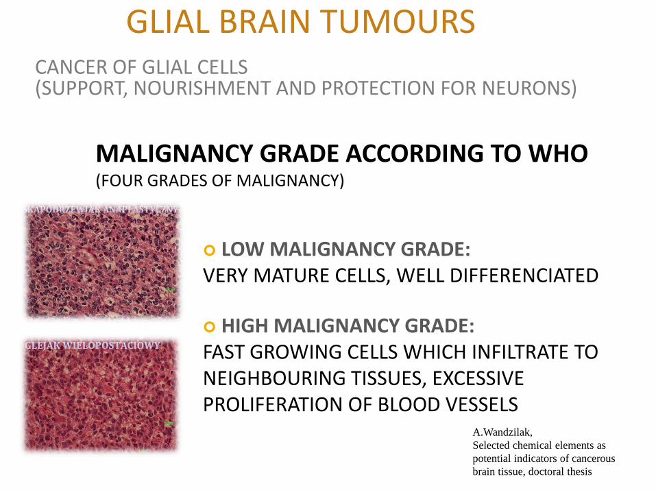
GLIAL BRAIN TUMOURS
10/21
CANCER OF GLIAL CELLS (SUPPORT, NOURISHMENT AND PROTECTION FOR NEURONS)
MALIGNANCY GRADE ACCORDING TO WHO (FOUR GRADES OF MALIGNANCY)
LOW MALIGNANCY GRADE: VERY MATURE CELLS, WELL DIFFERENCIATED
HIGH MALIGNANCY GRADE: FAST GROWING CELLS WHICH INFILTRATE TO NEIGHBOURING TISSUES, EXCESSIVE PROLIFERATION OF BLOOD VESSELS
A.Wandzilak,
Selected chemical elements as
potential indicators of cancerous
brain tissue, doctoral thesis

CHEMICAL ELEMENTAL IMAGING WITH
THE USE OF X-RAY FLUORESCENCE
MICROSPECTROSCOPY
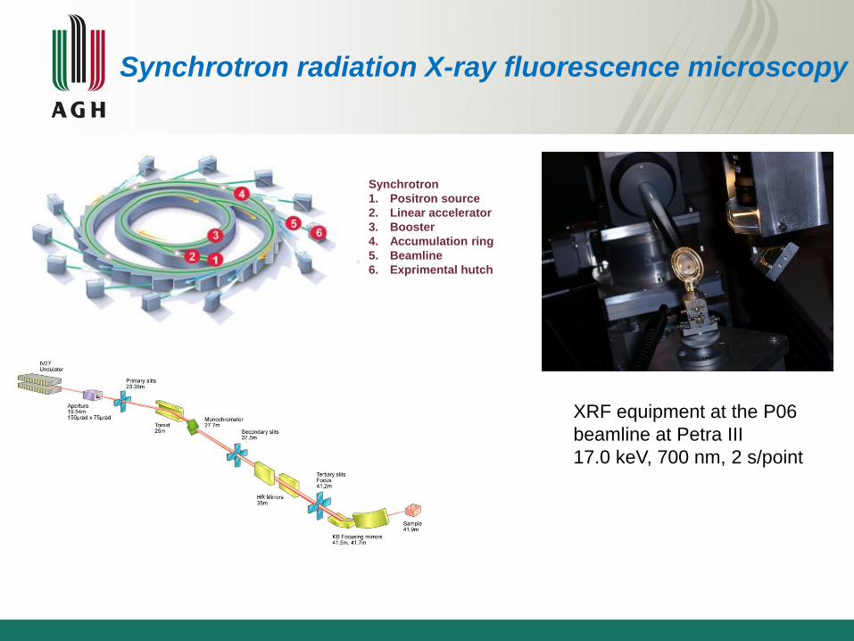
XRF equipment at the P06
beamline at Petra III
17.0 keV, 700 nm, 2 s/point
Synchrotron
1. Positron source
2. Linear accelerator
3. Booster
4. Accumulation ring
5. Beamline
6. Exprimental hutch
Synchrotron radiation X-ray fluorescence microscopy

XRF BEAMLINE at ELETTRA
• RuB4C monochromator
• pixel size: 120 x 250 µm2
• 30 mm2 SDD
• ulitra thin polymer window
• high vacuum (2·10-7 mbar)
• 45°/45° geometry
• 5 s / pixel
• PyMCA for data analysis
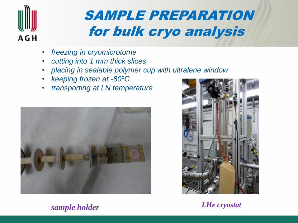
sample holder
SAMPLE PREPARATION
for bulk cryo analysis
S LHe cryostat
• freezing in cryomicrotome
• cutting into 1 mm thick slices
• placing in sealable polymer cup with ultralene window
• keeping frozen at -80ºC.
• transporting at LN temperature

Sample holder for cryo XRF microscopy

cutting into 5m thick sections
(cryomicrotome)
Tissue - shock freezing
histopathology biochemical analyses
Silicon nitride membrane
freeze-drying at -80 °C
SAMPLE PREPARATION
for biochemical micro-imaging

Types of ovarian tumors used in elemental studies
Sample
labeling
Type of tumor Number of
analysed areas
1 Control 6
2 Bening tumor 4
3 Bordeline tumor 7
4 Cancer 12
5 Stroma 3
Ł. Chmura, et al. Journal of Physiology
and Pathomorphology, submitted for
publication

ARCHITECTURE OF OVARIAN CANCER TISSUES
A) Solid tumours – solid sheets of epithelial cells
B) Borderline tumours – single layer of epithelial cells grown
into stromal cells

EXAMPLE of XRF IMAGING
endometroid adenocarcinoma 8 x 9.4 mm
NMF: Ca, Cu, Fe, Mn, Si, Zn excluded
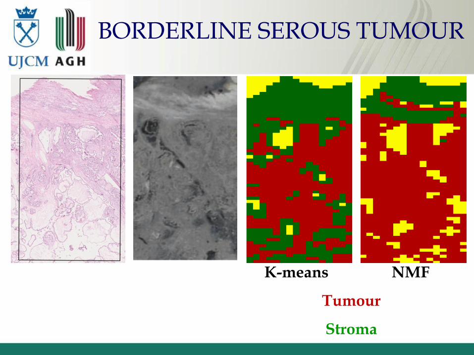
BORDERLINE SEROUS TUMOUR
K-means NMF
Tumour
Stroma

Optical microscope
image of malignant tissue
stained with the use of
hematoxylin-eosin
Maps of elemental distribution in malignant
tissue and optical microscope image of tissue.
Data presented in μg/cm2. X-Y coordinates in μm

The scatter plot of observations in the space of discriminant variables for different types of
ovarian cancer (two factors)
DF1 = −2, 498K + 1, 501S + 0, 824Cl − 0, 596Fe
DF2 = 2, 673S − 2, 952Cl + 1, 191Br + 0, 904Rb − 0, 690Zn
Multivariate Discriminant Analysis
Chemical elemental analysis of mean concentrations of elements
in brain cancers
A.Wandzilak et al.
Metallomics, 5 (2013) 1547-1553
M.Lankosz, et al
Spectrochimica Acta Part B, 101 (2014)
98–105

Examined material
• Neoplasma benignum
• Oligodendroglioma, II grade WHO
• Astrocytoma diffusum, II-III grade WHO
• Oligodendroglioma anaplasticum, III grade WHO
• Glioblastoma multiforme, IV grade WHO
• Control tissues
Astrocytoma
diffusum

XRF spectrum
Astrocytoma diffusum XRF sum spectrum probed from 15 352 points

Distribution of Ca, P, S, Fe, Cu and Zn in a section of
diffuse astrocytoma

Cancerous vs. healthy tissue
0,00
0,01
0,02
0,03
0,06
0,13
0,25
0,50
1,00
2,00
4,00
8,00
P S Cl K Ca Fe Cu Zn
ma
ss d
ep
osit p
er
un
it a
rea
, µ
g/c
m2
control III, OA IV, GM
Mean content of elements in healthy and cancerous tissues

Classifier
1. Differences in the chemical composition of tissues with different cancer type
2. Elements of the greatest importance in the differentiation of cancer type
3. Model to identify the cancerous case by its chemical composition (the
fingerprint of cancer)

Surface densities of Cl, Fe and Br within blood vessel area

Changes in the surface density of Cl as a function of distance from a blood vessel.
Analysis of Fe, Cu and Zn chemical
environment and oxidation states in
brain cancers with the use of XANES
and EXAFS microspectroscopies
A.Wandzilak et al.
Metallomics, 5 (2013) 1547-1553
DLS Report 2012

XAS = XANES + EXAFS
32/21 XANES - X-RAY ABSORPTION NEAR EDGE STRUCTURE
EXAFS - EXTENDED X-RAY ABSORPTION FINE STRUCTURE
3.0
2.5
2.0
1.5
-ln(I
/I0)
800078007600740072007000
X-ray photon energy / eV
EXAFS XANES
Chemical shift Oxidation state
photoelectrons
MATTER WAVES
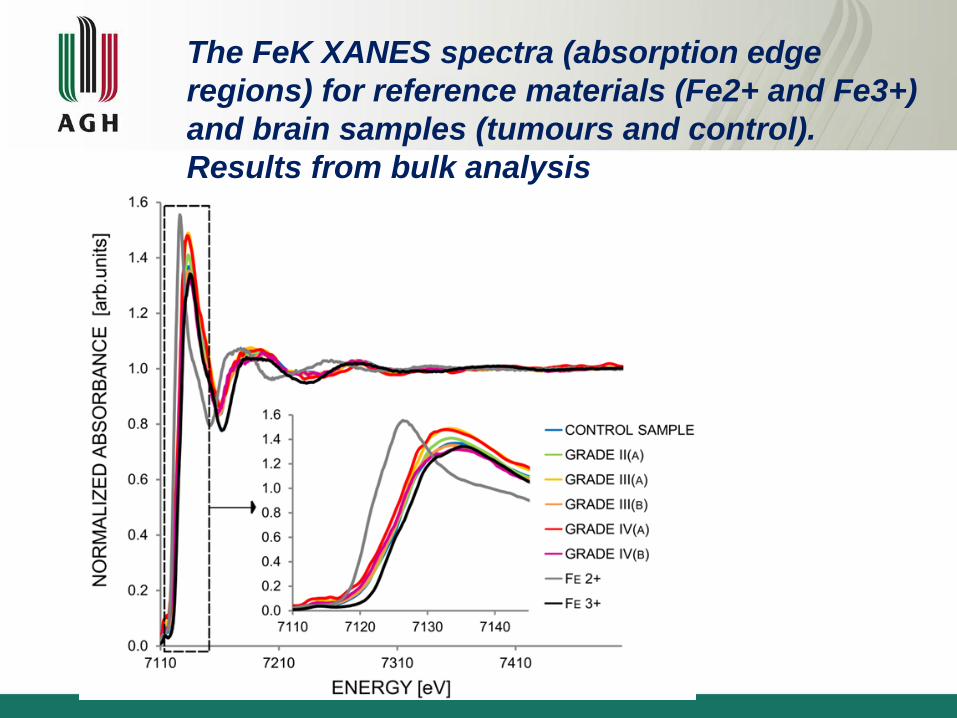
The FeK XANES spectra (absorption edge
regions) for reference materials (Fe2+ and Fe3+)
and brain samples (tumours and control).
Results from bulk analysis

Fe average oxidation state in neoplastic tissues
Absorption edge energies of Fe for various malignancy grade

35/25
Fourier transform of Fe EXAFS data for brain
tumor samples with various malignancy grades as a
function of radial coordinate

Fourier transform of Zn EXAFS data for brain tumor
samples with various malignancy grades as a function of
radial coordinate
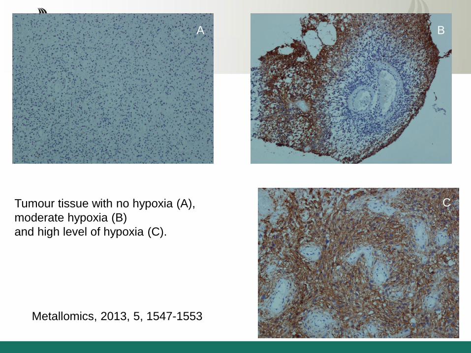
vessels are under hypoxia.
Tumour tissue with no hypoxia (A),
moderate hypoxia (B)
and high level of hypoxia (C).
A B
C
Metallomics, 2013, 5, 1547-1553
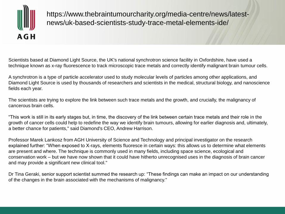
Scientists based at Diamond Light Source, the UK's national synchrotron science facility in Oxfordshire, have used a
technique known as x-ray fluorescence to track microscopic trace metals and correctly identify malignant brain tumour cells.
A synchrotron is a type of particle accelerator used to study molecular levels of particles among other applications, and
Diamond Light Source is used by thousands of researchers and scientists in the medical, structural biology, and nanoscience
fields each year.
The scientists are trying to explore the link between such trace metals and the growth, and crucially, the malignancy of
cancerous brain cells.
“This work is still in its early stages but, in time, the discovery of the link between certain trace metals and their role in the
growth of cancer cells could help to redefine the way we identify brain tumours, allowing for earlier diagnosis and, ultimately,
a better chance for patients," said Diamond's CEO, Andrew Harrison.
Professor Marek Lankosz from AGH University of Science and Technology and principal investigator on the research
explained further: “When exposed to X-rays, elements fluoresce in certain ways: this allows us to determine what elements
are present and where. The technique is commonly used in many fields, including space science, ecological and
conservation work – but we have now shown that it could have hitherto unrecognised uses in the diagnosis of brain cancer
and may provide a significant new clinical tool."
Dr Tina Geraki, senior support scientist summed the research up: “These findings can make an impact on our understanding
of the changes in the brain associated with the mechanisms of malignancy."
https://www.thebraintumourcharity.org/media-centre/news/latest-
news/uk-based-scientists-study-trace-metal-elements-ide/

Summary
The MDA based on the elemental composition of tissue (SRXRF)
may be a potentially valuable method in assisting the differentiation
and/or classification (diagnosis) of ovarian and brain tumors
including doubtful cases.
The external hybridization of images obtained from optical
microscopy of stained tissue, SR XRF elemental microscopy and IR
micro spectroscopy should be improved
The techniques based on SR for physicochemical characterization
of tissue samples (XANES, EXAFS) should be performed in cryo
conditions
XANES and EXAFS enable analysis of oxidation states and
chemical environment of Fe, Cu and Zn in tumors cells. Methods for
modelling of chemical environment and identification of proteins
binding Fe, Cu and Zn in cancer cells should be improved

Cooperation
1. Chair of Pathomorphology, CM UJ
Dariusz Adamek, Edyta Radwańska, Łukasz Chmura
Department of Gynecology and Oncology, CM UJ
Robert Jach
2. P06 at Petra III:
G. Falkenberg, M. Alfeld and U. Bösenberg
3. CEMO at DORIS III, P64 at Petra III
E. Welter, K. Apple
4. I18 at Diamond
T. Geraki, F. Mosselmann
5. BM23 at ESRF
Olivier Mathon, Sacura Pacarelli
6. ID21 at ESRF M. Salome, H. Castillo-Michel, B. Hesse and
G. Veronesi
7. FLUO at ANKA
Rolf Simon

Faculty of Physics and Applied Computer Sciences
Aleksandra Wandzilak
Paweł Wróbel
Mateusz Czyżycki
Maria Grzelak
Daria Krauze
Beata Ostachowicz
Zdzisław Stęgowski
Magdalena Szczerbowska-Boruchowska

DESY
I-20160422EC, I-20160038EC, I-20140190EC,
I-20140109EC, I-20120172EC
ESRF
MD935, MD726, MD676
DIAMOND
SP7553
ANKA
A2014-024-006633
Proposals

Acknowledgements:
The research leading to these results has received fundings from:
Diamond Light Source Ltd,, Didcot Oxfordshire,
European Synchrotron Radiation Facility, Grenoble, France,
Synchrotron Light Source ANKA
Photo Science DESY, Hamburg, Germany
Ministry of Science and Higher Education (Warsaw, Poland)
grant no W116/IAEA/2014, W57/IAEA/2015
IAEA Research Contract No. 18199 (2014-2018).

Thank you for your attention